

Engineering coordinated lymphatic–vascular responses with Haversian-mimetic IL-6-releasing scaffolds for bone regeneration

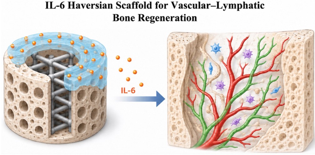
Bone regeneration requires coordinated vascularization and microenvironmental remodeling; however, most bone biomaterials have primarily emphasized angiogenesis and have less explicitly addressed lymphatic-associated responses. Here, inspired by developmental bone formation and emerging evidence implicating the interleukin-6 (IL-6)–CXCL12 axis in regenerative processes, we developed a biomimetic platform to promote bone repair, accompanied by coordinated blood-vascular and lymphatic-associated responses. A 3D-printed Haversian-mimetic scaffold with architecturally defined microchannels was fabricated to support guided tissue infiltration and vascular organization. To provide instructive biochemical signaling, a gelatin methacrylate (GelMA)-based coating enabled sustained, low-dose delivery of IL-6. Developmental immunofluorescence analysis revealed pronounced spatial association between CXCL12 and osteocalcin-positive osteogenic regions, providing a biological rationale for engaging this axis as a regeneration-associated cue. In vitro studies demonstrated enhanced osteogenic differentiation and concomitant upregulation of angiogenic and lymphatic-associated markers. Transcriptomic profiling further indicated activation of gene programs associated with extracellular matrix remodeling, vascular-related signaling, and tissue regeneration. In a critical-sized defect model, the IL-6-functionalized Haversian-mimetic scaffold promoted new bone deposition and was accompanied by increased CD31-positive vascular structures, lymphatic vessel endothelial hyaluronan receptor 1 (LYVE1)-positive lymphatic-associated structures, and CXCL12 expression. Collectively, this work demonstrates a strategy that integrates architectural mimicry with cytokine signaling to enhance bone regeneration while explicitly evaluating lymphatic-associated responses as a design parameter for next-generation bone biomaterials.
- Fu X, Luo Z, Guo Y, et al. Microenvironment-responsive multifunctional enzyme-linked hydrogel for diabetic bone defect regeneration. Nat Commun. 2025;16(1):10275. doi: 10.1038/s41467-025-65165-5
- Furuhashi K, Kakiuchi M, Ueda R, et al. Bone marrow niches orchestrate stem-cell hierarchy and immune tolerance. Nature. 2025;638(8049):206-215. doi: 10.1038/s41586-024-08352-6
- Biswas L, Chen J, De Angelis J, et al. Lymphatic vessels in bone support regeneration after injury. Cell. 2023;186(2):382-397.e24. doi: 10.1016/j.cell.2022.12.031
- Liang C, Gao Y, Li J, et al. Bioengineered human pre-vascularized microtissues reconstruct vascular niche and regenerative microenvironment to enhance functional skeletal muscle regeneration. Biomaterials. 2026;326:123645. doi: 10.1016/j.biomaterials.2025.123645
- Clark JR, Al Maruf DSA, Tomaskovic-Crook E, et al. Mechanobiologically-optimized non-resorbable artificial bone for patient-matched scaffold-guided bone regeneration. Nat Commun. 2025;16(1):9422. doi: 10.1038/s41467-025-64466-z
- Zhang M, Lin R, Wang X, et al. 3D printing of Haversian bone-mimicking scaffolds for multicellular delivery in bone regeneration. Sci Adv. 2020;6(12):eaaz6725. doi: 10.1126/sciadv.aaz6725
- Zhang H, Wu C. 3D printing of biomaterials for vascularized and innervated tissue regeneration. Int J Bioprint. 2023;9(3):706. doi: 10.18063/ijb.706
- Roohani-Esfahani SI, Newman P, Zreiqat H. Design and Fabrication of 3D printed Scaffolds with a Mechanical Strength Comparable to Cortical Bone to Repair Large Bone Defects. Sci Rep. 2016;6:19468. doi: 10.1038/srep19468
- Ng WL, Bartolo P. 3D bioprinting of tissues and organs for systemic diseases and localized injuries. Mil Med Res. 2026;13(1):100006. doi: 10.1016/j.mmr.2026.100006
- Qian J, Qin H, Su E, et al. Angiogenesis-osteogenesis coupling and anti-osteoclastogenesis zoledronate intermixed calcium silicate metal-organic/inorganic hybrid coating on biodegradable zinc-based intramedullary nails for osteoporotic fracture healing. Bioact Mater. 2025;44:46-67. doi: 10.1016/j.bioactmat.2024.09.041
- Zhang J, Tong D, Song H, et al. Osteoimmunity-Regulating Biomimetically Hierarchical Scaffold for Augmented Bone Regeneration. Adv Mater. 2022;34(36):e2202044. doi: 10.1002/adma.202202044
- Liu H, Wei J, Xiao S, et al. Regulation Energy Metabolism of Fiber Scaffolds Orchestrates Osteoimmunomodulation and Angio/Osteogenesis. Small. 2025;21(18):e2409747. doi: 10.1002/smll.202409747
- Wu H, Yang L, Luo R, et al. A drug-free cardiovascular stent functionalized with tailored collagen supports in-situ healing of vascular tissues. Nat Commun. 2024;15(1):735. doi: 10.1038/s41467-024-44902-2
- Zheng Y, Cong L, Zhao L, et al. Lymphatic platelet thrombosis limits bone repair by precluding lymphatic transporting DAMPs. Nat Commun. 2025;16(1):829. doi: 10.1038/s41467-025-56147-8
- Zhu C, Li Z, Qiu X, et al. A Functional 3D Biomimetic Osteon Accelerates Bone Regeneration. Adv Funct Mater. 2025;35(35):2500603. doi: 10.1002/adfm.202500603
- Cao H, Shi K, Long J, et al. PDGF-BB improves cortical bone quality through restoring the osteogenic microenvironment in the steroid-associated osteonecrosis of rabbits. J Orthop Translat. 2025;52:97-115. doi: 10.1016/j.jot.2025.03.010
- Ha Y, Ma X, Li S, et al. Bone Microenvironment-Mimetic Scaffolds with Hierarchical Microstructure for Enhanced Vascularization and Bone Regeneration. Adv Funct Mater. 2022;32(20):2200011. doi: 10.1002/adfm.202200011
- Lu G, Xu Y, Liu Q, et al. An instantly fixable and self-adaptive scaffold for skull regeneration by autologous stem cell recruitment and angiogenesis. Nat Commun. 2022;13(1):2499. doi: 10.1038/s41467-022-30243-5
- Zhang X, Jiang W, Wu X, et al. Divide-and-conquer strategy with engineered ossification center organoids for rapid bone healing through developmental cell recruitment. Nat Commun. 2025;16(1):6200. doi: 10.1038/s41467-025-61619-y
- Su Y, Wang H, Liu W, et al. Bioinspired Engineering of Streamlined Skeletal Interoception: Neural Bioprinted Piezoelectric Scaffolds for Neuro-Vascularized Bone Regeneration. Adv Sci. 2026;13(21):e24181. doi: 10.1002/advs.202524181
- Wang G, Liu R, Ma H, et al. Elevator-Like Hollow Channels in Porous Scaffolds Accelerate Vascularized Bone Regeneration via NETs-Fibrin-Mediated Macrophage Recruitment. Adv Sci. 2026;13(9):e15693. doi: 10.1002/advs.202515693
- Wu X, Wang F, Li R, et al. Enzyme-Programmable DNA-PEG Hydrogel Spatiotemporally Regulates Bone Regeneration Microenvironment. Adv Mater. 2026;38(6):e14461. doi: 10.1002/adma.202514461
- Abaurrea A, Araujo AM, Caffarel MM. The Role of the IL-6 Cytokine Family in Epithelial-Mesenchymal Plasticity in Cancer Progression. Int J Mol Sci. 2021;22(15):8334. doi: 10.3390/ijms22158334
- Hu X, Li J, Fu M, Zhao X, Wang W. The JAK/STAT signaling pathway: from bench to clinic. Signal Transduct Target Ther. 2021;6(1):402. doi: 10.1038/s41392-021-00791-1
- Naba A. Mechanisms of assembly and remodelling of the extracellular matrix. Nat Rev Mol Cell Biol. 2024;25(11):865-885. doi: 10.1038/s41580-024-00767-3
- Pittenger MF, Discher DE, Péault BM, Phinney DG, Hare JM, Caplan AI. Mesenchymal stem cell perspective: cell biology to clinical progress. NPJ Regen Med. 2019;4:22. doi: 10.1038/s41536-019-0083-6
- Novoseletskaya ES, Evdokimov PV, Efimenko AY. Extracellular matrix-induced signaling pathways in mesenchymal stem/stromal cells. Cell Commun Signal. 2023;21(1):244. doi: 10.1186/s12964-023-01252-8
- Barnett SN, Cujba AM, Yang L, et al. An organotypic atlas of human vascular cells. Nat Med. 2024;30(12):3468-3481. doi: 10.1038/s41591-024-03376-x
- Huang J, Liao C, Yang J, Zhang L. The role of vascular and lymphatic networks in bone and joint homeostasis and pathology. Front Endocrinol. 2024;15:1465816. doi: 10.3389/fendo.2024.1465816
- Wang L, You X, Zhang L, Zhang C, Zou W. Mechanical regulation of bone remodeling. Bone Res. 2022;10(1):16. doi: 10.1038/s41413-022-00190-4
- Hu J, Liao H, Ma Z, et al. Focal Adhesion Kinase Signaling Mediated the Enhancement of Osteogenesis of Human Mesenchymal Stem Cells Induced by Extracorporeal Shockwave. Sci Rep. 2016;6:20875. doi: 10.1038/srep20875
- Ma C, Du T, Niu X, Fan Y. Biomechanics and mechanobiology of the bone matrix. Bone Res. 2022;10(1):59. doi: 10.1038/s41413-022-00223-y
- Hu Z, Zhao X, Wu Z, et al. Lymphatic vessel: origin, heterogeneity, biological functions, and therapeutic targets. Signal Transduct Target Ther. 2024;9(1):9. doi: 10.1038/s41392-023-01723-x
- Wei ST, Huang YC, Hsieh ML, et al. Atypical chemokine receptor ACKR3/CXCR7 controls postnatal vasculogenesis and arterial specification by mesenchymal stem cells via Notch signaling. Cell Death Dis. 2020;11(5):307. doi: 10.1038/s41419-020-2512-2
- Russell PS, Velivolu R, Maldonado Zimbrón VE, et al. Fluorescent Tracers for In Vivo Imaging of Lymphatic Targets. Front Pharmacol. 2022;13:952581. doi: 10.3389/fphar.2022.952581
- Zhang Q, Niu Y, Li Y, et al. Meningeal lymphatic drainage: novel insights into central nervous system disease. Signal Transduct Target Ther. 2025;10(1):142. doi: 10.1038/s41392-025-02177-z
- Mountziaris PM, Spicer PP, Kasper FK, Mikos AG. Harnessing and modulating inflammation in strategies for bone regeneration. Tissue Eng Part B Rev. 2011;17(6):393-402. doi: 10.1089/ten.TEB.2011.0182
- Mountziaris PM, Mikos AG. Modulation of the inflammatory response for enhanced bone tissue regeneration. Tissue Eng Part B Rev. 2008;14(2):179-186. doi: 10.1089/ten.teb.2008.0038
- Song P, Zhou D, Wang F, Li G, Bai L, Su J. Programmable biomaterials for bone regeneration. Mater Today Bio. 2024;29:101296. doi: 10.1016/j.mtbio.2024.101296