

Development and biological validation of a dexamethasone-loaded, polylactic acid-based 3D-printable composite filament for bone tissue engineering

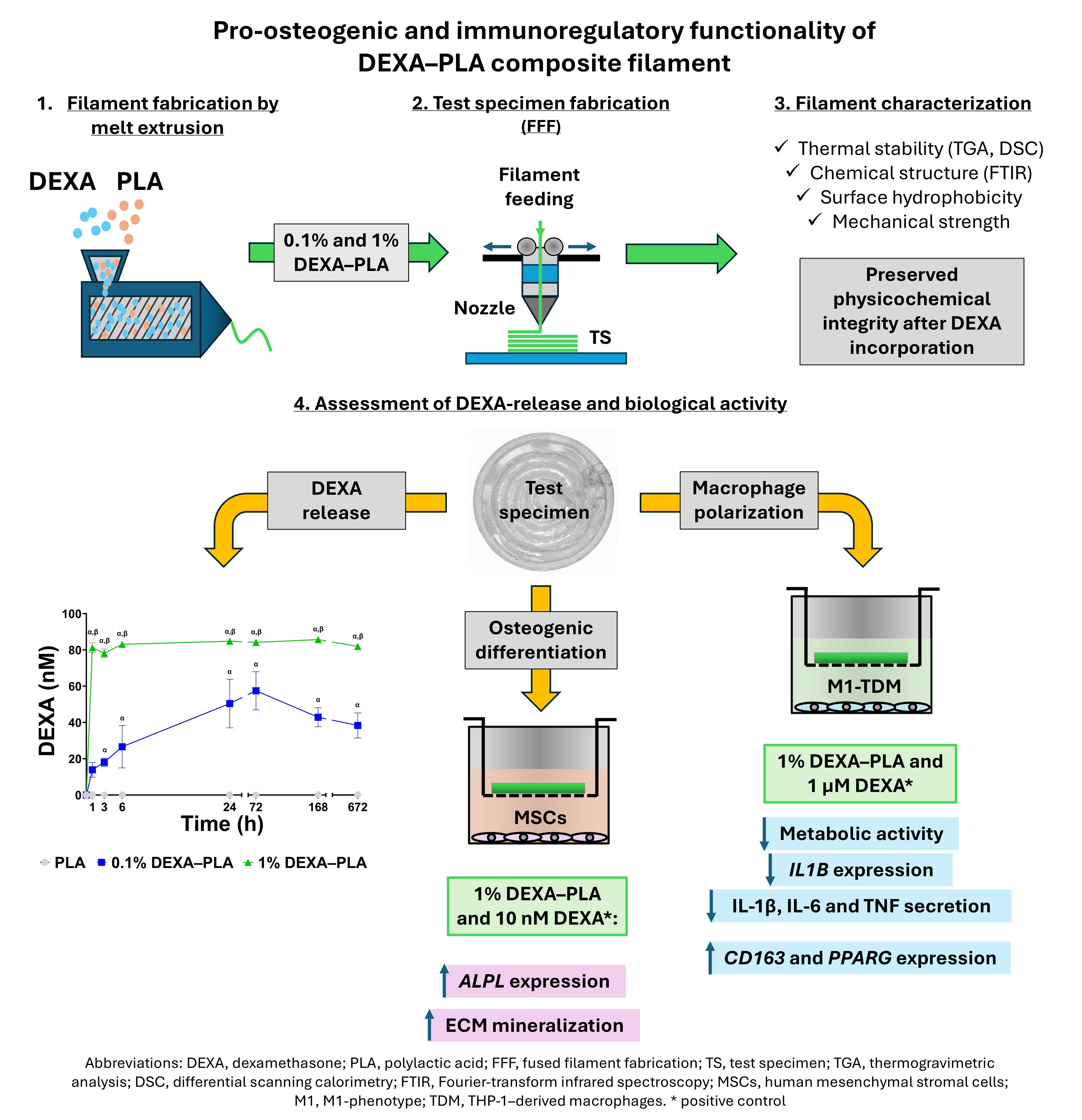
Critical-sized bone defects remain a major clinical challenge, motivating the development of biomaterial-based alternatives to autografting. Among additive manufacturing techniques, fused filament fabrication offers a scalable approach for producing patient-specific scaffolds with integrated bioactive functionality. This study aimed to fabricate and evaluate a dexamethasone (DEXA)-loaded polylactic acid (PLA) filament for controlled local drug delivery through fused filament fabrication. DEXA–PLA composite filaments containing 0.1% and 1% (w/w) DEXA were produced via melt extrusion and characterized in terms of their thermal, chemical, surface, and mechanical properties. Drug release was quantified over 28 days, and biological performance was evaluated using human mesenchymal stromal cells (MSCs) and THP-1-derived macrophages. DEXA incorporation preserved the physicochemical properties of PLA. The 0.1% formulation exhibited a transient release profile, whereas the 1% DEXA–PLA formulation provided sustained release over 28 days. MSC metabolic activity was unaffected by either formulation. However, the 1% DEXA–PLA formulation significantly enhanced osteogenic differentiation, as indicated by increased ALPL expression and extracellular matrix mineralization, with levels comparable to those observed in cells cultured in standard osteogenic medium. In M1 macrophages, the 1% formulation decreased metabolic activity and IL1B expression and reduced the secretion of IL-1β, IL-6, and TNF-α. It also increased the expression of the anti-inflammatory markers CD163 and PPARG. In conclusion, the 1% DEXA–PLA filament maintained material integrity and enabled sustained delivery of bioactive DEXA, promoting both osteogenic and immunomodulatory responses. These findings demonstrate the feasibility of incorporating DEXA into a controlled-release system for bone tissue engineering applications.
- Oryan A, Alidadi S, Moshiri A, Maffulli N. Bone regenerative medicine: classic options, novel strategies, and future directions. J Orthop Surg Res. 2014;9:18. doi: 10.1186/1749-799X-9-18
- Tsang STJ, van Rensburg AJ, van Heerden J, et al. The management of critical bone defects: outcomes of a systematic approach. Eur J Orthop Surg Traumatol. 2024;34(6):3225-3231. doi: 10.1007/s00590-024-04050-1
- O'Brien FJ. Biomaterials & scaffolds for tissue engineering. Mater Today. 2011;14(3):88-95. doi: 10.1016/S1369-7021(11)70058-X
- Martin I, Simmons PJ, Williams DF. Manufacturing challenges in regenerative medicine. Sci Transl Med. 2014;6(232):232fs16. doi: 10.1126/scitranslmed.3008558
- Grivet-Brancot A, Boffito M, Ciardelli G. Use of polyesters in fused deposition modeling for biomedical applications. Macromol Biosci. 2022;22(10):2200039. doi: 10.1002/mabi.202200039
- Neijhoft J, Ijpma FFA. Advances of 3D printing technologies in orthopaedic trauma and surgical training: a transformative approach. Eur J Trauma Emerg Surg. 2024;50(1):1-2. doi: 10.1007/s00068-024-02464-4
- Bose S, Roy M, Bandyopadhyay A. Recent advances in bone tissue engineering scaffolds. Trends Biotechnol. 2012;30(10):546-554. doi: 10.1016/j.tibtech.2012.07.005
- Costa PF, Puga AM, Díaz-Gomez L, et al. Additive manufacturing of scaffolds with dexamethasone controlled release for enhanced bone regeneration. Int J Pharm. 2015;496(2):541-550. doi: 10.1016/j.ijpharm.2015.10.055
- Almeida CR, Serra T, Oliveira MI, Planell JA, Barbosa MA, Navarro M. Impact of 3-D printed PLA- and chitosan-based scaffolds on human monocyte/macrophage responses: unraveling the effect of 3-D structures on inflammation. Acta Biomater. 2014;10(2):613-622. doi: 10.1016/j.actbio.2013.10.035
- Serra T, Ortiz-Hernandez M, Engel E, Planell JA, Navarro M. Relevance of PEG in PLA-based blends for tissue engineering 3D-printed scaffolds. Mater Sci Eng C Mater Biol Appl. 2014;38:55-62. doi: 10.1016/j.msec.2014.01.003
- Shaqour B, Samaro A, Verleije B, et al. Production of drug delivery systems using fused filament fabrication: a systematic review. Pharmaceutics. 2020;12(6):517. doi: 10.3390/pharmaceutics12060517
- Lima AC, Puga AM, Mano JF, Concheiro A, Alvarez-Lorenzo C. Free and copolymerized γ-cyclodextrins regulate the performance of dexamethasone-loaded dextran microspheres for bone regeneration. J Mater Chem B. 2014;2(30):4943-4956. doi: 10.1039/c3tb21665a
- Coutinho AE, Chapman KE. The anti-inflammatory and immunosuppressive effects of glucocorticoids, recent developments and mechanistic insights. Mol Cell Endocrinol. 2011;335(1):2-13. doi: 10.1016/j.mce.2010.04.005
- Kósa JP, Kis A, Bácsi K, et al. The protective role of bone morphogenetic protein-8 in the glucocorticoid-induced apoptosis on bone cells. Bone. 2011;48(5):1052-1057. doi: 10.1016/j.bone.2011.01.017
- Jaiswal N, Haynesworth SE, Caplan AI, Bruder SP. Osteogenic differentiation of purified, culture-expanded human mesenchymal stem cells in vitro. J Cell Biochem. 1997;64(2):295-312. doi: 10.1002/(SICI)1097-4644(199702)64:2<295::AID-JCB12>3.0.CO;2-I
- Lane NE. An update on glucocorticoid-induced osteoporosis. Rheum Dis Clin North Am. 2001;27(1):235-253. doi: 10.1016/S0889-857X(05)70196-4
- Walsh S, Jordan GR, Jefferiss C, Stewart K, Beresford JN. High concentrations of dexamethasone suppress the proliferation but not the differentiation or further maturation of human osteoblast precursors in vitro: relevance to glucocorticoid-induced osteoporosis. Rheumatology (Oxford). 2001;40(1):74-83. doi: 10.1093/rheumatology/40.1.74
- DeConde AS, Sidell D, Lee M, et al. Bone morphogenetic protein-2–impregnated biomimetic scaffolds successfully induce bone healing in a marginal mandibular defect. Laryngoscope. 2013;123(5):1149-1155. doi: 10.1002/lary.23782
- Zhang ZZ, Zhang HZ, Zhang ZY. 3D printed poly(ε-caprolactone) scaffolds function with simvastatin-loaded poly(lactic-co-glycolic acid) microspheres to repair load-bearing segmental bone defects. Exp Ther Med. 2019;17(1):79-90. doi: 10.3892/etm.2018.6947
- Pérez-Davila S, Potel-Alvarellos C, Carballo R, et al. Vancomycin-loaded 3D-printed polylactic acid-hydroxyapatite scaffolds for bone tissue engineering. Polymers (Basel). 2023;15(21):4250. doi: 10.3390/polym15214250
- Lee JH, Baik JM, Yu YS, et al. Development of a heat labile antibiotic eluting 3D printed scaffold for the treatment of osteomyelitis. Sci Rep. 2020;10(1):7554. doi: 10.1038/s41598-020-64573-5
- Cichos S, Schätzlein E, Wiesmann-Imilowski N, et al. A new 3D-printed polylactic acid-bioglass composite for bone tissue engineering induces angiogenesis in vitro and in ovo. Int J Bioprint. 2023;9(5):751. doi: 10.18063/ijb.751
- Schätzlein E, Kicker C, Söhling N, et al. 3D-printed PLA-bioglass scaffolds with controllable calcium release and MSC adhesion for bone tissue engineering. Polymers (Basel). 2022;14(12):2389. doi: 10.3390/polym14122389
- Söhling N, Al Zoghool S, Schätzlein E, et al. In vitro evaluation of a 20% bioglass-containing 3D printable PLA composite for bone tissue engineering. Int J Bioprint. 2022;8(4):602. doi: 10.18063/ijb.v8i4.602
- Li X, Wang Y, Wang Z, et al. Composite PLA/PEG/nHA/dexamethasone scaffold prepared by 3D printing for bone regeneration. Macromol Biosci. 2018;18(6):1800068. doi: 10.1002/mabi.201800068
- Farto-Vaamonde X, Auriemma G, Aquino RP, et al. Post-manufacture loading of filaments and 3D printed PLA scaffolds with prednisolone and dexamethasone for tissue regeneration applications. Eur J Pharm Biopharm. 2019;141:100-110. doi: 10.1016/j.ejpb.2019.05.018
- Chen CH, Dash BS, Ting WC, et al. Bone tissue engineering with adipose-derived stem cells in polycaprolactone/graphene oxide/dexamethasone 3D-printed scaffolds. ACS Biomater Sci Eng. 2024;10(10):6425-6440. doi: 10.1021/acsbiomaterials.4c00774
- Moyadee C, Chaloeijitkul K, Wadaugsorn K, et al. Influence of molecular weight on functional performance and migration of PLA food-contact films. Polym Eng Sci. 2026;66(6):4422-4430. doi: 10.1002/pen.70510
- Backes EH, Pires LDN, Costa LC, Passador FR, Pessan LA. Analysis of the degradation during melt processing of PLA/biosilicate composites. J Compos Sci. 2019;3(2):52. doi: 10.3390/jcs3020052
- Neijhoft J, Weslati H, Eras V, et al. Granulate-to-filament: an extrusion-mixed PLA–human bone material system for 3D-printed bone scaffolds. J Funct Biomater. 2026;17(4):187. doi: 10.3390/jfb17040187
- dos Santos J, Balbinot GDS, Buchner S, et al. 3D printed matrix solid forms: can the drug solubility and dose customisation affect their controlled release behaviour? Int J Pharm X. 2023;5:100153. doi: 10.1016/j.ijpx.2022.100153
- International Organization for Standardization. Plastics—Thermogravimetry (TG) of Polymers—Part 1: General Principles. ISO 11358-1:2022. Geneva, Switzerland: International Organization for Standardization; 2022.
- International Organization for Standardization. Plastics—Differential Scanning Calorimetry (DSC)—Part 1: General Principles. ISO 11357-1:2023. Geneva, Switzerland: International Organization for Standardization; 2023.
- International Organization for Standardization. Plastics—Determination of Dynamic Mechanical Properties—Part 1: General Principles. ISO 6721-1:2019. Geneva, Switzerland: International Organization for Standardization; 2019.
- McKinney W. Data structures for statistical computing in Python. In: van der Walt S, Millman J, eds. Proceedings of the 9th Python in Science Conference. 2010:56-61. doi: 10.25080/Majora-92bf1922-00a
- Harris CR, Millman KJ, van der Walt SJ, et al. Array programming with NumPy. Nature. 2020;585(7825):357-362. doi: 10.1038/s41586-020-2649-2
- Erb D. pybaselines: a Python library of algorithms for the baseline correction of experimental data. Zenodo. Published 2022. doi: 10.5281/zenodo.7255880
- Hunter JD. Matplotlib: a 2D graphics environment. Comput Sci Eng. 2007;9(3):90-95. doi: 10.1109/MCSE.2007.55
- International Organization for Standardization. Plastics—Determination of Flexural Properties. ISO 178:2019. Geneva, Switzerland: International Organization for Standardization; 2019.
- Langenbach F, Handschel J. Effects of dexamethasone, ascorbic acid and β-glycerophosphate on the osteogenic differentiation of stem cells in vitro. Stem Cell Res Ther. 2013;4(5):117. doi: 10.1186/scrt328
- Park EK, Jung HS, Yang HI, et al. Optimized THP-1 differentiation is required for the detection of responses to weak stimuli. Inflamm Res. 2007;56(1):45-50. doi: 10.1007/s00011-007-6115-5
- Bianconi S, Leppik L, Oppermann E, et al. Direct current electrical stimulation shifts THP-1-derived macrophage polarization towards pro-regenerative M2 phenotype. Int J Mol Sci. 2024;25(13):7272. doi: 10.3390/ijms25137272
- Knijff EM, Breunis MN, van Geest MC, et al. A relative resistance of T cells to dexamethasone in bipolar disorder. Bipolar Disord. 2006;8(6):740-750. doi: 10.1111/j.1399-5618.2006.00359.x
- Vandesompele J, De Preter K, Pattyn F, et al. Accurate normalization of real-time quantitative RT-PCR data by geometric averaging of multiple internal control genes. Genome Biol. 2002;3(7):research0034. doi: 10.1186/gb-2002-3-7-research0034
- Livak KJ, Schmittgen TD. Analysis of relative gene expression data using real-time quantitative PCR and the 2−ΔΔCt method. Methods. 2001;25(4):402-408. doi: 10.1006/meth.2001.1262
- Wickramasinghe S, Do T, Tran P. FDM-based 3D printing of polymer and associated composite: a review on mechanical properties, defects and treatments. Polymers (Basel). 2020;12(7):1529. doi: 10.3390/polym12071529
- Kervran M, Vagner C, Cochez M, et al. Thermal degradation of polylactic acid (PLA)/polyhydroxybutyrate (PHB) blends: a systematic review. Polym Degrad Stab. 2022;201:109995. doi: 10.1016/j.polymdegradstab.2022.109995
- Svoboda R, Vrbenský R, Honzíček J, Chromčíková M. Thermal stability of dexamethasone—evaluation with regard to modern medicinal and pharmaceutical 3D-printing applications. Molecules. 2025;30(21):4234. doi: 10.3390/molecules30214234
- Abas M, Habib T, Noor S, et al. Parametric investigation and optimization to study the effect of process parameters on the dimensional deviation of fused deposition modeling of 3D printed parts. Polymers (Basel). 2022;14(17):3667. doi: 10.3390/polym14173667
- Feng Y, Ma P, Xu P, et al. The crystallization behavior of poly(lactic acid) with different types of nucleating agents. Int J Biol Macromol. 2018;106:955-962. doi: 10.1016/j.ijbiomac.2017.08.095
- Cristea M, Ionita D, Iftime MM. Dynamic mechanical analysis investigations of PLA-based renewable materials: how are they useful? Materials (Basel). 2020;13(22):5302. doi: 10.3390/ma13225302
- Leroy A, Ribeiro S, Grossiord C, et al. FTIR microscopy contribution for comprehension of degradation mechanisms in PLA-based implantable medical devices. J Mater Sci Mater Med. 2017;28(6):87. doi: 10.1007/s10856-017-5894-7
- Rasselet D, Ruellan A, Guinault A, et al. Oxidative degradation of polylactide (PLA) and its effects on physical and mechanical properties. Eur Polym J. 2014;50:109-116. doi: 10.1016/j.eurpolymj.2013.10.011
- Maia FR, Bidarra SJ, Granja PL, Barrias CC. Functionalization of biomaterials with small osteoinductive moieties. Acta Biomater. 2013;9(11):8773-8789. doi: 10.1016/j.actbio.2013.08.004
- Majrashi M, Kotowska A, Scurr D, et al. Sustained release of dexamethasone from 3D-printed scaffolds modulates macrophage activation and enhances osteogenic differentiation. ACS Appl Mater Interfaces. 2023;15(49):56623-56638. doi: 10.1021/acsami.3c09774
- Rampersad SN. Multiple applications of Alamar Blue as an indicator of metabolic function and cellular health in cell viability bioassays. Sensors (Basel). 2012;12(9):12347-12360. doi: 10.3390/s120912347
- Ma L, Feng X, Wang K, et al. Dexamethasone promotes mesenchymal stem cell apoptosis and inhibits osteogenesis by disrupting mitochondrial dynamics. FEBS Open Bio. 2020;10(2):211-220. doi: 10.1002/2211-5463.12771
- Freeman FE, Stevens HY, Owens P, Guldberg RE, McNamara LM. Osteogenic differentiation of mesenchymal stem cells by mimicking the cellular niche of the endochondral template. Tissue Eng Part A. 2016;22(19-20):1176-1190. doi: 10.1089/ten.TEA.2015.0339
- Alm JJ, Heino TJ, Hentunen TA, Väänänen HK, Aro HT. Transient 100 nM dexamethasone treatment reduces inter- and intraindividual variations in osteoblastic differentiation of bone marrow-derived human mesenchymal stem cells. Tissue Eng Part C Methods. 2012;18(9):658-666. doi: 10.1089/ten.TEC.2011.0675
- Hong L, Sultana H, Paulius K, Zhang G. Steroid regulation of proliferation and osteogenic differentiation of bone marrow stromal cells: a gender difference. J Steroid Biochem Mol Biol. 2009;114(3-5):180-185. doi: 10.1016/j.jsbmb.2009.02.001
- Xiao Y, Peperzak V, van Rijn L, Borst J, de Bruijn JD. Dexamethasone treatment during the expansion phase maintains stemness of bone marrow mesenchymal stem cells. J Tissue Eng Regen Med. 2010;4(5):374-386. doi: 10.1002/term.250
- Baron R, Kneissel M. WNT signaling in bone homeostasis and disease: from human mutations to treatments. Nat Med. 2013;19(2):179-192. doi: 10.1038/nm.3074
- Morimoto E, Li M, Khalid AB, et al. Glucocorticoids hijack RUNX2 to stimulate WIF1 for suppression of osteoblast growth and differentiation. J Cell Physiol. 2017;232(1):145-153. doi: 10.1002/jcp.25399
- de Melo Pereira D, Eischen-Loges M, Birgani ZT, Habibovic P. Proliferation and osteogenic differentiation of hMSCs on biomineralized collagen. Front Bioeng Biotechnol. 2020;8:554565. doi: 10.3389/fbioe.2020.554565
- Kim BB, Kim M, Park YH, et al. Short-term application of dexamethasone on stem cells derived from human gingiva reduces the expression of RUNX2 and β-catenin. J Int Med Res. 2017;45(3):993-1006. doi: 10.1177/0300060517701035
- Franceschi RT, Xiao G. Regulation of the osteoblast-specific transcription factor, Runx2: responsiveness to multiple signal transduction pathways. J Cell Biochem. 2003;88(3):446-454. doi: 10.1002/jcb.10369
- Koromila T, Baniwal SK, Song YS, et al. Glucocorticoids antagonize RUNX2 during osteoblast differentiation in cultures of ST2 pluripotent mesenchymal cells. J Cell Biochem. 2014;115(1):27-33. doi: 10.1002/jcb.24646
- Golub EE, Boesze-Battaglia K. The role of alkaline phosphatase in mineralization. Curr Opin Orthop. 2007;18(5):444-448. doi: 10.1097/bco.0b013e3282630851
- Millán JL. The role of phosphatases in the initiation of skeletal mineralization. Calcif Tissue Int. 2013;93(4):299-306. doi: 10.1007/s00223-012-9672-8
- Advani S, LaFrancis D, Bogdanovic E, Taxel P, Raisz LG, Kream BE. Dexamethasone suppresses in vivo levels of bone collagen synthesis in neonatal mice. Bone. 1997;20(1):41-46. doi: 10.1016/S8756-3282(96)00314-6
- Iu MF, Kaji H, Sowa H, et al. Dexamethasone suppresses Smad3 pathway in osteoblastic cells. J Endocrinol. 2005;185(1):131-138. doi: 10.1677/joe.1.05962
- Schlesinger PH, Blair HC, Beer Stolz D, et al. Cellular and extracellular matrix of bone, with principles of synthesis and dependency of mineral deposition on cell membrane transport. Am J Physiol Cell Physiol. 2020;318(1):C111-C124. doi: 10.1152/ajpcell.00120.2019
- Umrath F, Pfeifer A, Cen W, et al. How osteogenic is dexamethasone? Effect of the corticosteroid on the osteogenesis, extracellular matrix, and secretion of osteoclastogenic factors of jaw periosteum-derived mesenchymal stem/stromal cells. Front Cell Dev Biol. 2022;10:953516. doi: 10.3389/fcell.2022.953516
- Warmink K, Lijnzaad P, Schubert SA, et al. Mechanisms of dexamethasone-induced bone toxicity in developing bone: a single-cell perspective. JBMR Plus. 2025;9(10):ziaf146. doi: 10.1093/jbmrpl/ziaf146
- Si J, Wang C, Zhang D, et al. Osteopontin in bone metabolism and bone diseases. Med Sci Monit. 2020;26:e919159. doi: 10.12659/msm.919159
- Icer MA, Gezmen-Karadag M. The multiple functions and mechanisms of osteopontin. Clin Biochem. 2018;59:17-24. doi: 10.1016/j.clinbiochem.2018.07.003
- Shamekh MM, Lindqvist B, Nalvarte I. Dexamethasone drives macrophage repolarization linked to increased triple-negative breast cancer aggressiveness. Cell Death Dis. 2026;17:114. doi: 10.1038/s41419-025-08363-9
- Stifel U, Wolfschmitt EM, Vogt J, et al. Glucocorticoids coordinate macrophage metabolism through the regulation of the tricarboxylic acid cycle. Mol Metab. 2022;57:101424. doi: 10.1016/j.molmet.2021.101424
- Thong L, Sandby Thomas O, Ó Gallchobhair O, et al. Dexamethasone inhibits Mycobacterium tuberculosis–induced glycolysis but preserves antimicrobial function in primary human macrophages. Sci Rep. 2025;15(1):34310. doi: 10.1038/s41598-025-20188-2
- Viola A, Munari F, Sánchez-Rodríguez R, Scolaro T, Castegna A. The metabolic signature of macrophage responses. Front Immunol. 2019;10:1462. doi: 10.3389/fimmu.2019.01462
- Mosser DM, Edwards JP. Exploring the full spectrum of macrophage activation. Nat Rev Immunol. 2008;8(12):958-969. doi: 10.1038/nri2448
- Martinez FO, Gordon S. The M1 and M2 paradigm of macrophage activation: time for reassessment. F1000Prime Rep. 2014;6:13. doi: 10.12703/P6-13
- De Bosscher K, Schmitz ML, Vanden Berghe W, Plaisance S, Fiers W, Haegeman G. Glucocorticoid-mediated repression of nuclear factor-κB-dependent transcription involves direct interference with transactivation. Proc Natl Acad Sci USA. 1997;94(25):13504-13509. doi: 10.1073/pnas.94.25.13504
- De Bosscher K, Vanden Berghe W, Vermeulen L, Plaisance S, Boone E, Haegeman G. Glucocorticoids repress NF-κB-driven genes by disturbing the interaction of p65 with the basal transcription machinery, irrespective of coactivator levels in the cell. Proc Natl Acad Sci USA. 2000;97(8):3919-3924. doi: 10.1073/pnas.97.8.3919
- Jeon YJ, Han SH, Lee YW, Lee M, Yang KH, Kim HM. Dexamethasone inhibits IL-1β gene expression in LPS-stimulated RAW 264.7 cells by blocking NF-κB/Rel and AP-1 activation. Immunopharmacology. 2000;48(2):173-183. doi: 10.1016/S0162-3109(00)00199-5
- Schaer DJ, Boretti FS, Schoedon G, Schaffner A. Induction of the CD163-dependent haemoglobin uptake by macrophages as a novel anti-inflammatory action of glucocorticoids. Br J Haematol. 2002;119(1):239-243. doi: 10.1046/j.1365-2141.2002.03790.x
- Takenouchi T, Morozumi T, Wada E, et al. Dexamethasone enhances CD163 expression in porcine IPKM immortalized macrophages. In Vitro Cell Dev Biol Anim. 2021;57(1):10-16. doi: 10.1007/s11626-020-00535-5
- Iraji Asiabadi A, Esmaeil N, Zargar Kharazi A, et al. Harnessing IL-10-induced anti-inflammatory response in maturing macrophages in presence of electrospun dexamethasone-loaded PLLA scaffold. J Biomed Mater Res B Appl Biomater. 2024;112(6):e35411. doi: 10.1002/jbm.b.35411
- Peranteau WH, Zhang L, Muvarak N, et al. IL-10 overexpression decreases inflammatory mediators and promotes regenerative healing in an adult model of scar formation. J Invest Dermatol. 2008;128(7):1852-1860. doi: 10.1038/sj.jid.5701232
- Sziksz E, Pap D, Lippai R, et al. Fibrosis-related inflammatory mediators: role of the IL-10 cytokine family. Mediators Inflamm. 2015;2015:764641. doi: 10.1155/2015/764641
- Cao S, Zhang X, Edwards JP, Mosser DM. NF-κB1 (p50) homodimers differentially regulate pro- and anti-inflammatory cytokines in macrophages. J Biol Chem. 2006;281(36):26041-26050. doi: 10.1074/jbc.M602222200
- Saraiva M, O'Garra A. The regulation of IL-10 production by immune cells. Nat Rev Immunol. 2010;10(3):170-181. doi: 10.1038/nri2711
- Boutilier AJ, Elsawa SF. Macrophage polarization states in the tumor microenvironment. Int J Mol Sci. 2021;22(13):6995. doi: 10.3390/ijms22136995
- Li R, Ye JJ, Gan L, et al. Traumatic inflammatory response: pathophysiological role and clinical value of cytokines. Eur J Trauma Emerg Surg. 2024;50(4):1313-1330. doi: 10.1007/s00068-023-02388-5