

Bioprinted biomaterials for tendon repair and regeneration: Material systems and functional strategies

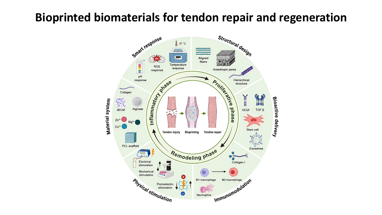
Tendon injuries often heal slowly because of the limited vascularity, low cellularity, and low metabolic activity of native tendon tissue. Newly formed tendon tissue is frequently characterized by disorganized collagen alignment, reduced mechanical performance, and scar-like remodeling, making it difficult to restore the structure and function of native tendon. Bioprinting enables precise control over material composition, spatial architecture, fiber orientation, and the distribution of cells and bioactive components, thereby providing a promising strategy for functional tendon regeneration. However, tendon is a highly load-bearing and anisotropic tissue, whereas most hydrogel-based bioinks, despite their favorable cytocompatibility and cell-loading capacity, often remain biomechanically weak, with insufficient tensile strength, limited fatigue stability, and inadequate long-term shape fidelity, making them insufficient to independently meet the biomechanical requirements of tendon repair. This review summarizes natural and modified natural bioinks, synthetic reinforcing materials, printed load-bearing frameworks, and inorganic or ion-releasing functional components for tendon bioprinting, with emphasis on printability, biocompatibility, mechanical reinforcement, degradation behavior, and structural stability. We further discuss functional strategies involving structural and topographical modulation, bioactive molecule delivery, immunomodulatory regulation, physical stimulation, and smart responsiveness. Future studies should prioritize multi-material cooperative printing, tendon–bone interface gradient construction, responsive bioink development, long-term in vivo functional evaluation, and translational validation to advance tendon bioprinting materials toward functional regenerative applications.
- Benjamin M, Kaiser E, Milz S. Structure-function relationships in tendons: a review. J Anat. 2008;212(3):211-228. doi: 10.1111/j.1469-7580.2008.00864.x
- Sharma P, Maffulli N. Tendon injury and tendinopathy: healing and repair. J Bone Joint Surg Am. 2005;87(1):187-202. doi: 10.2106/JBJS.D.01850
- Voleti PB, Buckley MR, Soslowsky LJ. Tendon healing: repair and regeneration. Annu Rev Biomed Eng. 2012;14(1):47-71. doi: 10.1146/annurev-bioeng-071811-150122
- Snedeker JG, Foolen J. Tendon injury and repair - A perspective on the basic mechanisms of tendon disease and future clinical therapy. Acta Biomater. 2017;63:18-36. doi: 10.1016/j.actbio.2017.08.032
- Darrieutort-Laffite C, Blanchard F, Soslowsky LJ, Le Goff B. Biology and physiology of tendon healing. Joint Bone Spine. 2024;91(5):105696. doi: 10.1016/j.jbspin.2024.105696
- Arvind V, Huang AH. Reparative and Maladaptive Inflammation in Tendon Healing. Front Bioeng Biotechnol. 2021;9:719047. doi: 10.3389/fbioe.2021.719047
- Chisari E, Rehak L, Khan WS, Maffulli N. The role of the immune system in tendon healing: a systematic review. Br Med Bull. 2020;133(1):49-64. doi: 10.1093/bmb/ldz040
- Wang Y, Lu X, Lu J, Hernigou P, Jin F. The role of macrophage polarization in tendon healing and therapeutic strategies: Insights from animal models. Front Bioeng Biotechnol. 2024;12:1366398. doi: 10.3389/fbioe.2024.1366398
- Bi Y, Ehirchiou D, Kilts TM, et al. Identification of tendon stem/progenitor cells and the role of the extracellular matrix in their niche. Nat Med. 2007;13(10):1219-1227. doi: 10.1038/nm1630
- Lu J, Chen H, Lyu K, et al. The Functions and Mechanisms of Tendon Stem/Progenitor Cells in Tendon Healing. Stem Cells Int. 2023;2023:1-8. doi: 10.1155/2023/1258024
- Jiang L, Lu J, Chen Y, et al. Mesenchymal stem cells: An efficient cell therapy for tendon repair (Review). Int J Mol Med. 2023;52(2). doi: 10.3892/ijmm.2023.5273
- Ruiz-Alonso S, Lafuente-Merchan M, Ciriza J, Saenz-Del-Burgo L, Pedraz JL. Tendon tissue engineering: Cells, growth factors, scaffolds and production techniques. J Control Release. 2021;333:448-486. doi: 10.1016/j.jconrel.2021.03.040
- Mandrycky C, Wang Z, Kim K, Kim DH. 3D bioprinting for engineering complex tissues. Biotechnol Adv. 2016;34(4):422-434. doi: 10.1016/j.biotechadv.2015.12.011
- Bai X, Yang Y, Chu J, Deng Y, Li M, Yang H. 3D bioprinting patient-specific grafts for tendon/ligament repair in motion: emerging trends and challenges. Front Bioeng Biotechnol. 2025;13:1643430. doi: 10.3389/fbioe.2025.1643430
- Park W, Gao G, Cho DW. Tissue-Specific Decellularized Extracellular Matrix Bioinks for Musculoskeletal Tissue Regeneration and Modeling Using 3D Bioprinting Technology. Int J Mol Sci. 2021;22(15):7837. doi: 10.3390/ijms22157837
- Rosset J, Olaniyanu E, Stein K, Almeida ND, França R. Exploring the frontier of 3D bioprinting for tendon regeneration: a review. Eng. 2024;5(3):1838-1849. doi: 10.3390/eng5030098
- Zhou H, Chen Y, Yan W, Chen X, Zi Y. Advances and challenges in biomaterials for tendon and enthesis repair. Bioact Mater. 2025;47:531-545. doi: 10.1016/j.bioactmat.2025.01.001
- Alvarez-Lorenzo C, Zarur M, Seijo-Rabina A, Blanco-Fernandez B, Rodriguez-Moldes I, Concheiro A. Physical stimuli-emitting scaffolds: The role of piezoelectricity in tissue regeneration. Mater Today Bio. 2023;22:100740. doi: 10.1016/j.mtbio.2023.100740
- Wu F, Nerlich M, Docheva D. Tendon injuries: Basic science and new repair proposals. EFORT Open Rev. 2017;2(7):332-342. doi: 10.1302/2058-5241.2.160075
- Leong NL, Kator JL, Clemens TL, James A, Enamoto-Iwamoto M, Jiang J. Tendon and Ligament Healing and Current Approaches to Tendon and Ligament Regeneration. J Orthop Res. 2020;38(1):7-12. doi: 10.1002/jor.24475
- Wong JK, Lui YH, Kapacee Z, Kadler KE, Ferguson MW, McGrouther DA. The cellular biology of flexor tendon adhesion formation: an old problem in a new paradigm. Am J Pathol. 2009;175(5):1938-1951. doi: 10.2353/ajpath.2009.090380
- Zhao X, Jiang S, Liu S, et al. Optimization of intrinsic and extrinsic tendon healing through controllable water-soluble mitomycin-C release from electrospun fibers by mediating adhesion-related gene expression. Biomaterials. 2015;61:61-74. doi: 10.1016/j.biomaterials.2015.05.012
- Chartier C, ElHawary H, Baradaran A, Vorstenbosch J, Xu L, Efanov JI. Tendon: Principles of Healing and Repair. Semin Plast Surg. 2021;35(3):211-215. doi: 10.1055/s-0041-1731632
- Hou J, Yang R, Vuong I, Li F, Kong J, Mao HQ. Biomaterials strategies to balance inflammation and tenogenesis for tendon repair. Acta Biomater. 2021;130:1-16. doi: 10.1016/j.actbio.2021.05.043
- Vasiliadis AV, Katakalos K. The Role of Scaffolds in Tendon Tissue Engineering. J Funct Biomater. 2020;11(4):78. doi: 10.3390/jfb11040078
- Wang Y, Li J. Current progress in growth factors and extracellular vesicles in tendon healing. Int Wound J. 2023;20(9):3871-3883. doi: 10.1111/iwj.14261
- Evrova O, Buschmann J. In vitro and in vivo effects of PDGF-BB delivery strategies on tendon healing: a review. Eur Cell Mater. 2017;34:15-39. doi: 10.22203/eCM.v034a02
- Crowe LAN, McLean M, Kitson SM, et al. S100A8 & S100A9: alarmin mediated inflammation in tendinopathy. Sci Rep. 2019;9(1):1463. doi: 10.1038/s41598-018-37684-3
- D'Addona A, Maffulli N, Formisano S, Rosa D. Inflammation in tendinopathy. Surgeon. 2017;15(5):297-302. doi: 10.1016/j.surge.2017.04.004
- Xu HT, Lee CW, Li MY, Wang YF, Yung PS, Lee OK. The shift in macrophages polarisation after tendon injury: A systematic review. J Orthop Translat. 2020;21:24-34. doi: 10.1016/j.jot.2019.11.009
- Russo V, El Khatib M, Prencipe G, et al. Tendon Immune Regeneration: Insights on the Synergetic Role of Stem and Immune Cells during Tendon Regeneration. Cells. 2022;11(3):434. doi: 10.3390/cells11030434
- Docheva D, Muller SA, Majewski M, Evans CH. Biologics for tendon repair. Adv Drug Deliv Rev. 2015;84:222-239. doi: 10.1016/j.addr.2014.11.015
- Maffulli N, Ewen SW, Waterston SW, Reaper J, Barrass V. Tenocytes from ruptured and tendinopathic achilles tendons produce greater quantities of type III collagen than tenocytes from normal achilles tendons. An in vitro model of human tendon healing. Am J Sports Med. 2000;28(4):499-505. doi: 10.1177/03635465000280040901
- Huang Z, Yin Z, Xu J, et al. Tendon Stem/Progenitor Cell Subpopulations and Their Implications in Tendon Biology. Front Cell Dev Biol. 2021;9:631272. doi: 10.3389/fcell.2021.631272
- Galloway MT, Lalley AL, Shearn JT. The role of mechanical loading in tendon development, maintenance, injury, and repair. J Bone Joint Surg Am. 2013;95(17):1620-1628. doi: 10.2106/JBJS.L.01004
- Killian ML, Cavinatto L, Galatz LM, Thomopoulos S. The role of mechanobiology in tendon healing. J Shoulder Elbow Surg. 2012;21(2):228-237. doi: 10.1016/j.jse.2011.11.002
- Murphy SV, Atala A. 3D bioprinting of tissues and organs. Nat Biotechnol. 2014;32(8):773-785. doi: 10.1038/nbt.2958
- Hospodiuk M, Dey M, Sosnoski D, Ozbolat IT. The bioink: A comprehensive review on bioprintable materials. Biotechnol Adv. 2017;35(2):217-239. doi: 10.1016/j.biotechadv.2016.12.006
- Malda J, Visser J, Melchels FP, et al. 25th anniversary article: Engineering hydrogels for biofabrication. Adv Mater. 2013;25(36):5011-5028. doi: 10.1002/adma.201302042
- Schwab A, Levato R, D'Este M, Piluso S, Eglin D, Malda J. Printability and Shape Fidelity of Bioinks in 3D Bioprinting. Chem Rev. 2020;120(19):11028-11055. doi: 10.1021/acs.chemrev.0c00084
- Paxton N, Smolan W, Bock T, Melchels F, Groll J, Jungst T. Proposal to assess printability of bioinks for extrusion-based bioprinting and evaluation of rheological properties governing bioprintability. Biofabrication. 2017;9(4):044107. doi: 10.1088/1758-5090/aa8dd8
- Ouyang L, Yao R, Zhao Y, Sun W. Effect of bioink properties on printability and cell viability for 3D bioplotting of embryonic stem cells. Biofabrication. 2016;8(3):035020. doi: 10.1088/1758-5090/8/3/035020
- Hull SM, Brunel LG, Heilshorn SC. 3D Bioprinting of Cell-Laden Hydrogels for Improved Biological Functionality. Adv Mater. 2022;34(2):e2103691. doi: 10.1002/adma.202103691
- Boularaoui S, Al Hussein G, Khan KA, Christoforou N, Stefanini C. An overview of extrusion-based bioprinting with a focus on induced shear stress and its effect on cell viability. Bioprinting. 2020;20:e00093. doi: 10.1016/j.bprint.2020.e00093
- Temirel M, Dabbagh SR, Tasoglu S. Shape Fidelity Evaluation of Alginate-Based Hydrogels through Extrusion-Based Bioprinting. J Funct Biomater. 2022;13(4):225. doi: 10.3390/jfb13040225
- Unagolla JM, Jayasuriya AC. Hydrogel-based 3D bioprinting: A comprehensive review on cell-laden hydrogels, bioink formulations, and future perspectives. Appl Mater Today. 2020;18:100479. doi: 10.1016/j.apmt.2019.100479
- Gopinathan J, Noh I. Recent trends in bioinks for 3D printing. Biomater Res. 2018;22(1):11. doi: 10.1186/s40824-018-0122-1
- Chen XB, Anvari-Yazdi AF, Duan X, et al. Biomaterials / bioinks and extrusion bioprinting. Bioact Mater. 2023;28:511-536. doi: 10.1016/j.bioactmat.2023.06.006
- GhavamiNejad A, Ashammakhi N, Wu XY, Khademhosseini A. Crosslinking Strategies for 3D Bioprinting of Polymeric Hydrogels. Small. 2020;16(35):e2002931. doi: 10.1002/smll.202002931
- Zhu S, He Z, Ji L, et al. Advanced Nanofiber-Based Scaffolds for Achilles Tendon Regenerative Engineering. Front Bioeng Biotechnol. 2022;10:897010. doi: 10.3389/fbioe.2022.897010
- Silva M, Ferreira FN, Alves NM, Paiva MC. Biodegradable polymer nanocomposites for ligament/tendon tissue engineering. J Nanobiotechnology. 2020;18(1):23. doi: 10.1186/s12951-019-0556-1
- Jiang X, Wu S, Kuss M, et al. 3D printing of multilayered scaffolds for rotator cuff tendon regeneration. Bioact Mater. 2020;5(3):636-643. doi: 10.1016/j.bioactmat.2020.04.017
- Zhang H, Zhou L, Zhang W. Control of scaffold degradation in tissue engineering: a review. Tissue Eng Part B Rev. 2014;20(5):492-502. doi: 10.1089/ten.TEB.2013.0452
- Middleton JC, Tipton AJ. Synthetic biodegradable polymers as orthopedic devices. Biomaterials. 2000;21(23):2335-2346. doi: 10.1016/s0142-9612(00)00101-0
- Lai Y, Xiao X, Huang Z, et al. Photocrosslinkable Biomaterials for 3D Bioprinting: Mechanisms, Recent Advances, and Future Prospects. Int J Mol Sci. 2024;25(23):12567. doi: 10.3390/ijms252312567
- Chen R, Chen F, Chen K, Xu J. Advances in the application of hydrogel-based scaffolds for tendon repair. Genes Dis. 2024;11(4):101019. doi: 10.1016/j.gendis.2023.04.039
- Benwood C, Chrenek J, Kirsch RL, et al. Natural Biomaterials and Their Use as Bioinks for Printing Tissues. Bioengineering. 2021;8(2):27. doi: 10.3390/bioengineering8020027
- Yang G, Rothrauff BB, Lin H, Yu S, Tuan RS. Tendon-Derived Extracellular Matrix Enhances Transforming Growth Factor-beta3-Induced Tenogenic Differentiation of Human Adipose-Derived Stem Cells. Tissue Eng Part A. 2017;23(3-4):166-176. doi: 10.1089/ten.TEA.2015.0498
- Al-Hakim Khalak F, Garcia-Villen F, Ruiz-Alonso S, Pedraz JL, Saenz-Del-Burgo L. Decellularized Extracellular Matrix-Based Bioinks for Tendon Regeneration in Three-Dimensional Bioprinting. Int J Mol Sci. 2022;23(21):12930. doi: 10.3390/ijms232112930
- Pires Figueiredo M, Rodriguez-Fernandez S, Copes F, Mantovani D. Review of collagen type I-based hydrogels: focus on composition-structure-properties relationships. NPJ Biomed Innov. 2025;2(1):16. doi: 10.1038/s44385-025-00018-w
- Sarrigiannidis SO, Rey JM, Dobre O, Gonzalez-Garcia C, Dalby MJ, Salmeron-Sanchez M. A tough act to follow: collagen hydrogel modifications to improve mechanical and growth factor loading capabilities. Mater Today Bio. 2021;10:100098. doi: 10.1016/j.mtbio.2021.100098
- Stepanovska J, Otahal M, Hanzalek K, Supova M, Matejka R. pH Modification of High-Concentrated Collagen Bioinks as a Factor Affecting Cell Viability, Mechanical Properties, and Printability. Gels. 2021;7(4):252. doi: 10.3390/gels7040252
- Jia J, Richards DJ, Pollard S, et al. Engineering alginate as bioink for bioprinting. Acta Biomater. 2014;10(10):4323-4331. doi: 10.1016/j.actbio.2014.06.034
- Piras CC, Smith DK. Multicomponent polysaccharide alginate-based bioinks. J Mater Chem B. 2020;8(36):8171-8188. doi: 10.1039/d0tb01005g
- Gonzalez-Fernandez T, Tenorio AJ, Campbell KT, Silva EA, Leach JK. Alginate-Based Bioinks for 3D Bioprinting and Fabrication of Anatomically Accurate Bone Grafts. Tissue Eng Part A. 2021;27(17-18):1168-1181. doi: 10.1089/ten.TEA.2020.0305
- Lee KY, Mooney DJ. Alginate: properties and biomedical applications. Prog Polym Sci. 2012;37(1):106-126. doi: 10.1016/j.progpolymsci.2011.06.003
- Boontheekul T, Kong HJ, Mooney DJ. Controlling alginate gel degradation utilizing partial oxidation and bimodal molecular weight distribution. Biomaterials. 2005;26(15):2455-2465. doi: 10.1016/j.biomaterials.2004.06.044
- Zhou H, Lu H. Advances in the Development of Anti-Adhesive Biomaterials for Tendon Repair Treatment. Tissue Eng Regen Med. 2021;18(1):1-14. doi: 10.1007/s13770-020-00300-5
- Miescher I, Schaffner N, Rieber J, et al. Hyaluronic acid/PEO electrospun tube reduces tendon adhesion to levels comparable to native tendons - An in vitro and in vivo study. Int J Biol Macromol. 2024;273(Pt 2):133193. doi: 10.1016/j.ijbiomac.2024.133193
- Chen H, Xue H, Zeng H, Dai M, Tang C, Liu L. 3D printed scaffolds based on hyaluronic acid bioinks for tissue engineering: a review. Biomater Res. 2023;27(1):137. doi: 10.1186/s40824-023-00460-0
- Luo Z, Wang Y, Xu Y, Wang J, Yu Y. Modification and crosslinking strategies for hyaluronic acid-based hydrogel biomaterials. Smart Med. 2023;2(4):e20230029. doi: 10.1002/SMMD.20230029
- Sun W, Gregory DA, Tomeh MA, Zhao X. Silk Fibroin as a Functional Biomaterial for Tissue Engineering. Int J Mol Sci. 2021;22(3):1499. doi: 10.3390/ijms22031499
- Kim SH, Yeon YK, Lee JM, et al. Precisely printable and biocompatible silk fibroin bioink for digital light processing 3D printing. Nat Commun. 2018;9(1):1620. doi: 10.1038/s41467-018-03759-y
- Ning X, Du R, Zhang M, et al. Three-dimensional bioprinted silk fibroin-hydroxypropyl cellulose scaffold loaded with tendon stem/progenitor cells for the prevention of heterotopic ossification following Achilles tendon injury. Int J Bioprint. 2025;0(0):025210203. doi: 10.36922/IJB025210203
- Zhao F, Cheng J, Sun M, et al. Digestion degree is a key factor to regulate the printability of pure tendon decellularized extracellular matrix bio-ink in extrusion-based 3D cell printing. Biofabrication. 2020;12(4):045011. doi: 10.1088/1758-5090/aba411
- Zhang H, Wang Y, Zheng Z, et al. Strategies for improving the 3D printability of decellularized extracellular matrix bioink. Theranostics. 2023;13(8):2562-2587. doi: 10.7150/thno.81785
- Zhao F, Cheng J, Zhang J, et al. Comparison of three different acidic solutions in tendon decellularized extracellular matrix bio-ink fabrication for 3D cell printing. Acta Biomater. 2021;131:262-275. doi: 10.1016/j.actbio.2021.06.026
- Yang G, Lin H, Rothrauff BB, Yu S, Tuan RS. Multilayered polycaprolactone/gelatin fiber-hydrogel composite for tendon tissue engineering. Acta Biomater. 2016;35:68-76. doi: 10.1016/j.actbio.2016.03.004
- Yang Q, Li J, Su W, et al. Electrospun aligned poly(epsilon-caprolactone) nanofiber yarns guiding 3D organization of tendon stem/progenitor cells in tenogenic differentiation and tendon repair. Front Bioeng Biotechnol. 2022;10:960694. doi: 10.3389/fbioe.2022.960694
- Baudequin T, Gaut L, Mueller M, et al. The Osteogenic and Tenogenic Differentiation Potential of C3H10T1/2 (Mesenchymal Stem Cell Model) Cultured on PCL/PLA Electrospun Scaffolds in the Absence of Specific Differentiation Medium. Materials (Basel). 2017;10(12):1387. doi: 10.3390/ma10121387
- Cai J, Xie X, Li D, et al. A novel knitted scaffold made of microfiber/nanofiber core-sheath yarns for tendon tissue engineering. Biomater Sci. 2020;8(16):4413-4425. doi: 10.1039/d0bm00816h
- Paxton JZ, Donnelly K, Keatch RP, Baar K. Engineering the bone-ligament interface using polyethylene glycol diacrylate incorporated with hydroxyapatite and the cell-adhesive peptide RGD. Tissue Eng Part A. 2009;15(6):1201-1209. doi: 10.1089/ten.tea.2008.0105
- Xu Z, Fang Y, Chen Y, Zhao Y, Wei W, Teng C. Hydrogel Development for Rotator Cuff Repair. Front Bioeng Biotechnol. 2022;10:851660. doi: 10.3389/fbioe.2022.851660
- Chen CH, Chang CH, Wang KC, et al. Enhancement of rotator cuff tendon-bone healing with injectable periosteum progenitor cells-BMP-2 hydrogel in vivo. Knee Surg Sports Traumatol Arthrosc. 2011;19(9):1597-1607. doi: 10.1007/s00167-010-1373-0
- Xie X, Wang Y, Li Z, et al. Recent advances in gradient biomimetic scaffolds for tendon-bone interface repair. Front Bioeng Biotechnol. 2025;13:1629816. doi: 10.3389/fbioe.2025.1629816
- Cheng P, Weng Z, Hamushan M, et al. High-purity magnesium screws modulate macrophage polarization during the tendon-bone healing process in the anterior cruciate ligament reconstruction rabbit model. Regen Biomater. 2022;9:rbac067. doi: 10.1093/rb/rbac067
- Gao X, Wu S, Yao Z, et al. Engineered decellularized tendon hydrogel with sustained zinc ion release orchestrates anti-inflammatory microenvironment and functional regeneration in Achilles tendinopathy. Mater Today Bio. 2025;34:102104. doi: 10.1016/j.mtbio.2025.102104
- Li J, Ke H, Wu D, et al. Bilayer book-like decellularized extracellular matrix scaffold with bioactive coatings for rotator cuff repair. Mater Today Bio. 2025;33:102038. doi: 10.1016/j.mtbio.2025.102038
- Wu C, Zhou Y, Xu M, et al. Copper-containing mesoporous bioactive glass scaffolds with multifunctional properties of angiogenesis capacity, osteostimulation and antibacterial activity. Biomaterials. 2013;34(2):422-433. doi: 10.1016/j.biomaterials.2012.09.066
- Liu Y, Xu J, Yuan J, et al. Gradient hydrogel with bioactive glass for tendon-bone interface regeneration: Enhancing biomechanical strength and synchronized tissue regeneration. Acta Biomater. 2025;204:643-656. doi: 10.1016/j.actbio.2025.07.072
- Du L, Qin C, Zhang H, et al. Multicellular Bioprinting of Biomimetic Inks for Tendon-to-Bone Regeneration. Adv Sci (Weinh). 2023;10(21):e2301309. doi: 10.1002/advs.202301309
- Tang Y, Wang Z, Xiang L, Zhao Z, Cui W. Functional biomaterials for tendon/ligament repair and regeneration. Regen Biomater. 2022;9:rbac062. doi: 10.1093/rb/rbac062
- Hu J, Liu S, Fan C. Applications of functionally-adapted hydrogels in tendon repair. Front Bioeng Biotechnol. 2023;11:1135090. doi: 10.3389/fbioe.2023.1135090
- Wang S, Ou Z, Xiao F, et al. Advanced bioactive materials and strategies for tendon repair and function restoration. J Orthop Translat. 2025;55:204-227. doi: 10.1016/j.jot.2025.08.012
- Wu W, Yang H, Li T, Xie Y, Huang G, Zhang W. Conductive and piezoelectric biomaterials: a comprehensive review of load-bearing soft tissue repair. Biomater Sci. 2025;13(14):3755-3771. doi: 10.1039/d5bm00368g
- Jiang Y, Zhu C, Ma X, Fan D. Smart hydrogel-based trends in future tendon injury repair: A review. Int J Biol Macromol. 2024;282(Pt 5):137092. doi: 10.1016/j.ijbiomac.2024.137092
- Caliari SR, Harley BAC. The effect of anisotropic collagen-GAG scaffolds and growth factor supplementation on tendon cell recruitment, alignment, and metabolic activity. Biomaterials. 2011;32(23):5330-5340. doi: 10.1016/j.biomaterials.2011.04.021
- Zheng Y, Han Q, Li D, Sheng F, Song Z, Wang J. Promotion of tendon growth into implant through pore-size design of a Ti-6Al-4V porous scaffold prepared by 3D printing. Mater Des. 2021;197:109219. doi: 10.1016/j.matdes.2020.109219
- Yao K, Lv S, Zhang X, et al. 3D printing of multiscale biomimetic scaffold for tendon regeneration. Adv Funct Mater. 2025;35(4):2413970. doi: 10.1002/adfm.202413970
- Li M, Wu Y, Yuan T, et al. Biofabrication of Composite Tendon Constructs with the Fibrous Arrangement, High Cell Density, and Enhanced Cell Alignment. ACS Appl Mater Interfaces. 2023;15(41):47989-48000. doi: 10.1021/acsami.3c10697
- Fang J, Wang X, Lai H, et al. Decoding the mechanical characteristics of the human anterior cruciate ligament entheses through graduated mineralization interfaces. Nat Commun. 2024;15(1):9253. doi: 10.1038/s41467-024-53542-5
- Killian ML. Growth and mechanobiology of the tendon-bone enthesis. Semin Cell Dev Biol. 2022;123:64-73. doi: 10.1016/j.semcdb.2021.07.015
- Kim W, Kwon DR, Lee H, et al. 3D bioprinted multi-layered cell constructs with gradient core-shell interface for tendon-to-bone tissue regeneration. Bioact Mater. 2025;43:471-490. doi: 10.1016/j.bioactmat.2024.10.002
- Chae S, Sun Y, Choi YJ, Ha DH, Jeon I, Cho DW. 3D cell-printing of tendon-bone interface using tissue-derived extracellular matrix bioinks for chronic rotator cuff repair. Biofabrication. 2021;13(3):035005. doi: 10.1088/1758-5090/abd159
- Kapoor A, Caporali EH, Kenis PJ, Stewart MC. Microtopographically patterned surfaces promote the alignment of tenocytes and extracellular collagen. Acta Biomater. 2010;6(7):2580-2599. doi: 10.1016/j.actbio.2009.12.047
- English A, Azeem A, Spanoudes K, et al. Substrate topography: A valuable in vitro tool, but a clinical red herring for in vivo tenogenesis. Acta Biomater. 2015;27:3-12. doi: 10.1016/j.actbio.2015.08.035
- Tsiapalis D, Rana S, Doulgkeroglou M, et al. The effect of aligned electrospun fibers and macromolecular crowding in tenocyte culture. Methods Cell Biol. 2020;157:225-247. doi: 10.1016/bs.mcb.2019.11.003
- Long Q, Liu C, Zheng H, et al. Enhancing Tendon Regeneration: Investigating the Impact of Topography on the Secretome of Adipose-Derived Stem Cells. Adv Sci (Weinh). 2025;12(18):e2417447. doi: 10.1002/advs.202417447
- Vermeulen S, Vasilevich A, Tsiapalis D, et al. Identification of topographical architectures supporting the phenotype of rat tenocytes. Acta Biomater. 2019;83:277-290. doi: 10.1016/j.actbio.2018.10.041
- Keselowsky BG, Collard DM, Garcia AJ. Surface chemistry modulates focal adhesion composition and signaling through changes in integrin binding. Biomaterials. 2004;25(28):5947-5954. doi: 10.1016/j.biomaterials.2004.01.062
- Place ES, Evans ND, Stevens MM. Complexity in biomaterials for tissue engineering. Nat Mater. 2009;8(6):457-470. doi: 10.1038/nmat2441
- Wang H, Sun Z, Wang S, et al. Chiral arginine modified electrospun membrane for enhancing tendon healing. Adv Funct Mater. 2024;34(38):2402845. doi: 10.1002/adfm.202402845
- Yao X, Hu W, Li Y, et al. Dual dynamic crosslinked hydrogel patch embodied with anti-bacterial and macrophage regulatory properties for synergistic prevention of peritendinous adhesion. Adv Funct Mater. 2024;34(34):2400660. doi: 10.1002/adfm.202400660
- Ouyang C, Tu T, Yu H, et al. One-Step Formed Janus Hydrogel with Time-Space Regulating Properties for Suture-Free and High-Quality Tendon Healing. Adv Sci (Weinh). 2025;12(13):e2411400. doi: 10.1002/advs.202411400
- Evrova O, Kellenberger D, Calcagni M, Vogel V, Buschmann J. Supporting Cell-Based Tendon Therapy: Effect of PDGF-BB and Ascorbic Acid on Rabbit Achilles Tenocytes in Vitro. Int J Mol Sci. 2020;21(2):458. doi: 10.3390/ijms21020458
- Li Y, Ge Z, Liu Z, et al. Integrating electrospun aligned fiber scaffolds with bovine serum albumin-basic fibroblast growth factor nanoparticles to promote tendon regeneration. J Nanobiotechnology. 2024;22(1):799. doi: 10.1186/s12951-024-03022-1
- Ruiz-Alonso S, Ordoyo-Pascual J, Lafuente-Merchan M, et al. Hydrogel bioink formulation for 3D bioprinting: Sustained delivery of PDGF-BB and VEGF in biomimetic scaffolds for tendon partial rupture repair. Int J Bioprint. 2024;0(0):2632. doi: 10.36922/ijb.2632
- Chen C, Shi Q, Li M, et al. Engineering an enthesis-like graft for rotator cuff repair: An approach to fabricate highly biomimetic scaffold capable of zone-specifically releasing stem cell differentiation inducers. Bioact Mater. 2022;16:451-471. doi: 10.1016/j.bioactmat.2021.12.021
- Kim HI, Park J, Zhu Y, Wang X, Han Y, Zhang D. Recent advances in extracellular vesicles for therapeutic cargo delivery. Exp Mol Med. 2024;56(4):836-849. doi: 10.1038/s12276-024-01201-6
- Yu H, Cheng J, Shi W, et al. Bone marrow mesenchymal stem cell-derived exosomes promote tendon regeneration by facilitating the proliferation and migration of endogenous tendon stem/progenitor cells. Acta Biomater. 2020;106:328-341. doi: 10.1016/j.actbio.2020.01.051
- Shen H, Yoneda S, Abu-Amer Y, Guilak F, Gelberman RH. Stem cell-derived extracellular vesicles attenuate the early inflammatory response after tendon injury and repair. J Orthop Res. 2020;38(1):117-127. doi: 10.1002/jor.24406
- Chen R, Ai L, Zhang J, Jiang D. Dendritic Cell-Derived Exosomes Promote Tendon Healing and Regulate Macrophage Polarization in Preventing Tendinopathy. Int J Nanomedicine. 2024;19:11701-11718. doi: 10.2147/IJN.S466363
- Xu H, Zhu Y, Hsiao AW, et al. Bioactive glass-elicited stem cell-derived extracellular vesicles regulate M2 macrophage polarization and angiogenesis to improve tendon regeneration and functional recovery. Biomaterials. 2023;294:121998. doi: 10.1016/j.biomaterials.2023.121998
- Dou Y, Zhai H, Li H, Xing H, Zhu C, Xuan Z. Endothelial cells-derived exosomes-based hydrogel improved tendinous repair via anti-inflammatory and tissue regeneration-promoting properties. J Nanobiotechnology. 2024;22(1):401. doi: 10.1186/s12951-024-02607-0
- Zhang X, Wu Y, Han K, et al. 3-Dimensional Bioprinting of a Tendon Stem Cell-Derived Exosomes Loaded Scaffold to Bridge the Unrepairable Massive Rotator Cuff Tear. Am J Sports Med. 2024;52(9):2358-2371. doi: 10.1177/03635465241255918
- Deng D, Wang W, Wang B, et al. Repair of Achilles tendon defect with autologous ASCs engineered tendon in a rabbit model. Biomaterials. 2014;35(31):8801-8809. doi: 10.1016/j.biomaterials.2014.06.058
- Kokubu S, Inaki R, Hoshi K, Hikita A. Adipose-derived stem cells improve tendon repair and prevent ectopic ossification in tendinopathy by inhibiting inflammation and inducing neovascularization in the early stage of tendon healing. Regen Ther. 2020;14:103-110. doi: 10.1016/j.reth.2019.12.003
- Shen W, Chen J, Yin Z, et al. Allogenous tendon stem/progenitor cells in silk scaffold for functional shoulder repair. Cell Transplant. 2012;21(5):943-958. doi: 10.3727/096368911X627453
- Zhang J, Han Q, Zhao X, et al. Recent advances in immunoregulatory biomaterials for tendon healing: From immune remodeling to functional regeneration. Mater Today Bio. 2026;38:103090. doi: 10.1016/j.mtbio.2026.103090
- Wei Y, Yun X, Guan Y, et al. Wnt3a-Modified Nanofiber Scaffolds Facilitate Tendon Healing by Driving Macrophage Polarization during Repair. ACS Appl Mater Interfaces. 2023;15(7):9010-9023. doi: 10.1021/acsami.2c20386
- Feng H, Zhang G, Xiong L, et al. Functionally graded scaffold with M2 macrophage-derived LncRNA-Encoded peptide: Mechanistic and therapeutic evaluation for rotator cuff repair. Bioact Mater. 2025;52:668-686. doi: 10.1016/j.bioactmat.2025.06.032
- Du L, Wu J, Han Y, Wu C. Immunomodulatory multicellular scaffolds for tendon-to-bone regeneration. Sci Adv. 2024;10(10):eadk6610. doi: 10.1126/sciadv.adk6610
- Fernandez-Yague MA, Trotier A, Demir S, et al. A self-powered piezo-bioelectric device regulates tendon repair-associated signaling pathways through modulation of mechanosensitive ion channels. Adv Mater. 2021;33(40):2008788. doi: 10.1002/adma.202008788
- Luo R, Xiong Y, Li J, et al. Piezoelectric Injectable Anti-Adhesive Hydrogel to Promote Endogenous Healing of Tendon Injuries. Adv Mater. 2025;37(40):e2501306. doi: 10.1002/adma.202501306
- Xu Y, Wang Q, Li Y, et al. Cyclic Tensile Strain Induces Tenogenic Differentiation of Tendon-Derived Stem Cells in Bioreactor Culture. Biomed Res Int. 2015;2015:790804. doi: 10.1155/2015/790804
- Park H, Nazhat SN, Rosenzweig DH. Mechanical activation drives tenogenic differentiation of human mesenchymal stem cells in aligned dense collagen hydrogels. Biomaterials. 2022;286:121606. doi: 10.1016/j.biomaterials.2022.121606
- Li J, Wang Z, Yang W, et al. Bionic Janus hydrogel drives infected Achilles tendon regeneration via mechano-immune spatiotemporal steering. Nat Commun. 2026;17(1):1805. doi: 10.1038/s41467-026-68514-0
- Wu R, Pang S, Lv W, et al. Injectable pH-responsive CI1040 delayed-release hydrogel for the treatment of tendon adhesion. Adv Funct Mater. 2024;34(30):2314731. doi: 10.1002/adfm.202314731
- Zhu YL, Gu SC, Lu BL, et al. ROS-responsive hydrogel loaded with capsaicin promotes tenogenic differentiation of tendon stem/progenitor cells and enhances tendon injury repair. Mater Today Bio. 2026;36:102707. doi: 10.1016/j.mtbio.2025.102707
- Zhou Z, Li S, Gong X. Polydopamine nanoparticles-based photothermal effect against adhesion formation in a rat model of Achilles tendon laceration repair. Int J Nanomedicine. 2023;18:1765-1776. doi: 10.2147/IJN.S393454
- Tan S, Cui C, Ma S, Zhu L, Liu X. Introducing nanocrystalline/amorphous heterostructures on laminated FeSiBCr to synchronously enhance absorption, expand absorption bandwidth and reduce matching thickness. Acta Phys-Chim Sin. 2026;42(7):100283. doi: 10.1016/j.actphy.2026.100283