

Engineering a biocompatible 3D bone marrow niche model for drug screening in the KMT2A-rearranged acute myeloid leukaemia cell line MOLM-13

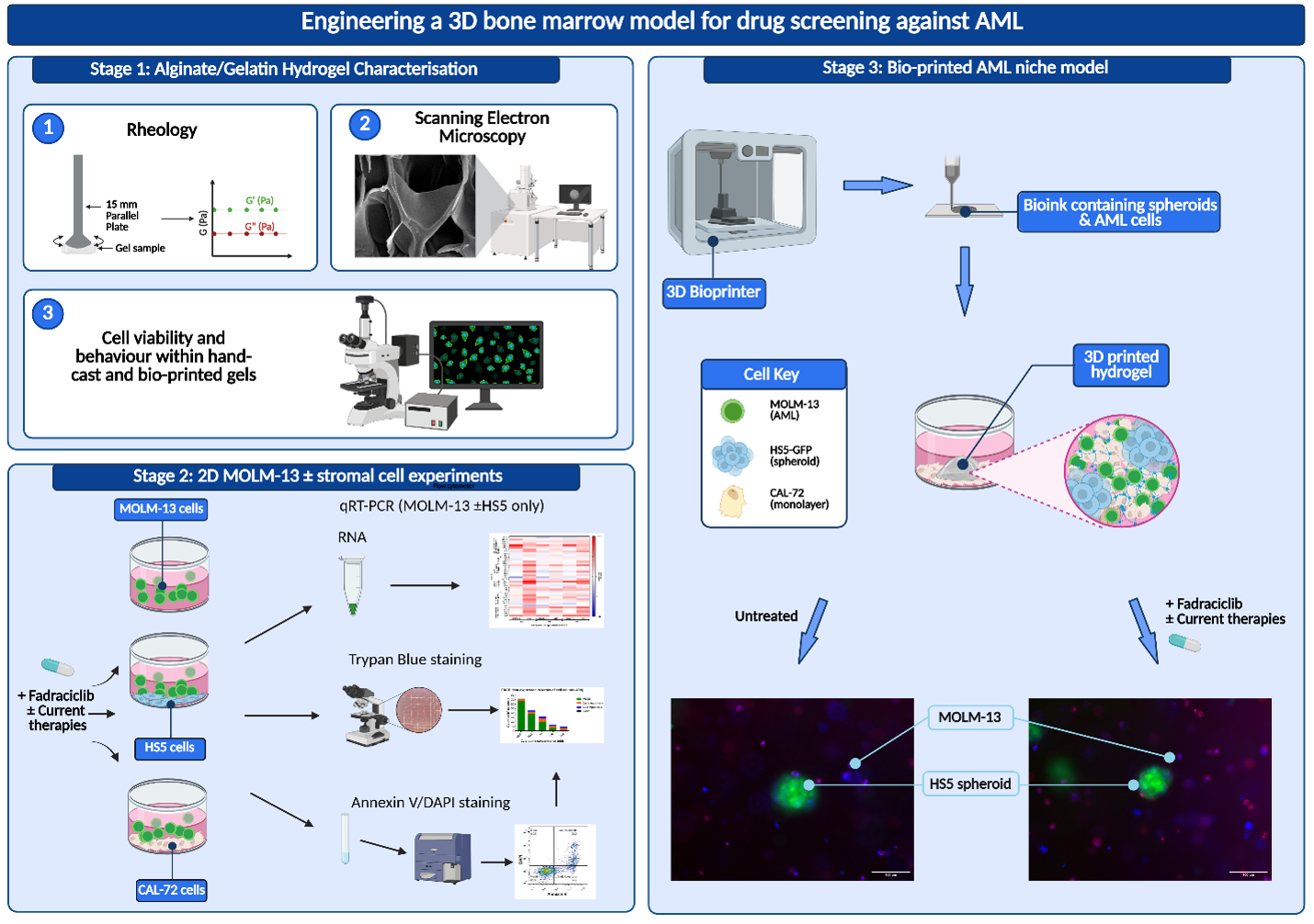
Three-dimensional (3D) bioprinting technology has the potential to fabricate highly reproducible, advanced biocompatible in vitro 3D models. It has been used successfully to emulate aspects of the bone marrow microenvironment to study leukaemia, including acute myeloid leukaemia (AML). AML is an aggressive haematological cancer which arises due to the accumulation of genetic mutations in haematopoietic stem and progenitor cells within the bone marrow, leading to the production of leukaemic stem cells (LSCs). Interactions between LSCs and bone marrow stroma induce LSC quiescence, protecting them from chemotherapy. In this study, we engineered a 3D-bioprinted co-culture bone marrow model containing MOLM-13 AML cells in co-culture with two main supportive cell types: bone marrow stromal cells (HS5) and osteoblast-like cells (CAL-72). This system was used to explore the efficacy of the cyclin-dependent kinase 2/9 inhibitor fadraciclib alone and in combination with current AML therapies. Characterisation of alginate/gelatin hydrogels revealed a highly porous matrix structure, with a hydrogel stiffness within the range of human bone marrow. These hydrogels sustained MOLM-13 cells and HS5 spheroid viability for up to seven days, highlighting the suitability of this system to model bone marrow. Next, the efficacy of fadraciclib alone and in combination with current chemotherapies was assessed in two-dimensional (2D) culture and in the final 3D-bioprinted model. These data revealed that fadraciclib induced MOLM-13 cell death in both 2D and 3D culture, with a reduced level of cell death observed in 3D culture. Together, these data reveal a reproducible human bone marrow niche system, with potential for screening of novel single and combination therapies.
- Jani CT, Ahmed A, Singh H, et al. Burden of AML, 1990-2019: Estimates From the Global Burden of Disease Study. JCO Glob Oncol. 2023;9:e2300229. doi: 10.1200/GO.23.00229
- Chen P, Liu X, Zhao Y, Hu Y, Guo J, Wang H. Global, national, and regional burden of acute myeloid leukemia among 60–89 years-old individuals: insights from a study covering the period 1990 to 2019. Front Public Health. 2024;11. doi: 10.3389/fpubh.2023.1329529
- El Chaer F, Bewersdorf JP, Stahl M, Zeidan AM. The global epidemiology of acute myeloid leukaemia. Nat Rev Clin Oncol. 2025;23(2):107-120. doi: 10.1038/s41571-025-01099-7
- Khwaja A, Bjorkholm M, Gale RE, et al. Acute myeloid leukaemia. Nat Rev Dis Primers. 2016;2(1):1. doi: 10.1038/nrdp.2016.10
- Allan DS, De Lisio M. Reversing pathological remodelling of the bone marrow microenvironment in acute myeloid leukemia. Stem Cell Investig. 2018;5(1):29. doi: 10.21037/sci.2018.09.02
- Barrera-Ramirez J, Lavoie JR, Maganti HB, et al. Micro-RNA Profiling of Exosomes from Marrow-Derived Mesenchymal Stromal Cells in Patients with Acute Myeloid Leukemia: Implications in Leukemogenesis. Stem Cell Rev. 2017;13(6):817-825. doi: 10.1007/s12015-017-9762-0
- Chandran P, Le Y, Li Y, et al. Mesenchymal stromal cells from patients with acute myeloid leukemia have altered capacity to expand differentiated hematopoietic progenitors. Leuk Res. 2015;39(4):486-493. doi: 10.1016/j.leukres.2015.01.013
- Ishikawa F, Yoshida S, Saito Y, et al. Chemotherapy-resistant human AML stem cells home to and engraft within the bone-marrow endosteal region. Nat Biotechnol. 2007;25(11):1315-1321. doi: 10.1038/nbt1350
- Raaijmakers MHGP, Mukherjee S, Guo S, et al. Bone progenitor dysfunction induces myelodysplasia and secondary leukemia. Nature. 2010;464(7290):852-857. doi: 10.1038/nature08851
- Waclawiczek A, Hamilton A, Rouault-Pierre K, et al. Mesenchymal niche remodeling impairs hematopoiesis via stanniocalcin 1 in acute myeloid leukemia. J Clin Invest. 2020;130(6):3038-3050. doi: 10.1172/JCI133187
- Yilmaz M, Wang F, Loghavi S, et al. Late relapse in acute myeloid leukemia (AML): clonal evolution or therapy-related leukemia? Blood Cancer J. 2019;9(2):2. doi: 10.1038/s41408-019-0170-3
- Kapałczyńska M, Kolenda T, Przybyła W, et al. 2D and 3D cell cultures – a comparison of different types of cancer cell cultures. Arch Med Sci. 2018;14(4):910-919. doi: 10.5114/aoms.2016.63743
- Bubnova X, Hope L, Wheadon H. Evaluation of current mouse in vivo models and advanced in vitro models for leukaemia research. Blood Rev. 2025;74:101318. doi: 10.1016/j.blre.2025.101318
- Alhattab DM, Isaioglou I, Alshehri S, et al. Fabrication of a three-dimensional bone marrow niche-like acute myeloid Leukemia disease model by an automated and controlled process using a robotic multicellular bioprinting system. Biomater Res. 2023;27(111):1-28. doi: 10.1186/s40824-023-00457-9
- Borella G, Da Ros A, Porcù E, et al. Acute Myeloid Leukemia (AML) in a 3D Bone Marrow Niche Showed High Performance for in Vitro and In Vivo Drug Screenings. Blood. 2019;134:544. doi: 10.1182/blood-2019-128382
- Bray LJ, Binner M, Körner Y, von Bonin M, Bornhäuser M, Werner C. A three-dimensional ex vivo tri-culture model mimics cell-cell interactions between acute myeloid leukemia and the vascular niche. Haematologica. 2017;102(7):1215-1226. doi: 10.3324/haematol.2016.157883
- de Janon A, Mantalaris A, Panoskaltsis N. Three-Dimensional Human Bone Marrow Organoids for the Study and Application of Normal and Abnormal Hematoimmunopoiesis. J Immunol. 2023;210(7):895-904. doi: 10.4049/jimmunol.2200836
- Gilchrist AE, Harley BAC. Engineered Tissue Models to Replicate Dynamic Interactions within the Hematopoietic Stem Cell Niche. Adv Healthc Mater. 2022;11(7):2102130. doi: 10.1002/adhm.202102130
- Xiao Y, McGuinness CAS, Doherty-Boyd WS, Salmeron-Sanchez M, Donnelly H, Dalby MJ. Current insights into the bone marrow niche: From biology in vivo to bioengineering ex vivo. Biomaterials. 2022;286:121568. doi: 10.1016/j.biomaterials.2022.121568
- Jiang T, Munguia-Lopez JG, Gu K, et al. Engineering bioprintable alginate/gelatin composite hydrogels with tunable mechanical and cell adhesive properties to modulate tumor spheroid growth kinetics. Biofabrication. 2019;12(1):015024. doi: 10.1088/1758-5090/ab3a5c
- Lewis EEL, Wheadon H, Lewis N, et al. A Quiescent, Regeneration-Responsive Tissue Engineered Mesenchymal Stem Cell Bone Marrow Niche Model via Magnetic Levitation. ACS Nano. 2016;10(9):8346-8354. doi: 10.1021/acsnano.6b02841
- Lewis NS, Lewis EE, Mullin M, Wheadon H, Dalby MJ, Berry CC. Magnetically levitated mesenchymal stem cell spheroids cultured with a collagen gel maintain phenotype and quiescence. J Tissue Eng. 2017;8:2041731417704428. doi: 10.1177/2041731417704428
- Anseth KS, Bowman CN, Brannon-Peppas L. Mechanical properties of hydrogels and their experimental determination. Biomaterials. 1996;17(17):1647-1657. doi: 10.1016/0142-9612(96)87644-7
- Giuseppe MD, Law N, Webb B, et al. Mechanical behaviour of alginate-gelatin hydrogels for 3D bioprinting. J Mech Behav Biomed Mater. 2018;79:150-157. doi: 10.1016/j.jmbbm.2017.12.018
- Mazzocchi A, Soker S, Skardal A. 3D bioprinting for high-throughput screening: Drug screening, disease modeling, and precision medicine applications. Appl Phys Rev. 2019;6(1):011302. doi: 10.1063/1.5056188
- Sanchez-Rubio A, Hope L, Barcelona-Estaje E, Jayawarna V, Williams J, Salmeron-Sanchez M. Gelatin vs GelMA in alginate-based bioinks as a platform for versatile 3D bioprintable in vitro systems. Biomater Adv. 2025;177:214408. doi: 10.1016/j.bioadv.2025.214408
- Agarwal T, Onesto V, Banerjee D, et al. 3D bioprinting in tissue engineering: current state-of-the-art and challenges towards system standardization and clinical translation. Biofabrication. 2025;17(4):042003. doi: 10.1088/1758-5090/ade47a
- Braham MVJ, Ahlfeld T, Akkineni AR, et al. Endosteal and Perivascular Subniches in a 3D Bone Marrow Model for Multiple Myeloma. Tissue Eng Part C Methods. 2018;24(5):300-312. doi: 10.1089/ten.tec.2017.0467
- Heinrich MA, Bansal R, Lammers T, Zhang YS, Michel Schiffelers R, Prakash J. 3D-Bioprinted Mini-Brain: A Glioblastoma Model to Study Cellular Interactions and Therapeutics. Adv Mater. 2019;31(14):1806590. doi: 10.1002/adma.201806590
- Badge AK, Dawande P, Bankar NJ, Noman O. Three-dimensional Bioprinting Platform for Chemotherapy Evaluation in Leukemia Patients: An Innovative Approach. J Appl Hematol. 2025;16(1):90-91. doi: 10.4103/joah.joah_15_25
- Rudzinska-Radecka M, Turos-Korgul L, Mukherjee D, Podszywalow-Bartnicka P, Piwocka K, Guzowski J. High-throughput formulation of reproducible 3D cancer microenvironments for drug testing in myeloid leukemia. Biofabrication. 2024;17(1):015035. doi: 10.1088/1758-5090/ad998d
- Zhou D, Chen L, Ding J, et al. A 3D engineered scaffold for hematopoietic progenitor/stem cell co-culture in vitro. Sci Rep. 2020;10(1):1. doi: 10.1038/s41598-020-68250-5
- Ribezzi D, Pinos R, Bonetti L, et al. Design of a novel bioink suitable for the 3D printing of lymphoid cells. Front Biomater Sci. 2023;2(1):1-12. doi: 10.3389/fbiom.2023.1081065
- Sbrana FV, Pinos R, Barbaglio F, et al. 3D Bioprinting Allows the Establishment of Long-Term 3D Culture Model for Chronic Lymphocytic Leukemia Cells. Front Immunol. 2021;12:639572. doi: 10.3389/fimmu.2021.639572
- Rodriguez C, Aleman J, George S, et al. Patient-Specific 3D Printed Multiple Myeloma Organoid Model for Niche Recapitulation and Prediction of Chemotherapy Response. Clin Lymphoma Myeloma Leuk. 2019;19:S336. doi: 10.1016/j.clml.2019.07.332
- Nair K, Gandhi M, Khalil S, et al. Characterization of cell viability during bioprinting processes. Biotechnol J. 2009;4(8):1168-1177. doi: 10.1002/biot.200900004
- Kačarević ŽP, Rider PM, Alkildani S, et al. An Introduction to 3D Bioprinting: Possibilities, Challenges and Future Aspects. Materials. 2018;11(11):2199. doi: 10.3390/ma11112199
- Y. Chung JH, Naficy S, Yue Z, et al. Bio-ink properties and printability for extrusion printing living cells. Biomater Sci. 2013;1(7):763-773. doi: 10.1039/C3BM00012E
- Ouyang L, Yao R, Zhao Y, Sun W. Effect of bioink properties on printability and cell viability for 3D bioplotting of embryonic stem cells. Biofabrication. 2016;8(3):035020. doi: 10.1088/1758-5090/8/3/035020
- Fonseca K, Raquel Maia F, A. Cruz F, et al. Enzymatic, physicochemical and biological properties of MMP-sensitive alginate hydrogels. Soft Matter. 2013;9(12):3283-3292. doi: 10.1039/C3SM27560D
- Schulz A, Gepp MM, Stracke F, von Briesen H, Neubauer JC, Zimmermann H. Tyramine-conjugated alginate hydrogels as a platform for bioactive scaffolds. J Biomed Mater Res A. 2019;107(1):114-121. doi: 10.1002/jbm.a.36538
- Contessi Negrini N, Angelova Volponi A, Sharpe PT, Celiz AD. Tunable Cross-Linking and Adhesion of Gelatin Hydrogels via Bioorthogonal Click Chemistry. ACS Biomater Sci Eng. 2021;7(9):4330-4346. doi: 10.1021/acsbiomaterials.1c00136
- Xie D, Bian J, Ni C, Zhao P, Pu Z, Yue J. Tuning Room-Temperature Injectability of Gelatin-Based Hydrogels via Introduction of Competitive Hydrogen Bonds. ACS Macro Lett. 2025;14(3):313-319. doi: 10.1021/acsmacrolett.5c00018
- He Y, Yang F, Zhao H, Gao Q, Xia B, Fu J. Research on the printability of hydrogels in 3D bioprinting. Sci Rep. 2016;6(1):1. doi: 10.1038/srep29977
- Chantkran W, Hsieh YC, Zheleva D, Frame S, Wheadon H, Copland M. Interrogation of novel CDK2/9 inhibitor fadraciclib (CYC065) as a potential therapeutic approach for AML. Cell Death Discov. 2021;7(1):1. doi: 10.1038/s41420-021-00496-y
- Frame S, Saladino C, MacKay C, et al. Fadraciclib (CYC065), a novel CDK inhibitor, targets key pro-survival and oncogenic pathways in cancer. PLOS ONE. 2020;15(7):e0234103. doi: 10.1371/journal.pone.0234103
- Matsuo Y, MacLeod R, Uphoff C, et al. Two acute monocytic leukemia (AML-M5a) cell lines (MOLM-13 and MOLM-14) with interclonal phenotypic heterogeneity showing MLL-AF9 fusion resulting from an occult chromosome insertion, ins(11;9)(q23;p22p23). Leukemia. 1997;11(9):1469-1477. doi: 10.1038/sj.leu.2400768
- Rao X, Huang X, Zhou Z, Lin X. An improvement of the 2ˆ(–delta delta CT) method for quantitative real-time polymerase chain reaction data analysis. Biostat Bioinforma Biomath. 2013;3(3):71-85.
- Jansen LE, Birch NP, Schiffman JD, Crosby AJ, Peyton SR. Mechanics of intact bone marrow. J Mech Behav Biomed Mater. 2015;50:299-307. doi: 10.1016/j.jmbbm.2015.06.023
- Zhang P, Zhang C, Li J, Han J, Liu X, Yang H. The physical microenvironment of hematopoietic stem cells and its emerging roles in engineering applications. Stem Cell Res Ther. 2019;10(1):327. doi: 10.1186/s13287-019-1422-7
- Bartl R, Bartl C, Bonél HM, von Tresckow E. Anatomy and Physiology of the Bone–Bone Marrow System. In: Bartl R, Bartl C, Bonél HM, von Tresckow E, eds. Bone Marrow Edema: Forms, Pathogenesis, Diagnosis, and Therapy. Berlin, Germany: Springer; 2025:1-15. doi: 10.1007/978-3-662-72321-0_1
- Tevis KM, Colson YL, Grinstaff MW. Embedded Spheroids as Models of the Cancer Microenvironment. Adv Biosyst. 2017;1(10):1700083. doi: 10.1002/adbi.201700083
- Zanoni M, Pignatta S, Arienti C, Bonafè M, Tesei A. Anticancer drug discovery using multicellular tumor spheroid models. Expert Opin Drug Discov. 2019;14(3):289-301. doi: 10.1080/17460441.2019.1570129
- Svozilová H, Plichta Z, Proks V, et al. RGDS-Modified Superporous Poly(2-Hydroxyethyl Methacrylate)-Based Scaffolds as 3D In Vitro Leukemia Model. Int J Mol Sci. 2021;22(5):5. doi: 10.3390/ijms22052376
- Vu TTT, Lim C, Lim M. Characterization of leukemic cell behaviors in a soft marrow mimetic alginate hydrogel. J Biomed Mater Res B Appl Biomater. 2012;100B(7):1980-1988. doi: 10.1002/jbm.b.32765
- Adamo A, Delfino P, Gatti A, et al. HS-5 and HS-27A Stromal Cell Lines to Study Bone Marrow Mesenchymal Stromal Cell-Mediated Support to Cancer Development. Front Cell Dev Biol. 2020;8:584232. doi: 10.3389/fcell.2020.584232
- Garrido SM, Appelbaum FR, Willman CL, Banker DE. Acute myeloid leukemia cells are protected from spontaneous and drug-induced apoptosis by direct contact with a human bone marrow stromal cell line (HS-5). Exp Hematol. 2001;29(4):448-457. doi: 10.1016/S0301-472X(01)00612-9
- Hou D, Wang B, You R, et al. Stromal cells promote chemoresistance of acute myeloid leukemia cells via activation of the IL-6/STAT3/OXPHOS axis. Ann Transl Med. 2020;8(21):1346. doi: 10.21037/atm-20-3191
- Miari KE, Williams MTS. Stromal bone marrow fibroblasts and mesenchymal stem cells support acute myeloid leukaemia cells and promote therapy resistance. Br J Pharmacol. 2023;181(2):216-237. doi: 10.1111/bph.16028
- Mohammadi S, Nikbakht M, Sajjadi SM, et al. Reciprocal Interactions of Leukemic Cells with Bone Marrow Stromal Cells Promote Enrichment of Leukemic Stem Cell Compartments in Response to Curcumin and Daunorubicin. Asian Pac J Cancer Prev. 2017;18(3):831-840. doi: 10.22034/APJCP.2017.18.3.831
- Wang Z, Chai C, Wang R, et al. Single-cell transcriptome atlas of human mesenchymal stem cells exploring cellular heterogeneity. Clin Transl Med. 2021;11(12):e650. doi: 10.1002/ctm2.650
- Yin JQ, Zhu J, Ankrum JA. Manufacturing of primed mesenchymal stromal cells for therapy. Nat Biomed Eng. 2019;3(2):2. doi: 10.1038/s41551-018-0325-8
- Keating GM. Azacitidine. Drugs. 2012;72(8):1111-1136. doi: 10.2165/11209430-000000000-00000
- Faruqi A, Tadi P. Cytarabine. In: StatPearls. Treasure Island, FL: StatPearls Publishing; 2023. Accessed December 7, 2023. http://www.ncbi.nlm.nih.gov/books/NBK557680/
- Bose P, Gandhi V, Konopleva M. Pathways and mechanisms of venetoclax resistance. Leuk Lymphoma. 2017;58(9):2026-2039. doi: 10.1080/10428194.2017.1283032
- Aldoss I, Yang D, Aribi A, et al. Efficacy of the combination of venetoclax and hypomethylating agents in relapsed/refractory acute myeloid leukemia. Haematologica. 2018;103(9):e404-e407. doi: 10.3324/haematol.2018.188094
- Cherry EM, Abbott D, Amaya M, et al. Venetoclax and azacitidine compared with induction chemotherapy for newly diagnosed patients with acute myeloid leukemia. Blood Adv. 2021;5(24):5565-5573. doi: 10.1182/bloodadvances.2021005538
- Grimm D, Bauer J, Wise P, et al. The role of SOX family members in solid tumours and metastasis. Semin Cancer Biol. 2020;67:122-153. doi: 10.1016/j.semcancer.2019.03.004
- Xu YR, Yang WX. SOX-mediated molecular crosstalk during the progression of tumorigenesis. Semin Cell Dev Biol. 2017;63:23-34. doi: 10.1016/j.semcdb.2016.07.028
- Hu J, Li K, Li Z, et al. Sex-determining region Y box-containing genes: regulators and biomarkers in gynecological cancers. Cancer Biol Med. 2019;16(3):462-474. doi: 10.20892/j.issn.2095-3941.2019.0062
- Wang J, Wan X, Le Q. Cross-regulation between SOX9 and the canonical Wnt signalling pathway in stem cells. Front Mol Biosci. 2023;10:1-12. doi: 10.3389/fmolb.2023.1250530
- Roecklein B, Torok-Storb B. Functionally distinct human marrow stromal cell lines immortalized by transduction with the human papilloma virus E6/E7 genes. Blood. 1995;85(4):997-1005. doi: 10.1182/blood.V85.4.997.bloodjournal854997
- Yu Z, Zhang W, Kone BC. Signal transducers and activators of transcription 3 (STAT3) inhibits transcription of the inducible nitric oxide synthase gene by interacting with nuclear factor κB. Biochem J. 2002;367(1):97-105. doi: 10.1042/bj20020588
- Fan Y, Li Y, Yao X, et al. Epithelial SOX9 drives progression and metastases of gastric adenocarcinoma by promoting immunosuppressive tumour microenvironment. Gut. 2023;72(4):624-637. doi: 10.1136/gutjnl-2021-326581
- Song S, Ajani JA, Honjo S, et al. Hippo Coactivator YAP1 Upregulates SOX9 and Endows Esophageal Cancer Cells with Stem-like Properties. Cancer Res. 2014;74(15):4170-4182. doi: 10.1158/0008-5472.CAN-13-3569
- Song S, Maru DM, Ajani JA, et al. Loss of TGF-β Adaptor β2SP Activates Notch Signaling and SOX9 Expression in Esophageal Adenocarcinoma. Cancer Res. 2013;73(7):2159-2169. doi: 10.1158/0008-5472.CAN-12-1962
- Rochet N, Leroy P, Far DF, Ollier L, Loubat A, Rossi B. CAL72: a human osteosarcoma cell line with unique effects on hematopoietic cells. Eur J Haematol. 2003;70(1):43-52. doi: 10.1034/j.1600-0609.2003.02766.x
- Bruserud O, Ryningen A, Wergeland L, Glenjen NI, Gjertsen BT. Osteoblasts increase proliferation and release of pro-angiogenic interleukin 8 by native human acute myelogenous leukemia blasts. Haematologica. 2004;89(4):391-402. doi: 10.3324/%x
- Hatfield KJ, Hovland R, Øyan AM, et al. Release of angiopoietin-1 by primary human acute myelogenous leukemia cells is associated with mutations of nucleophosmin, increased by bone marrow stromal cells and possibly antagonized by high systemic angiopoietin-2 levels. Leukemia. 2008;22(2):287-293. doi: 10.1038/sj.leu.2404985
- Reikvam H, Nepstad I, Bruserud Ø, Hatfield KJ. Pharmacologic targeting of the PI3K/mTOR pathway alters the release of angioregulatory mediators both from primary human acute myeloid leukemia cells and their neighboring stromal cells. Oncotarget. 2013;4(6):830-843. doi: 10.18632/oncotarget.971
- Roboz GJ, Guzman M. Acute Myeloid Leukemia Stem Cells: Seek and Destroy. Expert Rev Hematol. 2009;2(6):663-672. doi: 10.1586/ehm.09.53
- Sison EAR, Rau RE, McIntyre E, Li L, Small D, Brown P. MLL-rearranged acute lymphoblastic leukaemia stem cell interactions with bone marrow stroma promote survival and therapeutic resistance that can be overcome with CXCR4 antagonism. Br J Haematol. 2013;160(6):785-797. doi: 10.1111/bjh.12205
- Bessy T, Itkin T, Passaro D. Bioengineering the Bone Marrow Vascular Niche. Front Cell Dev Biol. 2021;9. Accessed December 31, 2023. https://www.frontiersin.org/articles/10.3389/fcell.2021.645496
- Aroguz AZ, Baysal K, Adiguzel Z, Baysal BM. Alginate/Polyoxyethylene and Alginate/Gelatin Hydrogels: Preparation, Characterization, and Application in Tissue Engineering. Appl Biochem Biotechnol. 2014;173(2):433-448. doi: 10.1007/s12010-014-0851-0
- Duan B, Hockaday LA, Kang KH, Butcher JT. 3D Bioprinting of heterogeneous aortic valve conduits with alginate/gelatin hydrogels. J Biomed Mater Res A. 2013;101A(5):1255-1264. doi: 10.1002/jbm.a.34420
- Cheng J, Lin F, Liu H, et al. Rheological Properties of Cell-Hydrogel Composites Extruding Through Small-Diameter Tips. J Manuf Sci Eng. 2008;130(0210141-5). doi: 10.1115/1.2896215
- Mondal A, Gebeyehu A, Miranda M, et al. Characterization and printability of Sodium alginate -Gelatin hydrogel for bioprinting NSCLC co-culture. Sci Rep. 2019;9:19914. doi: 10.1038/s41598-019-55034-9
- Yao R, Zhang R, Luan J, Lin F. Alginate and alginate/gelatin microspheres for human adipose-derived stem cell encapsulation and differentiation. Biofabrication. 2012;4(2):025007. doi: 10.1088/1758-5082/4/2/025007
- Miao F, Liu T, Zhang X, et al. Engineered bone tissues using biomineralized gelatin methacryloyl/sodium alginate hydrogels. J Biomater Sci Polym Ed. 2022;33(2):137-154. doi: 10.1080/09205063.2021.1980360
- Zuo X, Zhang H, Zhou T, et al. Spheroids of Endothelial Cells and Vascular Smooth Muscle Cells Promote Cell Migration in Hyaluronic Acid and Fibrinogen Composite Hydrogels. Research. 2020;2020:8970480. doi: 10.34133/2020/8970480
- Shang ZY, Sun H, Xiang HF, et al. Three-dimensional porous composite scaffolds for in vitro marrow microenvironment simulation to screen leukemia drug. Biomed Mater. 2020;15(3):035016. doi: 10.1088/1748-605X/ab74e2
- Jensen C, Teng Y. Is It Time to Start Transitioning From 2D to 3D Cell Culture? Front Mol Biosci. 2020;7:33. doi: 10.3389/fmolb.2020.00033
- Peter B, Eisenwort G, Sadovnik I, et al. BRD4 degradation blocks expression of MYC and multiple forms of stem cell resistance in Ph+ chronic myeloid leukemia. Am J Hematol. 2022;97(9):1215-1225. doi: 10.1002/ajh.26650
- El Achi H, Kanagal-Shamanna R. Biomarkers in Acute Myeloid Leukemia: Leveraging Next Generation Sequencing Data for Optimal Therapeutic Strategies. Front Oncol. 2021;11. doi: 10.3389/fonc.2021.748250
- Myeloid gene panel analysis. Manchester University NHS Foundation Trust. Accessed March 26, 2026. https://mft.nhs.uk/nwglh/test-information/cancer/haematology-oncology/national-genomic-test-directory-services-available/acute-myeloid-leukaemia/myeloid-gene-panel-analysis/
- Hiller T, Berg J, Elomaa L, et al. Generation of a 3D Liver Model Comprising Human Extracellular Matrix in an Alginate/Gelatin-Based Bioink by Extrusion Bioprinting for Infection and Transduction Studies. Int J Mol Sci. 2018;19(10):10. doi: 10.3390/ijms19103129
- Hong S, Song JM. 3D bioprinted drug-resistant breast cancer spheroids for quantitative in situ evaluation of drug resistance. Acta Biomater. 2022;138:228-239. doi: 10.1016/j.actbio.2021.10.031
- Heltmann-Meyer S, Fleischer S, Kadam S, et al. Ink comparison for extrusion-based bioprinting in the context of breast cancer and melanoma models. Int J Bioprinting. 2025;11(2):249. doi: 10.36922/ijb.5828
- Handorf AM, Zhou Y, Halanski MA, Li WJ. Tissue stiffness dictates development, homeostasis, and disease progression. Organogenesis. 2015;11(1):1-15. doi: 10.1080/15476278.2015.1019687