
Cancer as a syntrophic cellular consortium established by fatty acid oxidation–primed mitochondria and carrier macrophages

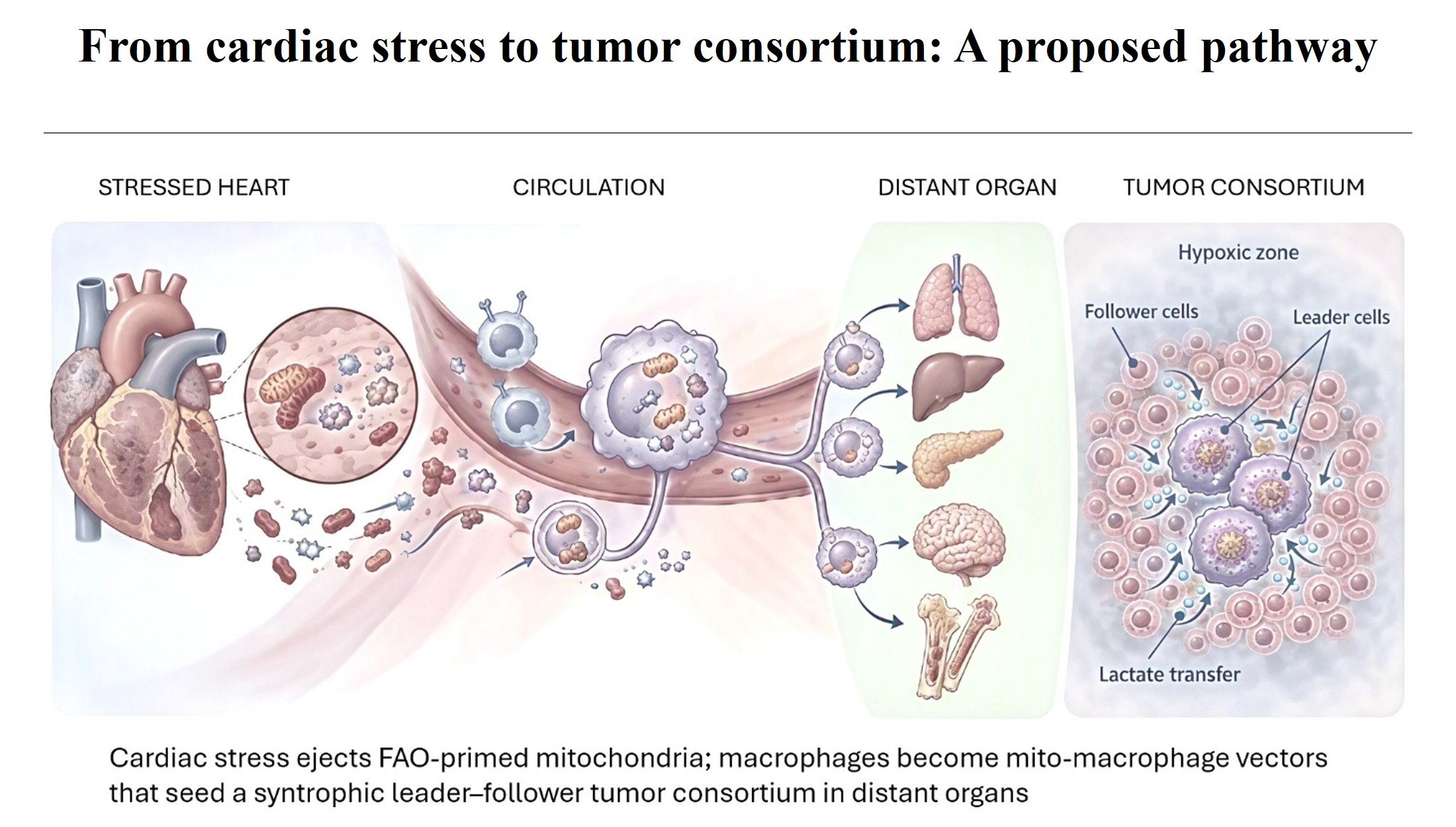
The escalating clinical link between cardiovascular disease and subsequent cancer suggests a systemic etiology beyond the sole purview of somatic mutation. This article reviews emerging evidence through a novel lens: cancer as a syntrophic cellular consortium, where metabolic stratification, driven by fatty acid oxidation (FAO)-primed mitochondria and mediated by carrier macrophages, establishes functionally distinct, metabolically interdependent cellular subpopulations. Stressed cardiomyocytes eject metabolically “primed” mitochondria. These organelles are engulfed by macrophages, transforming them into pathogenic “mito-macrophages” that act as mobile systemic carriers. These mito-macrophages instigate malignancy through mitochondrial transfer to recipient cells, inducing profound metabolic and epigenetic reprogramming. Within individual cells, the transferred mitochondria establish distinct intracellular oxidation zones fueled by FAO, juxtaposed with hypoxia zones favoring glycolysis. This primary compartmentalization scales up within the evolving tumor: cells from oxidation zones adopt a “leader” phenotype, characterized by high FAO, metabolic flexibility, and invasive capacity, while cells from hypoxia zones become “follower” phenotypes, relying on glycolysis and contributing to bulk tumor growth. Critical intercellular signaling pathways between these emerging “leader” and “follower” populations are central to establishing, maintaining, and propagating this obligate, syntrophic cross-feeding hierarchy, ensuring tumor progression. This framework bridges cardiac stress and distal cancer, elucidating how metabolic and epigenetic imprinting dictates cellular roles within a structured malignant consortium. Reconceiving cancer as such a syntrophic system offers new avenues for therapeutic intervention, targeting metabolic instigators and the signaling axes that define its consortium partners. This framework synthesizes established biological mechanisms into a novel, testable hypothesis and is presented as such. Direct experimental evidence for several of the proposed linkages—particularly long-distance macrophage-mediated trafficking of cardiomyocyte-derived mitochondria, and the FAO-primed state—remains to be established.
- Hanahan D, Weinberg RA. Hallmarks of cancer: the next Generation. Cell. 2011;144(5):646-674. doi: 10.1016/j.cell.2011.02.013
- Kalluri R. The biology and function of fibroblasts in cancer. Nat Rev Cancer. 2016;16(9):582-598. doi: 10.1038/nrc.2016.73
- Soto AM, Sonnenschein C. The tissue organization field theory of cancer: A testable replacement for the somatic mutation theory. BioEssays. 2011;33(5):332-340. doi: 10.1002/bies.201100025
- Jones PA, Baylin SB. The Epigenomics of Cancer. Cell. 2007;128(4):683-692. doi: 10.1016/j.cell.2007.01.029
- Huang S, Soto AM, Sonnenschein C. The end of the genetic paradigm of cancer. PLOS Biol. 2025;23(3):e3003052. doi: 10.1371/journal.pbio.3003052
- Frank SA. How cancer arises: Genetics releases, plasticity creates, genetics stabilizes. Proc Natl Acad Sci USA. 2025;122(31):e2505377122. doi: 10.1073/pnas.2505377122
- Bertero E, Canepa M, Maack C, Ameri P. Linking heart failure to cancer: Background evidence and research perspectives. Circulation. 2018;138(7):735-742. doi: 10.1161/circulationaha.118.033603
- Malmborg M, Christiansen CB, Schmiegelow MD, Torp-Pedersen C, Gislason G, Schou M. Incidence of new onset cancer in patients with a myocardial infarction: a nationwide cohort study. BMC Cardiovasc Disord. 2018;18(1):198. doi: 10.1186/s12872-018-0932-z
- Jaiswal S, Ebert BL. Clonal hematopoiesis in human aging and disease. Science. 2019;366(6465):eaan4673. doi: 10.1126/science.aan4673
- Viale A, Corti D, Draetta GF. Tumors and mitochondrial respiration: A neglected connection. Cancer Res. 2015;75(18):3687-3691. doi: 10.1158/0008-5472.can-15-0491
- Demicco M, Liu XZ, Leithner K, Fendt SM. Metabolic heterogeneity in cancer. Nat Metab. 2024;6(1):18-38. doi: 10.1038/s42255-023-00963-z
- Nakajima EC, Van Houten B. Metabolic symbiosis in cancer: Refocusing the Warburg lens. Mol Carcinog. 2012;52(5):329-337. doi: 10.1002/mc.21863
- Feng S, Hu Y, Li S, et al. Mitochondrial transfer in the tumor microenvironment: Mechanisms, immunometabolic consequences, and therapeutic implications. Crit Rev Oncol/Hematol. 2026;227:105530. doi: 10.1016/j.critrevonc.2026.105530
- Crewe C, Funcke JB, Li S, et al. Extracellular vesicle-based interorgan transport of mitochondria from energetically stressed adipocytes. Cell Metab. 2021;33(9):1853-1868.e11. doi: 10.1016/j.cmet.2021.08.002
- Al Amir Dache Z, Otandault A, Tanos R, et al. Blood contains circulating cell-free respiratory competent mitochondria. FASEB J. 2020;34(3):3616-3630. doi: 10.1096/fj.201901917rr
- Liu D, Gao Y, Liu J, et al. Intercellular mitochondrial transfer as a means of tissue revitalization. Signal Transduct Target Ther. 2021;6(1):65. doi: 10.1038/s41392-020-00440-z
- Boudreau LH, Duchez AC, Cloutier N, et al. Platelets release mitochondria serving as substrate for bactericidal group IIA-secreted phospholipase A2 to promote inflammation. Blood. 2014;124(14):2173-2183. doi: 10.1182/blood-2014-05-573543
- Islam MN, Das SR, Emin MT, et al. Mitochondrial transfer from bone-marrow–derived stromal cells to pulmonary alveoli protects against acute lung injury. Nat Med. 2012;18(5):759-765. doi: 10.1038/nm.2736
- Hayakawa K, Esposito E, Wang X, et al. Transfer of mitochondria from astrocytes to neurons after stroke. Nature. 2016;535(7613):551-555. doi: 10.1038/nature18928
- Lopaschuk GD, Ussher JR, Folmes CDL, Jaswal JS, Stanley WC. Myocardial fatty acid metabolism in health and disease. Physiol Rev. 2010;90(1):207-258. doi: 10.1152/physrev.00015.2009
- Dorn GW. Mitochondrial dynamism and heart disease: Changing shape and shaping change. EMBO Mol Med. 2015;7(7):865-877. doi: 10.15252/emmm.201404575
- Sun N, Barta H, Chaudhuri S, et al. Mitophagy mitigates mitochondrial fatty acid β-oxidation deficient cardiomyopathy. Nat Commun. 2025;16(1):5465. doi: 10.1038/s41467-025-60670-z
- Bergmann O, Zdunek S, Felker A, et al. Dynamics of Cell Generation and Turnover in the Human Heart. Cell. 2015;161(7):1566-1575. doi: 10.1016/j.cell.2015.05.026
- Derks W, Bergmann O. Polyploidy in Cardiomyocytes: Roadblock to heart regeneration? Circ Res. 2020;126(4):552-565. doi: 10.1161/circresaha.119.315408
- Nicolás-Ávila JA, Lechuga-Vieco AV, Esteban-Martínez L, et al. A network of macrophages supports mitochondrial homeostasis in the heart. Cell. 2020;183(1):94-109.e23. doi: 10.1016/j.cell.2020.08.031
- Divakaruni AS, Rogers GW, Murphy AN. Measuring mitochondrial function in permeabilized cells using the Seahorse XF analyzer or a Clark-type oxygen electrode. Curr Protoc Toxicol. 2014;60(1):25.2.1-25.2.16. doi: 10.1002/0471140856.tx2502s60
- Brand MD, Nicholls DG. Assessing mitochondrial dysfunction in cells. Biochemical Journal. 2011;435(2):297-312. doi: 10.1042/BJ20110162
- Klionsky DJ, Abdel-Aziz AK, Abdelfatah S, et al. Guidelines for the use and interpretation of assays for monitoring autophagy (4th edition). Autophagy. 2021;17(1):1-382. doi: 10.1080/15548627.2020.1797280
- Chen YR, Zweier JL. Cardiac Mitochondria and Reactive Oxygen Species Generation. Circ Res. 2014;114(3):524-537. doi: 10.1161/circresaha.114.300559
- Murphy MP. How mitochondria produce reactive oxygen species. Biochem J. 2009;417(1):1-13. doi: 10.1042/bj20081386
- Ali MA, Gioscia-Ryan R, Yang D, Sutton NR, Tyrrell DJ. Cardiovascular aging: spotlight on mitochondria. Am J Physiol-Heart Circ Physiol. 2024;326(2):H317-H333. doi: 10.1152/ajpheart.00632.2023
- Miyamoto S. Autophagy and cardiac aging. Cell Death Differ. 2019;26(4):653-664. doi: 10.1038/s41418-019-0286-9
- McGarry JD, Brown NF. The mitochondrial carnitine palmitoyltransferase system - From concept to molecular analysis. Eur J Biochem. 1997;244(1):1-14. doi: 10.1111/j.1432-1033.1997.00001.x
- Houten SM, Wanders RJA. A general introduction to the biochemistry of mitochondrial fatty acid β-oxidation. J Inherit Metab Dis. 2010;33(5):469-477. doi: 10.1007/s10545-010-9061-2
- Lopaschuk GD, Karwi QG, Tian R, Wende AR, Abel ED. Cardiac energy metabolism in heart failure. Circ Res. 2021;128(10):1487-1513. doi: 10.1161/CIRCRESAHA.121.318241
- Cardoso AC, Lam NT, Savla JJ, et al. Mitochondrial substrate utilization regulates cardiomyocyte cell-cycle progression. Nat Metab. 2020;2(2):167-178. doi: 10.1038/s42255-020-0169-x
- Sabharwal SS, Schumacker PT. Mitochondrial ROS in cancer: initiators, amplifiers or an Achilles’ heel? Nat Rev Cancer. 2014;14(11):709-721. doi: 10.1038/nrc3803
- Jakubzick C, Gautier EL, Gibbings SL, et al. Minimal differentiation of classical monocytes as they survey steady-state tissues and transport antigen to lymph nodes. Immunity. 2013;39(3):599-610. doi: 10.1016/j.immuni.2013.08.007
- Epelman S, Lavine KJ, Beaudin AE, et al. Embryonic and adult-derived resident cardiac macrophages are maintained through distinct mechanisms at steady state and during inflammation. Immunity. 2014;40(1):91-104. doi: 10.1016/j.immuni.2013.11.019
- Bajpai G, Schneider C, Wong N, et al. The human heart contains distinct macrophage subsets with divergent origins and functions. Nat Med. 2018;24(8):1234-1245. doi: 10.1038/s41591-018-0059-x
- Molawi K, Wolf Y, Kandalla PK, et al. Progressive replacement of embryo-derived cardiac macrophages with age. J Exp Med. 2014;211(11):2151-2158. doi: 10.1084/jem.20140639
- De Maeyer RPH, Chambers ES. The impact of ageing on monocytes and macrophages. Immunol Lett. 2021;230:1-10. doi: 10.1016/j.imlet.2020.12.003
- Ginhoux F, Schultze JL, Murray PJ, Ochando J, Biswas SK. New insights into the multidimensional concept of macrophage ontogeny, activation and function. Nat Immunol. 2016;17(1):34-40. doi: 10.1038/ni.3324
- Murray PJ, Allen JE, Biswas SK, et al. Macrophage activation and polarization: Nomenclature and experimental guidelines. Immunity. 2014;41(1):14-20. doi: 10.1016/j.immuni.2014.06.008
- Xue J, Schmidt SV, Sander J, et al. Transcriptome-based network analysis reveals a spectrum model of human macrophage activation. Immunity. 2014;40(2):274-288. doi: 10.1016/j.immuni.2014.01.006
- Chawla A. Control of macrophage activation and function by PPARs. Circ Res. 2010;106(10):1559-1569. doi: 10.1161/circresaha.110.216523
- Van den Bossche J, Baardman J, Otto NA, et al. Mitochondrial dysfunction prevents repolarization of inflammatory macrophages. Cell Rep. 2016;17(3):684-696. doi: 10.1016/j.celrep.2016.09.008
- Pello OM, De Pizzol M, Mirolo M, et al. Role of c-MYC in alternative activation of human macrophages and tumor-associated macrophage biology. Blood. 2012;119(2):411-421. doi: 10.1182/blood-2011-02-339911
- Molgora M, Esaulova E, Vermi W, et al. TREM2 modulation remodels the tumor myeloid landscape enhancing anti-PD-1 immunotherapy. Cell. 2020;182(4):886-900.e17. doi: 10.1016/j.cell.2020.07.013
- Oka T, Hikoso S, Yamaguchi O, et al. Mitochondrial DNA that escapes from autophagy causes inflammation and heart failure. Nature. 2012;485(7397):251-255. doi: 10.1038/nature10992
- Arts RJW, Carvalho A, La Rocca C, et al. Immunometabolic pathways in BCG-induced trained immunity. Cell Rep. 2016;17(10):2562-2571. doi: 10.1016/j.celrep.2016.11.011
- Cheng SC, Quintin J, Cramer RA, et al. mTOR- and HIF-1α-mediated aerobic glycolysis as metabolic basis for trained immunity. Science. 2014;345(6204):1250684. doi: 10.1126/science.1250684
- Mantovani A, Sozzani S, Locati M, Allavena P, Sica A. Macrophage polarization: tumor-associated macrophages as a paradigm for polarized M2 mononuclear phagocytes. Trends Immunol. 2002;23(11):549-555. doi: 10.1016/S1471-4906(02)02302-5
- Yu E, Calvert PA, Mercer JR, et al. Mitochondrial DNA damage can promote atherosclerosis independently of reactive oxygen species through effects on smooth muscle cells and monocytes and correlates with higher-risk plaques in humans. Circulation. 2014;128(7):702-712. doi: 10.1161/circulationaha.113.002271
- Yu EPK, Bennett MR. Mitochondrial DNA damage and atherosclerosis. Trends Endocrinol Metab. 2014;25(9):481-487. doi: 10.1016/j.tem.2014.06.008
- Tabas I, Bornfeldt KE. Macrophage phenotype and function in different stages of atherosclerosis. Circ Res. 2016;118(4):653-667. doi: 10.1161/circresaha.115.306256
- Tapia-Vieyra JV, Delgado-Coello B, Mas-Oliva J. Atherosclerosis and cancer; A resemblance with far-reaching implications. Arch Med Res. 2017;48(1):12-26. doi: 10.1016/j.arcmed.2017.03.005
- Kidwell CU, Casalini JR, Pradeep S, et al. Transferred mitochondria accumulate reactive oxygen species, promoting proliferation. eLife. 2023;12:e85494. doi: 10.7554/elife.85494
- Weinhäuser I, Pereira-Martins DA, Almeida LY, et al. M2 macrophages drive leukemic transformation by imposing resistance to phagocytosis and improving mitochondrial metabolism. Sci Adv. 2023;9(15):eadf8522. doi: 10.1126/sciadv.adf8522
- Brestoff JR, Singh KK, Aquilano K, et al. Recommendations for mitochondria transfer and transplantation nomenclature and characterization. Nat Metab. 2025;7(1):53-67. doi: 10.1038/s42255-024-01200-x
- Li H, Sun W, Gong W, Han Y. Transfer and fates of damaged mitochondria: role in health and disease. FEBS J. 2024;291(24):5342-5364. doi: 10.1111/febs.17119
- Davis CO, Kim KY, Bushong EA, et al. Transcellular degradation of axonal mitochondria. Proc Natl Acad Sci USA. 2014;111(26):9633-9638. doi: 10.1073/pnas.1404651111
- Longo N, Frigeni M, Pasquali M. Carnitine transport and fatty acid oxidation. Biochim Biophys Acta (BBA) - Mol Cell Res. 2016;1863(10):2422-2435. doi: 10.1016/j.bbamcr.2016.01.023
- Bonnefont JP, Djouadi F, Prip-Buus C, Gobin S, Munnich A, Bastin J. Carnitine palmitoyltransferases 1 and 2: biochemical, molecular and medical aspects. Mol Aspects Med. 2004;25(5-6):495-520. doi: 10.1016/j.mam.2004.06.004
- Foster DW. Malonyl-CoA: the regulator of fatty acid synthesis and oxidation. J Clin Investig. 2012;122(6):1958-1959. doi: 10.1172/JCI63967
- Shao D, Kolwicz SC Jr, Wang P, et al. Increasing fatty acid oxidation prevents high-fat diet-induced cardiomyopathy through regulating Parkin-mediated mitophagy. Circulation. 2020;142(10):983-997. doi: 10.1161/CIRCULATIONAHA.119.043319
- Cuthbert KD, Dyck JRB. Malonyl-CoA decarboxylase is a major regulator of myocardial fatty acid oxidation. Curr Hypertens Rep. 2005;7(6):407-411. doi: 10.1007/s11906-005-0034-z
- Merrill GF, Kurth EJ, Hardie DG, Winder WW. AICA riboside increases AMP-activated protein kinase, fatty acid oxidation, and glucose uptake in rat muscle. Am J Physiol Endocrinol Metab. 1997;273(6):E1107-E1112. doi: 10.1152/ajpendo.1997.273.6.E1107
- Gomes AP, Price NL, Ling AJY, et al. Declining NAD+ induces a pseudohypoxic state disrupting nuclear-mitochondrial communication during aging. Cell. 2013;155(7):1624-1638. doi: 10.1016/j.cell.2013.11.037
- Selak MA, Armour SM, MacKenzie ED, et al. Succinate links TCA cycle dysfunction to oncogenesis by inhibiting HIF-α prolyl hydroxylase. Cancer Cell. 2005;7(1):77-85. doi: 10.1016/j.ccr.2004.11.022
- King A, Selak MA, Gottlieb E. Succinate dehydrogenase and fumarate hydratase: linking mitochondrial dysfunction and cancer. Oncogene. 2006;25(34):4675-4682. doi: 10.1038/sj.onc.1209594
- Kim JW, Tchernyshyov I, Semenza GL, Dang CV. HIF-1-mediated expression of pyruvate dehydrogenase kinase: a metabolic switch required for cellular adaptation to hypoxia. Cell Metab. 2006;3(3):177-185. doi: 10.1016/j.cmet.2006.02.002
- Papandreou I, Cairns RA, Fontana L, Lim AL, Denko NC. HIF-1 mediates adaptation to hypoxia by actively downregulating mitochondrial oxygen consumption. Cell Metab. 2006;3(3):187-197. doi: 10.1016/j.cmet.2006.01.012
- Sonveaux P, Végran F, Schroeder T, et al. Targeting lactate-fueled respiration selectively kills hypoxic tumor cells in mice. J Clin Invest. 2008;118(12):3930-3942. doi: 10.1172/JCI36843
- Faubert B, Li KY, Cai L, et al. Lactate metabolism in human lung tumors. Cell. 2017;171(2):358-371.e9. doi: 10.1016/j.cell.2017.09.019
- Zhang D, Tang Z, Huang H, et al. Metabolic regulation of gene expression by histone lactylation. Nature. 2019;574(7779):575-580. doi: 10.1038/s41586-019-1678-1
- Liu R, Ren X, Park YE, et al. Nuclear GTPSCS functions as a lactyl-CoA synthetase to promote histone lactylation and gliomagenesis. Cell Metab. 2025;37(2):377-394.e9. doi: 10.1016/j.cmet.2024.11.005
- Colegio OR, Chu NQ, Szabo AL, et al. Functional polarization of tumour-associated macrophages by tumour-derived lactic acid. Nature. 2014;513(7519):559-563. doi: 10.1038/nature13490
- Vitale I, Manic G, Coussens LM, Kroemer G, Galluzzi L. Macrophages and metabolism in the tumor microenvironment. Cell Metab. 2019;30(1):36-50. doi: 10.1016/j.cmet.2019.06.001
- Ding Y, Liu Z, Desai S, et al. Receptor tyrosine kinase ErbB2 translocates into mitochondria and regulates cellular metabolism. Nat Commun. 2012;3:1271. doi: 10.1038/ncomms2236
- Brooks GA. The science and translation of lactate shuttle theory. Cell Metab. 2018;27(4):757-785. doi: 10.1016/j.cmet.2018.03.008
- Hui S, Ghergurovich JM, Morscher RJ, et al. Glucose feeds the TCA cycle via circulating lactate. Nature. 2017;551(7678):115-118. doi: 10.1038/nature24057
- Cruzat V, Macedo Rogero M, Noel Keane K, Curi R, Newsholme P. Glutamine: metabolism and immune function, supplementation and clinical translation. Nutrients. 2018;10(11):1564. doi: 10.3390/nu10111564
- Jha AK, Huang SC, Sergushichev A, et al. Network integration of parallel metabolic and transcriptional data reveals metabolic modules that regulate macrophage polarization. Immunity. 2015;42(3):419-430. doi: 10.1016/j.immuni.2015.02.005
- Tannahill GM, Curtis AM, Adamik J, et al. Succinate is an inflammatory signal that induces IL-1β through HIF-1α. Nature. 2013;496(7444):238-242. doi: 10.1038/nature11986
- Altman BJ, Stine ZE, Dang CV. From Krebs to clinic: glutamine metabolism to cancer therapy. Nat Rev Cancer. 2016;16(10):619-634. doi: 10.1038/nrc.2016.71
- Argilés JM, Busquets S, Stemmler B, López-Soriano FJ. Cancer cachexia: understanding the molecular basis. Nat Rev Cancer. 2014;14(11):754-762. doi: 10.1038/nrc3829
- Fearon K, Strasser F, Anker SD, et al. Definition and classification of cancer cachexia: an international consensus. Lancet Oncol. 2011;12(5):489-495. doi: 10.1016/S1470-2045(10)70218-7
- Engelen MPKJ, Safar AM, Bartter T, Koeman F, Deutz NEP. Reduced arginine availability and nitric oxide synthesis in cancer is related to impaired endogenous arginine synthesis. Clin Sci. 2016;130(14):1185-1195. doi: 10.1042/CS20160233
- Keshet R, Szlosarek P, Carracedo A, Erez A. Rewiring urea cycle metabolism in cancer to support anabolism. Nat Rev Cancer. 2018;18(10):634-645. doi: 10.1038/s41568-018-0054-z
- Spinelli JB, Yoon H, Ringel AE, Jeanfavre S, Clish CB, Haigis MC. Metabolic recycling of ammonia via glutamate dehydrogenase supports breast cancer biomass. Science. 2017;358(6365):941-946. doi: 10.1126/science.aam9305
- Currie E, Schulze A, Zechner R, Walther TC, Farese RV Jr. Cellular fatty acid metabolism and cancer. Cell Metab. 2013;18(2):153-161. doi: 10.1016/j.cmet.2013.05.017
- Menendez JA, Lupu R. Fatty acid synthase and the lipogenic phenotype in cancer pathogenesis. Nat Rev Cancer. 2007;7(10):763-777. doi: 10.1038/nrc2222
- Furuta E, Pai SK, Zhan R, et al. Fatty acid synthase gene is up-regulated by hypoxia via activation of Akt and sterol regulatory element binding protein-1. Cancer Res. 2008;68(4):1003-1011. doi: 10.1158/0008-5472.CAN-07-2489
- Snaebjornsson MT, Janaki-Raman S, Schulze A. Greasing the wheels of the cancer machine: the role of lipid metabolism in cancer. Cell Metab. 2020;31(1):62-76. doi: 10.1016/j.cmet.2019.11.010
- Grandhi S, Bosworth C, Maddox W, et al. Heteroplasmic shifts in tumor mitochondrial genomes reveal tissue-specific signals of relaxed and positive selection. Hum Mol Genet. 2017;26(15):2912-2922. doi: 10.1093/hmg/ddx172
- Jazwinski SM. The retrograde response: when mitochondrial quality control is not enough. Biochim Biophys Acta. 2013;1833(2):400-409. doi: 10.1016/j.bbamcr.2012.02.010
- Fu Y, Sacco O, DeBitetto E, et al. Mitochondrial DNA breaks activate an integrated stress response to reestablish homeostasis. Mol Cell. 2023;83(20):3740-3753.e9. doi: 10.1016/j.molcel.2023.09.026
- Mick E, Titov DV, Skinner OS, Sharma R, Jourdain AA, Mootha VK. Distinct mitochondrial defects trigger the integrated stress response depending on the metabolic state of the cell. eLife. 2020;9:e49178. doi: 10.7554/eLife.49178
- Tameire F, Verginadis II, Leli NM, et al. ATF4 couples MYC-dependent translational activity to bioenergetic demands during tumour progression. Nat Cell Biol. 2019;21(7):889-899. doi: 10.1038/s41556-019-0347-9
- Yoon JC, Ling AJY, Isik M, et al. GLTSCR2/PICT1 links mitochondrial stress and Myc signaling. Proc Natl Acad Sci USA. 2014;111(10):3781-3786. doi: 10.1073/pnas.1400705111.
- Stine ZE, Walton ZE, Altman BJ, Hsieh AL, Dang CV. MYC, Metabolism, and Cancer. Cancer Discov. 2015;5(10):1024-1039. doi: 10.1158/2159-8290.cd-15-0507
- Cheung EC, Vousden KH. The role of ROS in tumour development and progression. Nat Rev Cancer. 2022;22(5):280-297. doi: 10.1038/s41568-021-00435-0
- Li ZY, Yang Y, Ming M, Liu B. Mitochondrial ROS generation for regulation of autophagic pathways in cancer. Biochem Biophys Res Commun. 2011;414(1):5-8. doi: 10.1016/j.bbrc.2011.09.046
- Hopkins BD, Goncalves MD, Cantley LC. Insulin–PI3K signalling: an evolutionarily insulated metabolic driver of cancer. Nat Rev Endocrinol. 2020;16(5):276-283. doi: 10.1038/s41574-020-0329-9
- Cruz AL, Barreto EA, Fazolini NPB, Viola JPB, Bozza PT. Lipid droplets: platforms with multiple functions in cancer hallmarks. Cell Death Dis. 2020;11(2):105. doi: 10.1038/s41419-020-2297-3
- LeBleu VS, O’Connell JT, Gonzalez Herrera KN, et al. PGC-1α mediates mitochondrial biogenesis and oxidative phosphorylation in cancer cells to promote metastasis. Nat Cell Biol. 2014;16(10):992-1003. doi: 10.1038/ncb3039
- Pascual G, Dominguez D, Elosúa-Bayes M, et al. Dietary palmitic acid promotes a prometastatic memory via Schwann cells. Nature. 2021;599(7885):485-490. doi: 10.1038/s41586-021-04075-0
- Reavis HD, Chen HI, Drapkin R. Tumor innervation: cancer has some nerve. Trends Cancer. 2020;6(12):1059-1067. doi: 10.1016/j.trecan.2020.07.005
- Hoover G, Gilbert S, Curley O, et al. Nerve-to-cancer transfer of mitochondria during cancer metastasis. Nature. 2025;644(8075):252-262. doi: 10.1038/s41586-025-09176-8
- Wei W, Schön KR, Elgar G, et al. Nuclear-embedded mitochondrial DNA sequences in 66,083 human genomes. Nature. 2022;611(7934):105-114. doi: 10.1038/s41586-022-05288-7
- Alexandrov LB, Nik-Zainal S, Wedge DC, et al. Signatures of mutational processes in human cancer. Nature. 2013;500(7463):415-421. doi: 10.1038/nature12477
- Burns MB, Temiz NA, Harris RS. Evidence for APOBEC3B mutagenesis in multiple human cancers. Nat Genet. 2013;45(9):977-983. doi: 10.1038/ng.2701
- Petljak M, Dananberg A, Chu K, et al. Mechanisms of APOBEC3 mutagenesis in human cancer cells. Nature. 2022;607(7920):799-807. doi: 10.1038/s41586-022-04972-y
- Stavrou S, Ross SR. APOBEC3 proteins in viral immunity. J Immunol. 2015;195(10):4565-4570. doi: 10.4049/jimmunol.1501504
- Stevens TL, Cohen HM, Garbincius JF, Elrod JW. Mitochondrial calcium uniporter channel gatekeeping in cardiovascular disease. Nat Cardiovasc Res. 2024;3(5):500-514. doi: 10.1038/s44161-024-00463-7
- Pathak T, Gueguinou M, Walter V, et al. Dichotomous role of the human mitochondrial Na+/Ca2+/Li+ exchanger NCLX in colorectal cancer growth and metastasis. eLife. 2020;9:e59686. doi: 10.7554/eLife.59686
- Thiery JP, Acloque H, Huang RYJ, Nieto MA. Epithelial-mesenchymal transitions in development and disease. Cell. 2009;139(5):871-890. doi: 10.1016/j.cell.2009.11.007
- Kikuchi K, Gupta V, Wang J, et al. tcf21+ epicardial cells adopt non-myocardial fates during zebrafish heart development and regeneration. Development. 2011;138(14):2895-2902. doi: 10.1242/dev.067041
- Lepilina A, Coon AN, Kikuchi K, et al. A dynamic epicardial injury response supports progenitor cell activity during zebrafish heart regeneration. Cell. 2006;127(3):607-619. doi: 10.1016/j.cell.2006.08.052
- Porrello ER, Mahmoud AI, Simpson E, et al. Transient regenerative potential of the neonatal mouse heart. Science. 2011;331(6020):1078-1080. doi: 10.1126/science.1200708
- Li X, Wu F, Günther S, et al. Inhibition of fatty acid oxidation enables heart regeneration in adult mice. Nature. 2023;622(7983):619-626. doi: 10.1038/s41586-023-06585-5
- Wang H, Helin K. Roles of H3K4 methylation in biology and disease. Trends Cell Biol. 2025;35(2):115-128. doi: 10.1016/j.tcb.2024.06.001
- Hurlstone AF, Haramis APG, Wienholds E, et al. The Wnt/β-catenin pathway regulates cardiac valve formation. Nature. 2003;425(6958):633-637. doi: 10.1038/nature02028
- Bailey C, Pich O, Thol K, et al. Origins and impact of extrachromosomal DNA. Nature. 2024;635(8037):193-200. doi: 10.1038/s41586-024-08107-3
- Wu S, Turner KM, Nguyen N, et al. Circular ecDNA promotes accessible chromatin and high oncogene expression. Nature. 2019;575(7784):699-703. doi: 10.1038/s41586-019-1763-5
- Zhu Y, Gujar AD, Wong CH, et al. Oncogenic extrachromosomal DNA functions as mobile enhancers to globally amplify chromosomal transcription. Cancer Cell. 2021;39(5):694-707.e7. doi: 10.1016/j.ccell.2021.03.006
- Puente BN, Kimura W, Muralidhar SA, et al. The oxygen-rich postnatal environment induces cardiomyocyte cell-cycle arrest through DNA damage response. Cell. 2014;157(3):565-579. doi: 10.1016/j.cell.2014.03.032
- Semenza GL. Hypoxia-inducible factors in physiology and medicine. Cell. 2012;148(3):399-408. doi: 10.1016/j.cell.2012.01.021
- Vander Heiden MG, Cantley LC, Thompson CB. Understanding the Warburg effect: the metabolic requirements of cell proliferation. Science. 2009;324(5930):1029-1033. doi: 10.1126/science.1160809
- Ahuja P, Sdek P, MacLellan WR. Cardiac myocyte cell cycle control in development, disease, and regeneration. Physiol Rev. 2007;87(2):521-544. doi: 10.1152/physrev.00032.2006
- Mahmoud AI, Kocabas F, Muralidhar SA, et al. Meis1 regulates postnatal cardiomyocyte cell cycle arrest. Nature. 2013;497(7448):249-253. doi: 10.1038/nature12054
- Sherr CJ. Principles of tumor suppression. Cell. 2004;116(2):235-246. doi: 10.1016/S0092-8674(03)01075-4
- Egeblad M, Werb Z. New functions for the matrix metalloproteinases in cancer progression. Nat Rev Cancer. 2002;2(3):161-174. doi: 10.1038/nrc745
- Yahalom-Ronen Y, Rajchman D, Sarig R, Geiger B, Tzahor E. Reduced matrix rigidity promotes neonatal cardiomyocyte dedifferentiation, proliferation and clonal expansion. eLife. 2015;4:e07455. doi: 10.7554/eLife.07455
- Filippi MD, Ghaffari S. Mitochondria in the maintenance of hematopoietic stem cells: new perspectives and opportunities. Blood. 2019;133(18):1943-1952. doi: 10.1182/blood-2018-10-808873
- Hinge A, He J, Bartram J, et al. Asymmetrically segregated mitochondria provide cellular memory of hematopoietic stem cell replicative history and drive HSC attrition. Cell Stem Cell. 2020;26(3):420-430. doi: 10.1016/j.stem.2020.01.016
- Guo X, Can C, Liu W, et al. Mitochondrial transfer in hematological malignancies. Biomark Res. 2023;11(1):89. doi: 10.1186/s40364-023-00529-x
- Akhter W, Nakhle J, Vaillant L, et al. Transfer of mesenchymal stem cell mitochondria to CD4+ T cells contributes to repress Th1 differentiation by downregulating T-bet expression. Stem Cell Res Ther. 2023;14(1):12. doi: 10.1186/s13287-022-03219-x
- Yin X, Grove L, Datta NS, Long MW, Prochownik EV. C-myc overexpression and p53 loss cooperate to promote genomic instability. Oncogene. 1999;18(5):1177-1184. doi: 10.1038/sj.onc.1202410
- Franke TF, Hornik CP, Segev L, Shostak GA, Sugimoto C. PI3K/Akt and apoptosis: size matters. Oncogene. 2003;22(56):8983-8998. doi: 10.1038/sj.onc.1207115
- Shay JW, Wright WE. Telomeres and telomerase: three decades of progress. Nat Rev Genet. 2019;20(5):299-309. doi: 10.1038/s41576-019-0099-1
- Indran IR, Hande MP, Pervaiz S. hTERT overexpression alleviates intracellular ROS production, improves mitochondrial function, and inhibits ROS-mediated apoptosis in cancer cells. Cancer Res. 2011;71(1):266-276. doi: 10.1158/0008-5472.CAN-10-1588
- Iyer SS, He Q, Janczy JR, et al. Mitochondrial cardiolipin is required for Nlrp3 inflammasome activation. Immunity. 2013;39(2):311-323. doi: 10.1016/j.immuni.2013.08.001
- Ikeda H, Kawase K, Nishi T, et al. Immune evasion through mitochondrial transfer in the tumour microenvironment. Nature. 2025;638:225-236. doi: 10.1038/s41586-024-08439-0
- Latorre-Pellicer A, Moreno-Loshuertos R, Lechuga-Vieco AV, et al. Mitochondrial and nuclear DNA matching shapes metabolism and healthy ageing. Nature. 2016;535(7613):561-565. doi: 10.1038/nature18618
- Tabish TA, Hamblin MR. Mitochondria-targeted nanoparticles (mitoNANO): an emerging therapeutic shortcut for cancer. Biomater Biosyst. 2021;3:100023. doi: 10.1016/j.bbiosy.2021.100023
- Kagan VE, Tyurin VA, Jiang J, et al. Cytochrome c acts as a cardiolipin oxygenase required for release of proapoptotic factors. Nat Chem Biol. 2005;1(4):223-232. doi: 10.1038/nchembio727
- Gammage PA, Rorbach J, Vincent AI, Rebar EJ, Minczuk M. Mitochondrially targeted ZFNs for selective degradation of pathogenic mitochondrial genomes bearing large-scale deletions or point mutations. EMBO Mol Med. 2014;6(4):458-466. doi: 10.1002/emmm.201303672
- Mok BY, de Moraes MH, Zeng J, et al. A bacterial cytidine deaminase toxin enables CRISPR-free mitochondrial base editing. Nature. 2020;583(7817):631-637. doi: 10.1038/s41586-020-2477-4
- Smith RA, Murphy MP. Animal and human studies with the mitochondria-targeted antioxidant MitoQ. Ann N Y Acad Sci. 2010;1201(1):96-103. doi: 10.1111/j.1749-6632.2010.05627.x
- Trachootham D, Alexandre J, Huang P. Targeting cancer cells by ROS-mediated mechanisms: a radical therapeutic approach? Nat Rev Drug Discov. 2009;8(7):579-591. doi: 10.1038/nrd2803
- Liu R, Ospanova S, Perry RJ. The impact of variance in carnitine palmitoyltransferase-1 expression on breast cancer prognosis is stratified by clinical and anthropometric factors. PLoS One. 2023;18(2):e0281252. doi: 10.1371/journal.pone.0281252
- Samudio I, Harmancey R, Fiegl M, et al. Pharmacologic inhibition of fatty acid oxidation sensitizes human leukemia cells to apoptosis induction. J Clin Invest. 2010;120(1):142-156. doi: 10.1172/JCI38942
- Shao H, Mohamed EM, Xu GG, et al. Carnitine palmitoyltransferase 1A functions to repress FoxO transcription factors to allow cell cycle progression in ovarian cancer. Oncotarget. 2016;7(4):3832-3846. doi: 10.18632/oncotarget.6757
- Yuan L, Jiang H, Jia Y, et al. Fatty acid oxidation supports lymph node metastasis of cervical cancer via acetyl-CoA-mediated stemness. Adv Sci (Weinh). 2024;11(21):2308422. doi: 10.1002/advs.202308422
- Koelwyn GJ, Newman AA, Afonso MS, et al. Myocardial infarction accelerates breast cancer via innate immune reprogramming. Nat Med. 2020;26(9):1452-1458. doi: 10.1038/s41591-020-0964-7
- Meijers WC, Maglione M, Bakker SJL, et al. Heart failure stimulates tumor growth by circulating factors. Circulation. 2018;138(7):678-691. doi: 10.1161/CIRCULATIONAHA.117.030816