

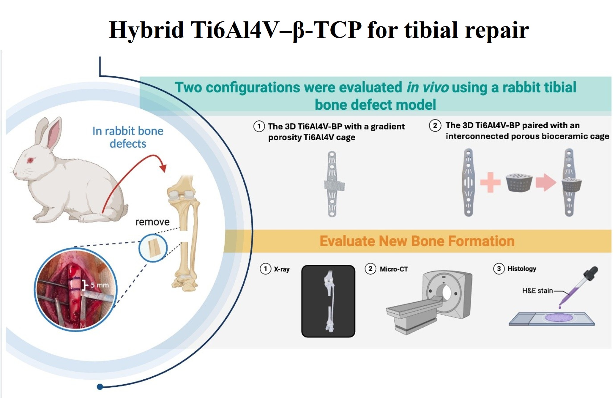
Computed tomography and finite element analysis-optimized Ti6Al4V bone plate combined with a porous 3D β-tricalcium phosphate cage for repairing segmental tibial defects in rabbits

Segmental bone defects caused by high-energy trauma remain a major clinical challenge because they often require mechanical stabilization and biological regeneration support for successful repair. Although autologous bone grafting is the clinical gold standard, its application is limited by donor-site morbidity and restricted graft availability. Metallic fixation devices provide mechanical stability, but metal-only implants do not actively support bone regeneration and may cause stress shielding or insufficient biological integration. To address these limitations, this study developed and evaluated two implant strategies for repairing rabbit tibial segmental defects. Computed tomography (CT) and computer-aided design (CAD) were used to establish a reproducible 5-mm rabbit tibial segmental defect model and guide implant design. Finite element analysis (FEA) optimized an integrated 3D-printed Ti6Al4V bone plate–cage construct with gradient porosity (50–70%) to reduce stress concentration while maintaining lightweight support. The cage construct was evaluated alongside a hybrid system consisting of a 3D-printed Ti6Al4V bone plate paired with an interconnected porous 3D β-tricalcium phosphate (β-TCP) cage fabricated by digital light processing using a negative-temperature-responsive slurry. Owing to its interconnected porous architecture, relatively slow degradation profile, and compressive properties comparable to cancellous bone, the porous β-TCP cage provided favorable structural support and maintained defect-site stability during early-stage regeneration. At 90 days, ex vivo micro-CT and histological analyses in the hybrid group confirmed bone ingrowth, vascularization, and osteogenic tissue infiltration. Metal-induced imaging artifacts and histological sampling constraints in the Ti6Al4V cage construct limited direct comparison of regenerative outcomes between the two groups. These findings support the feasibility of combining essential early mechanical fixation with an osteoconductive porous β-TCP cage for segmental tibial defect repair.
- Grün W, Hansen EJJ, Andreassen GS, Clarke-Jenssen J, Madsen JE. Functional outcomes and health-related quality of life after reconstruction of segmental bone loss in femur and tibia using the induced membrane technique. Arch Orthop Trauma Surg. 2023;143(8):4587-4596. doi: 10.1007/s00402-022-04714-9
- Konda SR, Gage M, Fisher N, Egol KA. Segmental bone defect treated with the induced membrane technique. J Orthop Trauma. 2017;31 Suppl 3:S21-S22. doi: 10.1097/BOT.0000000000000899
- Moura LB, Carvalho PH de A, Xavier CB, et al. Autogenous non-vascularized bone graft in segmental mandibular reconstruction: a systematic review. Int J Oral Maxillofac Surg. 2016;45(11):1388-1394. doi: 10.1016/j.ijom.2016.05.004
- Mohammadi SS, Phull SS, Bal BS, Towler MR. Bone void filler materials for augmentation in comminuted fractures: a comprehensive review. J Orthop Surg Res. 2025;20(1):449. doi: 10.1186/s13018-025-05857-2
- Sumner DR. Long-term implant fixation and stress-shielding in total hip replacement. J Biomech. 2015;48(5):797-800. doi: 10.1016/j.jbiomech.2014.12.021
- Qu H, Fu H, Han Z, Sun Y. Biomaterials for bone tissue engineering scaffolds: a review. RSC Adv. 2019;9(45):26252- 26262. doi: 10.1039/c9ra05214c
- Łuczak JW, Palusińska M, Matak D, et al. The future of bone repair: Emerging technologies and biomaterials in bone regeneration. Int J Mol Sci. 2024;25(23):12766. doi: 10.3390/ijms252312766
- Marin E, Lanzutti A. Biomedical applications of titanium alloys: A comprehensive review. Materials (Basel). 2023;17(1):114. doi: 10.3390/ma17010114
- Dewidar M, Mohamed HF, Jae-Kyoo Lim. A new approach for manufacturing a high porosity Ti-6Al-4V scaffolds for biomedical applications. J Mater Sci Technol. 2008;24(6):931.
- Wang R, Ni S, Ma L, Li M. Porous construction and surface modification of titanium-based materials for osteogenesis: A review. Front Bioeng Biotechnol. 2022;10:973297. doi: 10.3389/fbioe.2022.973297
- Brochu BM, Sturm SR, Kawase De Queiroz Goncalves JA, et al. Advances in bioceramics for bone regeneration: A narrative review. Biomimetics (Basel). 2024;9(11):690. doi: 10.3390/biomimetics9110690
- Lin CW, Su YF, Lee CY, et al. 3D printed bioceramics fabricated using negative thermoresponsive hydrogels and silicone oil sealing to promote bone formation in calvarial defects. Ceram Int. 2021;47(4):5464-5476. doi: 10.1016/j.ceramint.2020.10.129
- Lee CY, Nedunchezian S, Lin SY, et al. Bilayer osteochondral graft in rabbit xenogeneic transplantation model comprising sintered 3D-printed bioceramic and human adipose-derived stem cells laden biohydrogel. J Biol Eng. 2023;17(1):74. doi: 10.1186/s13036-023-00389-x
- Fu YC, Chen CH, Wang CZ, et al. Preparation of porous bioceramics using reverse thermo-responsive hydrogels in combination with rhBMP-2 carriers: in vitro and in vivo evaluation. J Mech Behav Biomed Mater. 2013;27:64-76. doi: 10.1016/j.jmbbm.2013.06.009
- Bose S, Vahabzadeh S, Bandyopadhyay A. Bone tissue engineering using 3D printing. Mater Today (Kidlington). 2013;16(12):496-504. doi: 10.1016/j.mattod.2013.11.017
- Pan CT, Hsu WH, Cheng YS, Wen ZH, Chen WF. A new design of porosity gradient Ti-6Al-4V encapsulated hydroxyapatite dual materials composite scaffold for bone defects. Micromachines (Basel). 2021;12(11):1294. doi: 10.3390/mi12111294
- Miltenberg B, Puzzitiello RN, Ruelos VCB, et al. Incidence of complications and revision surgery after high tibial osteotomy: A systematic review. Am J Sports Med. 2024;52(1):258-268. doi: 10.1177/03635465221142868
- Nguyen LH, Annabi N, Nikkhah M, et al. Vascularized bone tissue engineering: approaches for potential improvement. Tissue Eng Part B Rev. 2012;18(5):363-382. doi: 10.1089/ten.TEB.2012.0012
- Shi F, Xiao D, Zhang C, Zhi W, Liu Y, Weng J. The effect of macropore size of hydroxyapatite scaffold on the osteogenic differentiation of bone mesenchymal stem cells under perfusion culture. Regen Biomater. 2021;8(6):rbab050. doi: 10.1093/rb/rbab050
- Bacakova L, Filova E, Parizek M, Ruml T, Svorcik V. Modulation of cell adhesion, proliferation and differentiation on materials designed for body implants. Biotechnol Adv. 2011;29(6):739-767. doi: 10.1016/j.biotechadv.2011.06.004
- Zhou H, Lee J. Nanoscale hydroxyapatite particles for bone tissue engineering. Acta Biomater. 2011;7(7):2769-2781. doi: 10.1016/j.actbio.2011.03.019
- Habibovic P, Yuan H, van der Valk CM, Meijer G, van Blitterswijk CA, de Groot K. 3D microenvironment as essential element for osteoinduction by biomaterials. Biomaterials. 2005;26(17):3565-3575. doi: 10.1016/j.biomaterials.2004.09.056
- Wang X, Nie Z, Chang J, Lu ML, Kang Y. Multiple channels with interconnected pores in a bioceramic scaffold promote bone tissue formation. Sci Rep. 2021;11(1):20447. doi: 10.1038/s41598-021-00024-z
- Park JY, Jung YN, Jang KJ, et al. Effect of axis change on shrinkage rate of 3D-printed bioceramic Zirconia fabricated via digital light processing. Biomimetics (Basel). 2025;10(3):140. doi: 10.3390/biomimetics10030140
- Hussain M, Xia M, Ren X, et al. Digital light processing 3D printing of ceramic materials: a review on basic concept, challenges, and applications. Int J Adv Manuf Technol. 2024;130:2241-2267. doi: 10.1007/s00170-023-12847-3
- Sim JH, Koo BK, Jung M, Kim DS. Study on debinding and sintering processes for ceramics fabricated using digital light processing (DLP) 3D printing. Processes (Basel). 2022;10(11):2467. doi: 10.3390/pr10112467
- Barzanouni H, Houreh AB, Solati-Hashjin M, Barzanouni A, Shafieian M. 3D-Printed PLA: β-TCP scaffolds: fabrication and evaluation for bone regeneration in load-bearing defects. Polym Adv Technol. 2025;36(12):e70466. doi: 10.1002/pat.70466
- Guo W, Li P, Pang Y, et al. Biomimetic TPMS porous hydroxyapatite bone scaffolds doped with bioactive glass: digital light processing additive manufacturing, microstructure and performance. Compos Part A Appl Sci Manuf. 2025;193:108870. doi: 10.1016/j.compositesa.2025.108870
- Li Y, Li J, Jiang S, et al. The design of strut/TPMS-based pore geometries in bioceramic scaffolds guiding osteogenesis and angiogenesis in bone regeneration. Mater Today Bio. 2023;20:100667. doi: 10.1016/j.mtbio.2023.100667
- Gu Y, Sun Y, Shujaat S, Braem A, Politis C, Jacobs R. 3D-printed porous Ti6Al4V scaffolds for long bone repair in animal models: a systematic review. J Orthop Surg Res. 2022;17(1):68. doi: 10.1186/s13018-022-02960-6
- Wang X, Lin M, Kang Y. Engineering porous β-tricalcium phosphate (β-TCP) scaffolds with multiple channels to promote cell migration, proliferation, and angiogenesis. ACS Appl Mater Interfaces. 2019;11(9):9223-9232. doi: 10.1021/acsami.8b22041
- Ducheyne P, Bianco PD, Kim C. Bone tissue growth enhancement by calcium phosphate coatings on porous titanium alloys: the effect of shielding metal dissolution product. Biomaterials. 1992;13(9):617-624. doi: 10.1016/0142-9612(92)90030-r
- Lutzweiler G, Ndreu Halili A, Engin Vrana N. The overview of porous, bioactive scaffolds as instructive biomaterials for tissue regeneration and their clinical translation. Pharmaceutics. 2020;12(7):602. doi: 10.3390/pharmaceutics12070602
- Ivanovski S, Breik O, Carluccio D, Alayan J, Staples R, Vaquette C. 3D printing for bone regeneration: challenges and opportunities for achieving predictability. Periodontol 2000. 2023;93(1):358-384. doi: 10.1111/prd.12525
- Sadat-Shojai M, Asadnia M, Shahsavani MB, Yousefi MM. Bone regenerative medicine: An emerging field with opportunities and challenges. J Am Ceram Soc. 2025;108(11):e20508. doi: 10.1111/jace.20508
- Yuan B, Liu P, Zhao R, et al. Functionalized 3D-printed porous titanium scaffold induces in situ vascularized bone regeneration by orchestrating bone microenvironment. J Mater Sci Technol. 2023;153:92-105. doi: 10.1016/j.jmst.2022.12.033
- Liu J, Jin F, Zheng ML, et al. Cell behavior on 3D Ti-6Al-4V scaffolds with different porosities. ACS Appl Bio Mater. 2019;2(2):697-703. doi: 10.1021/acsabm.8b00550
- Ma L, Wang X, Zhou Y, et al. Biomimetic Ti-6Al-4V alloy/gelatin methacrylate hybrid scaffold with enhanced osteogenic and angiogenic capabilities for large bone defect restoration. Bioact Mater. 2021;6(10):3437-3448. doi: 10.1016/j.bioactmat.2021.03.010
- Van Bael S, Chai YC, Truscello S, et al. The effect of pore geometry on the in vitro biological behavior of human periosteum-derived cells seeded on selective laser-melted Ti6Al4V bone scaffolds. Acta Biomater. 2012;8(7):2824- 2834. doi: 10.1016/j.actbio.2012.04.001
- Arabnejad S, Johnston B, Tanzer M, Pasini D. Fully porous 3D printed titanium femoral stem to reduce stress-shielding following total hip arthroplasty. J Orthop Res. 2017;35(8):1774-1783. doi: 10.1002/jor.23445
- LeGeros RZ. Properties of osteoconductive biomaterials: calcium phosphates. Clin Orthop Relat Res. 2002;395(395):81- 98. doi: 10.1097/00003086-200202000-00009
- Hutmacher DW. Scaffolds in tissue engineering bone and cartilage. Biomaterials. 2000;21(24):2529-2543. doi: 10.1016/s0142-9612(00)00121-6
- Bose S, Roy M, Bandyopadhyay A. Recent advances in bone tissue engineering scaffolds. Trends Biotechnol. 2012;30(10):546-554. doi: 10.1016/j.tibtech.2012.07.005
- Wong SK, Yee MMF, Chin KY, Ima-Nirwana S. A review of the application of natural and synthetic scaffolds in bone regeneration. J Funct Biomater. 2023;14(5):286. doi: 10.3390/jfb14050286