

Ta/PEEK cages with enhanced hydrophilicity and cell adhesion improve intervertebral fusion in a rabbit model of extreme lateral intervertebral fusion

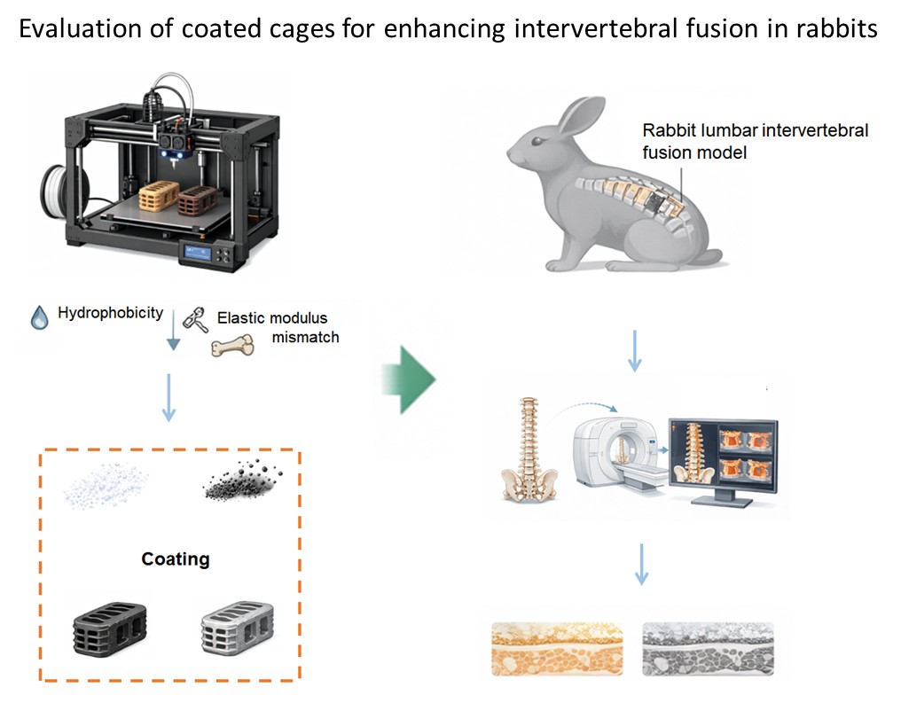
Conventional polyether ether ketone (PEEK) and titanium (Ti) cages for intervertebral fusion exhibit inherent limitations in bioactivity and elastic modulus, necessitating surface modification to enhance their performance. This study developed bioactive cages by fabricating hydroxyapatite-coated Ti (HA/Ti) and tantalum-coated PEEK (Ta/PEEK) cages and evaluated their physicochemical properties, cellular responses, and intervertebral fusion performance in a rabbit extreme lateral intervertebral fusion model. Both coatings improved surface wettability and maintained the intrinsic mechanical properties of the substrates. Ta/PEEK showed superior coating adhesion and enhanced expression of bone marrow-derived mesenchymal stem cell adhesion-related markers, whereas HA/Ti more strongly promoted osteogenic differentiation and matrix mineralization in vitro. In vivo, both coated cages significantly improved intervertebral fusion compared with uncoated Ti and PEEK cages. HA/Ti induced greater new bone formation, while Ta/PEEK enhanced cell adhesion and angiogenesis, as indicated by increased cadherin-1, integrin α2, cluster of differentiation 31, and hypoxia-inducible factor 1α expression. These findings suggest that Ta/PEEK cages provide a mechanically compatible and biologically active strategy for improving intervertebral fusion and represent a promising platform for next-generation printed spinal implants.
- Kotheeranurak V, Jitpakdee K, Lin GX, et al. Subsidence of Interbody Cage Following Oblique Lateral Interbody Fusion: An Analysis and Potential Risk Factors. Global Spine J. 2023;13(7):1981-1991. doi: 10.1177/21925682211067210
- Xuan W, Cheng Q, Gao Y, Song Z, Gao Z. Early-stage lumbar paraspinal muscle injury: endoscopic versus transforaminal lumbar interbody fusion: a retrospective comparative analysis. J Orthop Surg Res. 2025;20(1):841. doi: 10.1186/s13018-025-06235-8
- Zhou H, Bian H, Zhang Y, et al. Evaluation of Indirect Decompression Effect After Extreme Lateral Lumbar Interbody Fusion Using Three-Dimensional Volumetric Measurements-A Retrospective Study. Orthop Surg. 2025;17(9):2558-2569. doi: 10.1111/os.70108
- Wu H, Shan Z, Zhao F, Cheung JPY. Poor Bone Quality, Multilevel Surgery, and Narrow and Tall Cages Are Associated with Intraoperative Endplate Injuries and Late-onset Cage Subsidence in Lateral Lumbar Interbody Fusion: A Systematic Review. Clin Orthop Relat Res. 2022;480(1):163-188. doi: 10.1097/corr.0000000000001915
- Burkhard MD, Guven AE, Demopoulos B, et al. 3D-Printed Titanium: Game-Changer for Standalone Lateral Lumbar Interbody Fusion? An Analysis of Risk Factors for Revision Surgery. Global Spine J. 2025:21925682251383879. doi: 10.1177/21925682251383879
- Schnake KJ, Fleiter N, Hoffmann C, et al. PLIF with titanium-coated versus uncoated PEEK cages: a prospective randomized clinical trial. Eur Spine J. 2021;30(1):114-121. doi: 10.1007/s00586-020-06642-x
- Jain S, Eltorai AE, Ruttiman R, Daniels AH. Advances in Spinal Interbody Cages. Orthop Surg. 2016;8(3):278-284. doi: 10.1111/os.12264
- Zhang Y, Yang J, Wan W, et al. Evaluation of biological performance of 3D printed trabecular porous tantalum spine fusion cage in large animal models. J Orthop Translat. 2025;50:185-195. doi: 10.1016/j.jot.2024.10.010
- He X, Li Y, Zou D, Zu H, Li W, Zheng Y. An overview of magnesium-based implants in orthopaedics and a prospect of its application in spine fusion. Bioact Mater. 2024;39:456-478. doi: 10.1016/j.bioactmat.2024.04.026
- Zhang H, Gao G, Wang L, et al. Innovative 3D-printed porous piezoelectric poly(vinylidene fluoride) cages with accelerated spinal fusion. Mater Today Bio. 2025;35:102333. doi: 10.1016/j.mtbio.2025.102333
- Wu H, Dong H, Tang Z, et al. Electrical stimulation of piezoelectric BaTiO3 coated Ti6Al4V scaffolds promotes anti-inflammatory polarization of macrophages and bone repair via MAPK/JNK inhibition and OXPHOS activation. Biomaterials. 2023;293:121990. doi: 10.1016/j.biomaterials.2022.121990
- Zheng H, Cheng F, Guo D, He X, Zhou L, Zhang Q. Nanoenzyme-Reinforced Multifunctional Scaffold Based on Ti(3)C(2)Tx MXene Nanosheets for Promoting Structure-Functional Skeletal Muscle Regeneration via Electroactivity and Microenvironment Management. Nano Lett. 2023;23(16):7379-7388. doi: 10.1021/acs.nanolett.3c01784
- Luo L, Zheng W, Li J, et al. 3D-Printed Titanium Trabecular Scaffolds with Sustained Release of Hypoxia-Induced Exosomes for Dual-Mimetic Bone Regeneration. Adv Sci (Weinh). 2025;12(23):e2500599. doi: 10.1002/advs.202500599
- Lin R, Wang Z, Li Z, Gu L. A two-phase and long-lasting multi-antibacterial coating enables titanium biomaterials to prevent implants-related infections. Mater Today Bio. 2022;15:100330. doi: 10.1016/j.mtbio.2022.100330
- Coppola B, Montanaro L, Palmero P. DLP Fabrication of Zirconia Scaffolds Coated with HA/β-TCP Layer: Role of Scaffold Architecture on Mechanical and Biological Properties. J Funct Biomater. 2022;13(3):148. doi: 10.3390/jfb13030148
- Sun T, Huang H, Zhao Y, Li Z, Wang H, Zhou G. Low-Temperature Deposited Amorphous Poly(aryl ether ketone) Hierarchically Porous Scaffolds with Strontium-Doped Mineralized Coating for Bone Defect Repair. Adv Healthc Mater. 2024;13(23):e2400927. doi: 10.1002/adhm.202400927
- Durham JW 3rd, Rabiei A. Deposition, Heat Treatment And Characterization of Two Layer Bioactive Coatings on Cylindrical PEEK. Surf Coat Technol. 2016;301:106-113. doi: 10.1016/j.surfcoat.2015.12.045
- Torstrick FB, Lin ASP, Potter D, et al. Porous PEEK improves the bone-implant interface compared to plasma-sprayed titanium coating on PEEK. Biomaterials. 2018;185:106-116. doi: 10.1016/j.biomaterials.2018.09.009
- Li J, Zhao B, Wang W, Xu Y, Wu H, Zhang W. Improved intervertebral fusion in LLIF rabbit model with a novel titanium cage. Spine J. 2024;24(6):1109-1120. doi: 10.1016/j.spinee.2023.12.011
- Guillot R, Pignot-Paintrand I, Lavaud J, et al. Assessment of a polyelectrolyte multilayer film coating loaded with BMP-2 on titanium and PEEK implants in the rabbit femoral condyle. Acta Biomater. 2016;36:310-322. doi: 10.1016/j.actbio.2016.03.010
- Zhang J, Ma T, Liu X, Zhang X, Meng W, Wu J. Multifunctional surface of the nano-morphic PEEK implant with enhanced angiogenic, osteogenic and antibacterial properties. Regen Biomater. 2024;11:rbae067. doi: 10.1093/rb/rbae067
- An J, Shi X, Zhang J, et al. Dual aldehyde cross-linked hyaluronic acid hydrogels loaded with PRP and NGF biofunctionalized PEEK interfaces to enhance osteogenesis and vascularization. Mater Today Bio. 2024;24:100928. doi: 10.1016/j.mtbio.2023.100928
- Adl Amini D, Okano I, Oezel L, et al. Evaluation of cage subsidence in standalone lateral lumbar interbody fusion: novel 3D-printed titanium versus polyetheretherketone (PEEK) cage. Eur Spine J. 2021;30(8):2377-2384. doi: 10.1007/s00586-021-06912-2
- Mahjoubi H, Buck E, Manimunda P, et al. Surface phosphonation enhances hydroxyapatite coating adhesion on polyetheretherketone and its osseointegration potential. Acta Biomater. 2017;47:149-158. doi: 10.1016/j.actbio.2016.10.004
- Zhang W, Shi D, Huang S, Li S, Zeng M, Wei Y. Personalised 3D-printed bioactive peek bone plate scaffold for treating femoral defects. RSC Adv. 2025;15(7):5060-5072. doi: 10.1039/d4ra07573k
- Xu J, Wu D, Ge B, et al. Selective Laser Melting of the Porous Ta Scaffold with Mg-Doped Calcium Phosphate Coating for Orthopedic Applications. ACS Biomater Sci Eng. 2024;10(3):1435-1447. doi: 10.1021/acsbiomaterials.3c01503
- Zhu C, He M, Mao L, et al. Titanium interlayer-mediated hydroxyapatite-coated polyetheretherketone cage in transforaminal lumbar interbody fusion surgery. BMC Musculoskelet Disord. 2021;22(1):918. doi: 10.1186/s12891-021-04803-7
- Jia CQ, Zhang Z, Cao SQ, et al. A biomimetic gradient porous cage with a micro-structure for enhancing mechanical properties and accelerating osseointegration in spinal fusion. Bioact Mater. 2022;23:234-246. doi: 10.1016/j.bioactmat.2022.11.003
- Ragni E, Perucca Orfei C, Bidossi A, et al. Superior Osteo-Inductive and Osteo-Conductive Properties of Trabecular Titanium vs. PEEK Scaffolds on Human Mesenchymal Stem Cells: A Proof of Concept for the Use of Fusion Cages. Int J Mol Sci. 2021;22(5):2379. doi: 10.3390/ijms22052379
- Laubach M, Kobbe P, Hutmacher DW. Biodegradable interbody cages for lumbar spine fusion: Current concepts and future directions. Biomaterials. 2022;288:121699. doi: 10.1016/j.biomaterials.2022.121699
- Mobbs RJ, Phan K, Assem Y, Pelletier M, Walsh WR. Combination Ti/PEEK ALIF cage for anterior lumbar interbody fusion: Early clinical and radiological results. J Clin Neurosci. 2016;34:94-99. doi: 10.1016/j.jocn.2016.05.028
- Park PJ, Lehman RA. Optimizing the Spinal Interbody Implant: Current Advances in Material Modification and Surface Treatment Technologies. Curr Rev Musculoskelet Med. 2020;13(6):688-695. doi: 10.1007/s12178-020-09673-5
- Lu T, Wen J, Qian S, et al. Enhanced osteointegration on tantalum-implanted polyetheretherketone surface with bone-like elastic modulus. Biomaterials. 2015;51:173-183. doi: 10.1016/j.biomaterials.2015.02.018
- Hwangbo H, Lee J, Kim G. Mechanically and biologically enhanced 3D-printed HA/PLLA/dECM biocomposites for bone tissue engineering. Int J Biol Macromol. 2022;218:9-21. doi: 10.1016/j.ijbiomac.2022.07.040
- Blom AM, Petersen EB, Grieser-Yoder RA, Olinger CR, Fredericks DC. A Novel Nano-Synthetic Hydrophilic Bone Graft (OsteoFlo HydroFiber) Is a Non-inferior Alternative to Iliac Crest Autograft in a Rabbit Posterolateral Fusion Model. Global Spine J. 2026. doi: 10.1177/21925682261442308
- Jin YZ, Zheng GB, Cho M, Lee JH. Effect of Whitlockite as a new bone substitute for bone formation in spinal fusion and ectopic ossification animal model. Biomater Res. 2021;25(1):34. doi: 10.1186/s40824-021-00237-3
- Ryan DA, Cheng J, Masuda K, Cashman JR. Role of Curcuminoids and Tricalcium Phosphate Ceramic in Rat Spinal Fusion. Tissue Eng Part C Methods. 2020;26(11):577-589. doi: 10.1089/ten.TEC.2020.0217
- Sun J, Liu SS, Zou D, et al. A novel porous interbody fusion cage modified by microarc oxidation and hydrothermal treatment technology accelerate osseointegration and spinal fusion in sheep. RSC Adv. 2024;14(44):31966-31978. doi: 10.1039/d3ra08185k
- Tian L, Zhang Z, Tian B, Zhang X, Wang N. Study on antibacterial properties and cytocompatibility of EPL coated 3D printed PCL/HA composite scaffolds. RSC Adv. 2020;10(8):4805-4816. doi: 10.1039/c9ra10275b
- Liu B, Ma Z, Li J, et al. Experimental study of a 3D printed permanent implantable porous Ta-coated bone plate for fracture fixation. Bioact Mater. 2022;10:269-280. doi: 10.1016/j.bioactmat.2021.09.009
- Liu T, Li B, Chen G, Ye X, Zhang Y. Nano tantalum-coated 3D printed porous polylactic acid/beta-tricalcium phosphate scaffolds with enhanced biological properties for guided bone regeneration. Int J Biol Macromol. 2022;221:371-380. doi: 10.1016/j.ijbiomac.2022.09.003
- Tao Y, Jia M, Shao-Qiang Y, et al. A novel fluffy PLGA/HA composite scaffold for bone defect repair. J Mater Sci Mater Med. 2024;35(1):16. doi: 10.1007/s10856-024-06782-2
- Duan W, Chen C, Haque M, Hayes D, Lopez MJ. Polymer-mineral scaffold augments in vivo equine multipotent stromal cell osteogenesis. Stem Cell Res Ther. 2018;9(1):60. doi: 10.1186/s13287-018-0790-8
- Chi H, Chen G, He Y, et al. 3D-HA Scaffold Functionalized by Extracellular Matrix of Stem Cells Promotes Bone Repair. Int J Nanomedicine. 2020;15:5825-5838. doi: 10.2147/ijn.S259678
- Zhang K, Hu H, Sun Y, et al. The bio-functionalized membrane loaded with Ta/WH nanoparticles promote bone regeneration through neurovascular coupling. Colloids Surf B Biointerfaces. 2023;230:113506. doi: 10.1016/j.colsurfb.2023.113506
- Baldwin P, Li DJ, Auston DA, Mir HS, Yoon RS, Koval KJ. Autograft, Allograft, and Bone Graft Substitutes: Clinical Evidence and Indications for Use in the Setting of Orthopaedic Trauma Surgery. J Orthop Trauma. 2019;33(4):203-213. doi: 10.1097/bot.0000000000001420
- Gupta A, Kukkar N, Sharif K, Main BJ, Albers CE, El-Amin Iii SF. Bone graft substitutes for spine fusion: A brief review. World J Orthop. 2015;6(6):449-456. doi: 10.5312/wjo.v6.i6.449
- Zhang J, Tong D, Song H, et al. Osteoimmunity-Regulating Biomimetically Hierarchical Scaffold for Augmented Bone Regeneration. Adv Mater. 2022;34(36):e2202044. doi: 10.1002/adma.202202044
- Deshpande R, Shukla S, Kale A, Deshmukh N, Nisal A, Venugopalan P. Silk Fibroin Microparticle Scaffold for Use in Bone Void Filling: Safety and Efficacy Studies. ACS Biomater Sci Eng. 2022;8(3):1226-1238. doi: 10.1021/acsbiomaterials.1c01103