

Mesenchymal stem cell differentiation in constructs fabricated by three-dimensional bioprinting using a carboxyvinyl polymer-based support material

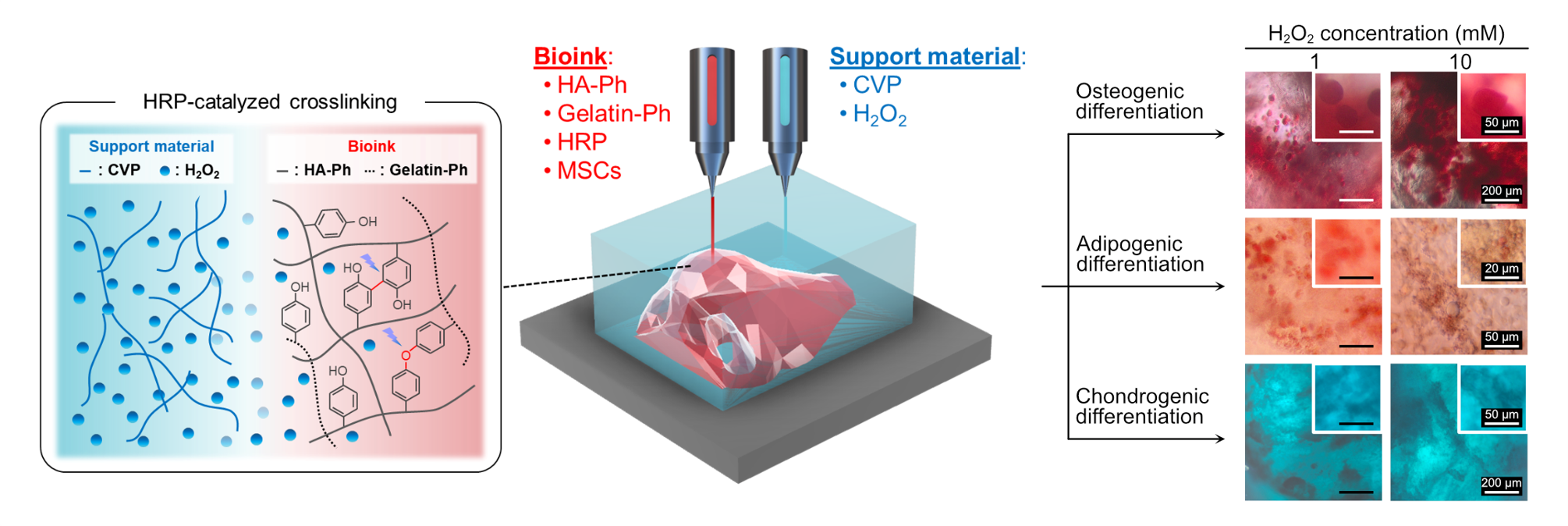
Three-dimensional (3D) bioprinting using a carboxyvinyl polymer (CVP) solution as a support material, alternately extruded with bioink, holds great promise for tissue engineering and regenerative medicine. This approach enables the fabrication of constructs that are sufficiently soft to preserve the functionality of enclosed cells. To evaluate the applicability of this support material and printing method for mesenchymal stem cells (MSCs), we examined the behavior of immortalized human bone marrow-derived MSCs (UE7T-13). This was carried out with constructs printed using bioinks containing 0.5 w/v% phenol-containing hyaluronic acid, 0.1 w/v% phenol-containing gelatin, and 10 U/mL horseradish peroxidase, which were crosslinked by hydrogen peroxide (H2O2) supplied from a support material composed primarily of CVP. During the 14 days of culture after printing, UE7T-13 cells enclosed in the printed constructs elongated and proliferated while maintaining high viability (>90%) and metabolic activity. Furthermore, antibody immunostaining revealed that the cells within the constructs maintained the characteristic MSC phenotype (CD44+ and CD45−). Additionally, following osteogenic, adipogenic, and chondrogenic induction, the cells successfully produced lineage-associated products within the constructs, and the amounts of these products were controlled by varying the H2O2 concentration (1–10 mM) in the support material. Overall, the use of CVP together with H2O2 in the support material, combined with 3D bioprinting, effectively minimized adverse effects on MSC-associated properties and differentiation potential, while providing a practical strategy for regulating lineage-specific differentiation. These results demonstrate that this method enhances the utility of horseradish peroxidase-containing bioinks and offers great potential for tissue engineering applications.
- Klebe RJ. Cytoscribing: a method for micropositioning cells and the construction of two- and three-dimensional synthetic tissues. Exp Cell Res. 1988;179(2):362-373. doi: 10.1016/0014-4827(88)90275-3
- Mironov V, Boland T, Trusk T, et al. Organ printing: computer-aided jet-based 3D tissue engineering. Trends Biotechnol. 2003;21(4):157-161. doi: 10.1016/S0167-7799(03)00033-7
- Nakamura M, Iwanaga S, Henmi C, et al. Biomatrices and biomaterials for future developments of bioprinting and biofabrication. Biofabrication. 2010;2(1):014110. doi: 10.1088/1758-5082/2/1/014110
- Holland I. Extrusion bioprinting: meeting the promise of human tissue biofabrication? Prog Biomed Eng (Bristol). 2025;7(2):023001. doi: 10.1088/2516-1091/adb254
- Kazemi M, Maralbashi S. Advances in 3D bioprinting for medical application: opportunities and challenges. Biomed Eng Online. 2025;25(1):11. doi: 10.1186/s12938-025-01498-y
- Frost BA, Sutliff BP, Thayer P, et al. Gradient Poly(ethylene glycol) Diacrylate and Cellulose Nanocrystals Tissue Engineering Composite Scaffolds via Extrusion Bioprinting. Front Bioeng Biotechnol. 2019;7:280. doi: 10.3389/fbioe.2019.00280
- Daly AC, Prendergast ME, Hughes AJ, et al. Bioprinting for the Biologist. Cell. 2021;184(1):18-32. doi: 10.1016/j.cell.2020.12.002
- Tejada Jacob G, Passamai VE, Katz S, et al. Hydrogels for extrusion-based bioprinting: General considerations. Bioprinting. 2022;27:e00212. doi: 10.1016/j.bprint.2022.e00212
- Shiwarski DJ, Hudson AR, Tashman JW, et al. Emergence of FRESH 3D printing as a platform for advanced tissue biofabrication. APL Bioeng. 2021;5(1):010904. doi: 10.1063/5.0032777
- Hinton TJ, Jallerat Q, Palchesko RN, et al. Three-dimensional printing of complex biological structures by freeform reversible embedding of suspended hydrogels. Sci Adv. 2015;1(9):e1500758. doi: 10.1126/sciadv.1500758
- Kotani T, Mubarok W, Hananouchi T. Horseradish Peroxidase-Mediated Bioprinting via Bioink Gelation by Alternately Extruded Support Material. ACS Biomater Sci Eng. 2023;9(10):5804-5812. doi: 10.1021/acsbiomaterials.3c00996
- Kotani T, Hananouchi T, Sakai S. Enhancing visible light-induced 3D bioprinting: alternating extruded support materials for bioink gelation. Biomed Mater. 2025;20(3):035005. doi: 10.1088/1748-605X/adc0d6
- Kurisawa M, Chung JE, Yang YY, Gao SJ, Uyama H. Injectable biodegradable hydrogels composed of hyaluronic acid-tyramine conjugates for drug delivery and tissue engineering. Chem Commun (Camb). 2005;(34):4312-4314. doi: 10.1039/b506989k
- Sakai S, Matsuyama T, Hirose K, et al. In situ simultaneous protein-polysaccharide bioconjugation and hydrogelation using horseradish peroxidase. Biomacromolecules. 2010;11(5):1370-1375. doi: 10.1021/bm1001608
- Fancy DA, Kodadek T. Chemistry for the analysis of protein-protein interactions: rapid and efficient cross-linking triggered by long wavelength light. Proc Natl Acad Sci USA. 1999;96(11):6020-6024. doi: 10.1073/pnas.96.11.6020
- Elvin CM, Brownlee AG, Huson MG, et al. The development of photochemically crosslinked native fibrinogen as a rapidly formed and mechanically strong surgical tissue sealant. Biomaterials. 2009;30(11):2059-2065. doi: 10.1016/j.biomaterials.2008.12.059
- Lee F, Chung JE, Kurisawa M. An injectable enzymatically crosslinked hyaluronic acid-hydrogel system with independent tuning of mechanical strength and gelation rate. Soft Matter. 2008;4(4):880-887. doi: 10.1039/b719557e
- Sakai S, Ohi H, Hotta T, et al. Differentiation potential of human adipose stem cells bioprinted with hyaluronic acid/gelatin-based bioink through microextrusion and visible light-initiated crosslinking. Biopolymers. 2018;109(2):e23080. doi: 10.1002/bip.23080
- Sakai S, Hirose K, Moriyama K, et al. Control of cellular adhesiveness in an alginate-based hydrogel by varying peroxidase and H(2)O(2) concentrations during gelation. Acta Biomater. 2010;6(4):1446-1452. doi: 10.1016/j.actbio.2009.10.004
- Sakai S, Kamei H, Mori T, et al. Visible Light-Induced Hydrogelation of an Alginate Derivative and Application to Stereolithographic Bioprinting Using a Visible Light Projector and Acid Red. Biomacromolecules. 2018;19(2):672- 679. doi: 10.1021/acs.biomac.7b01827
- Jin R, Moreira Teixeira LS, Dijkstra PJ, et al. Injectable chitosan-based hydrogels for cartilage tissue engineering. Biomaterials. 2009;30(13):2544-2551. doi: 10.1016/j.biomaterials.2009.01.020
- Hidaka M, Kojima M, Nakahata M, et al. Visible Light- Curable Chitosan Ink for Extrusion-Based and Vat Polymerization-Based 3D Bioprintings. Polymers (Basel). 2021;13(9):1382. doi: 10.3390/polym13091382
- Sakai S, Tsumura M, Inoue M, et al. Polyvinyl alcohol-based hydrogel dressing gellable on-wound via a co-enzymatic reaction triggered by glucose in the wound exudate. J Mater Chem B. 2013;1(38):5067-5075. doi: 10.1039/c3tb20780c
- Lim KS, Ramaswamy Y, Roberts JJ, et al. Promoting Cell Survival and Proliferation in Degradable Poly(vinyl alcohol)- Tyramine Hydrogels. Macromol Biosci. 2015;15(10):1423- 1432. doi: 10.1002/mabi.201500121
- Hu M, Kurisawa M, Deng R, et al. Cell immobilization in gelatin-hydroxyphenylpropionic acid hydrogel fibers. Biomaterials. 2009;30(21):3523-3531. doi: 10.1016/j.biomaterials.2009.03.004
- Sakai S, Ohi H, Taya M. Gelatin/Hyaluronic Acid Content in Hydrogels Obtained through Blue Light-Induced Gelation Affects Hydrogel Properties and Adipose Stem Cell Behaviors. Biomolecules. 2019;9(8):342. doi: 10.3390/biom9080342
- Ogushi Y, Sakai S, Kawakami K. Synthesis of enzymatically-gellable carboxymethylcellulose for biomedical applications. J Biosci Bioeng. 2007;104(1):30-33. doi: 10.1263/jbb.104.30
- Badali E, Hosseini M, Varaa N, et al. Production of uniform size cell-enclosing silk derivative vehicles through coaxial microfluidic device and horseradish crosslinking reaction. Eur Polym J. 2020;172:111237. doi: 10.1016/j.eurpolymj.2022.111237
- Mu X, Sahoo JK, Cebe P, et al. Photo-Crosslinked Silk Fibroin for 3D Printing. Polymers (Basel). 2020;12(12):2936. doi: 10.3390/polym12122936
- Prasetyaningrum PW, Mubarok W, Kotani T, et al. Development of starch-based support material alternately extruded with gelatin-based bioinks for 3D bioprinting application. Bioprinting. 2025;51:e00439. doi: 10.1016/j.bprint.2025.e00439
- Liang L, Li Z, Yao B, et al. Extrusion bioprinting of cellular aggregates improves mesenchymal stem cell proliferation and differentiation. Biomater Adv. 2023;149:213369. doi: 10.1016/j.bioadv.2023.213369
- Pittenger MF, Mackay AM, Beck SC, et al. Multilineage potential of adult human mesenchymal stem cells. Science. 1999;284(5411):143-147. doi: 10.1126/science.284.5411.143
- Mohand-Kaci F, Assoul N, Martelly I, et al. Optimized hyaluronic acid-hydrogel design and culture conditions for preservation of mesenchymal stem cell properties. Tissue Eng Part C Methods. 2013;19(4):288-298. doi: 10.1089/ten.TEC.2012.0144
- Caiado Decarli M, Ferreira HP, Sobreiro-Almeida R, et al. Embedding Bioprinting of Low Viscous, Photopolymerizable Blood-Based Bioinks in a Crystal Self-Healing Transparent Supporting Bath. Small Methods. 2025;9(1):e2400857. doi: 10.1002/smtd.202400857
- Ning L, Mehta R, Cao C, et al. Embedded 3D Bioprinting of Gelatin Methacryloyl-Based Constructs with Highly Tunable Structural Fidelity. ACS Appl Mater Interfaces. 2020;12(40):44563-44577. doi: 10.1021/acsami.0c15078
- Hinton TJ, Hudson A, Pusch K, et al. 3D Printing PDMS Elastomer in a Hydrophilic Support Bath via Freeform Reversible Embedding. ACS Biomater Sci Eng. 2016;2(10):1781-1786. doi: 10.1021/acsbiomaterials.6b00170
- Deng Z, Nakagawa M, Zheng S, Yu L, Iwasaki K, Honda Y. Induction of apoptosis for UE7T-13 cells using ultraviolet treatment. J Osaka Dent Univ. 2025;59(1):191-198. doi: 10.18905/jodu.59.1_191
- Chatree K, Sriboonaied P, Phetkong C, et al. Distinctions in bone matrix nanostructure, composition, and formation between osteoblast-like cells, MG-63, and human mesenchymal stem cells, UE7T-13. Heliyon. 2023;9(5):e15556. doi: 10.1016/j.heliyon.2023.e15556
- Takeda Y, Mori T, Imabayashi H, et al. Can the life span of human marrow stromal cells be prolonged by bmi-1, E6, E7, and/or telomerase without affecting cardiomyogenic differentiation? J Gene Med. 2004;6(8):833-845. doi: 10.1002/jgm.583
- Sakai S, Hirose K, Taguchi K, et al. An injectable, in situ enzymatically gellable, gelatin derivative for drug delivery and tissue engineering. Biomaterials. 2009;30(20):3371- 3377. doi: 10.1016/j.biomaterials.2009.03.030
- Sakai S, Kotani T, Harada R, et al. Development of phenol-grafted polyglucuronic acid and its application to extrusion-based bioprinting inks. Carbohydr Polym. 2022;277:118820. doi: 10.1016/j.carbpol.2021.118820
- Snyder J, Rin Son A, Hamid Q, et al. Mesenchymal stem cell printing and process regulated cell properties. Biofabrication. 2015;7(4):044106. doi: 10.1088/1758-5090/7/4/044106
- Dong JD, Gu YQ, Li CM, et al. Response of mesenchymal stem cells to shear stress in tissue-engineered vascular grafts. Acta Pharmacol Sin. 2009;30(5):530-536. doi: 10.1038/aps.2009.40
- Bai K, Huang Y, Jia X, et al. Endothelium oriented differentiation of bone marrow mesenchymal stem cells under chemical and mechanical stimulations. J Biomech. 2010;43(6):1176-1181. doi: 10.1016/j.jbiomech.2009.11.030
- Elvitigala K, Mubarok W, Sakai S. Hydrogels with Ultrasound-Treated Hyaluronic Acid Regulate CD44- Mediated Angiogenic Potential of Human Vascular Endothelial Cells In Vitro. Biomolecules. 2024;14(5):604. doi: 10.3390/biom14050604
- Mimura S, Kimura N, Hirata M, et al. Growth factor-defined culture medium for human mesenchymal stem cells. Int J Dev Biol. 2011;55(2):181-187. doi: 10.1387/ijdb.103232sm
- Demirel G, Cakil YD, Koltuk G, et al. The use of hyaluronic acid in a 3D biomimetic scaffold supports spheroid formation and the culture of cancer stem cells. Sci Rep. 2024;14(1):19560. doi: 10.1038/s41598-024-69047-6
- Kim Y, Kumar S. CD44-mediated adhesion to hyaluronic acid contributes to mechanosensing and invasive motility. Mol Cancer Res. 2014;12(10):1416-1429. doi: 10.1158/1541-7786.MCR-13-0629
- Sano M, Mine Y, Okazaki S, et al. Deep learning predicts osteogenic differentiation stages of human mesenchymal stem cells from phase-contrast microscopy images. Dent Mater J. 2025;44(5):557-563. doi: 10.4012/dmj.2025-015
- Edwards SD, Ganash M, Guan Z, et al. Enhanced osteogenesis of mesenchymal stem cells encapsulated in injectable microporous hydrogel. Sci Rep. 2024;14(1):14665. doi: 10.1038/s41598-024-65731-9
- Duarte Campos DF, Blaeser A, Buellesbach K, et al. Bioprinting Organotypic Hydrogels with Improved Mesenchymal Stem Cell Remodeling and Mineralization Properties for Bone Tissue Engineering. Adv Healthc Mater. 2016;5(11):1336-1345. doi: 10.1002/adhm.201501033
- Ogushi Y, Sakai S, Kawakami K. Adipose tissue engineering using adipose-derived stem cells enclosed within an injectable carboxymethylcellulose-based hydrogel. J Tissue Eng Regen Med. 2013;7(11):884-892. doi: 10.1002/term.1480
- Bian L, Hou C, Tous E, et al. The influence of hyaluronic acid hydrogel crosslinking density and macromolecular diffusivity on human MSC chondrogenesis and hypertrophy. Biomaterials. 2013;34(2):413-421. doi: 10.1016/j.biomaterials.2012.09.052
- Machour M, Hen N, Goldfracht I, et al. Print-and-Grow within a Novel Support Material for 3D Bioprinting and Post-Printing Tissue Growth. Adv Sci (Weinh). 2022;9(34):e2200882. doi: 10.1002/advs.202200882
- Poveda-Reyes S, Moulisova V, Sanmartin-Masia E, et al. Gelatin-Hyaluronic Acid Hydrogels with Tuned Stiffness to Counterbalance Cellular Forces and Promote Cell Differentiation. Macromol Biosci. 2016;16(9):1311-1324. doi: 10.1002/mabi.201500469
- McAndrews KM, McGrail DJ, Quach ND, et al. Spatially coordinated changes in intracellular rheology and extracellular force exertion during mesenchymal stem cell differentiation. Phys Biol. 2014;11(5):056004. doi: 10.1088/1478-3975/11/5/056004