

Progress, challenges, and clinical prospects of hydrogel-based bioinks in spinal fusion applications

Traditional spinal fusion cages and autologous bone grafts face well-recognized limitations, creating a growing clinical need for next-generation fusion technologies. Research has accelerated the development of bioactive materials, with hydrogels emerging as a particularly versatile class for bone repair. These materials can provide structural support while facilitating tissue regeneration through the incorporation of therapeutics that promote integration with native bone. The convergence of hydrogel design and bioprinting technologies may enable customizable, biologically active fusion constructs that function with the body’s repair mechanisms, allowing for more personalized and optimal treatment outcomes. This review summarizes current research on bioprinted hydrogel systems, focusing on materials selection, mechanical strength, degradability, bioink printability, and nanomaterial incorporation. It also discusses pre-clinical and computational approaches for assessing how scaffold design, material selection, printing parameters, printing strategies, and material flow affect mechanical behavior, material properties, and cell viability. Overall, the review highlights potential research avenues for developing new therapeutic platforms for spinal fusion applications.
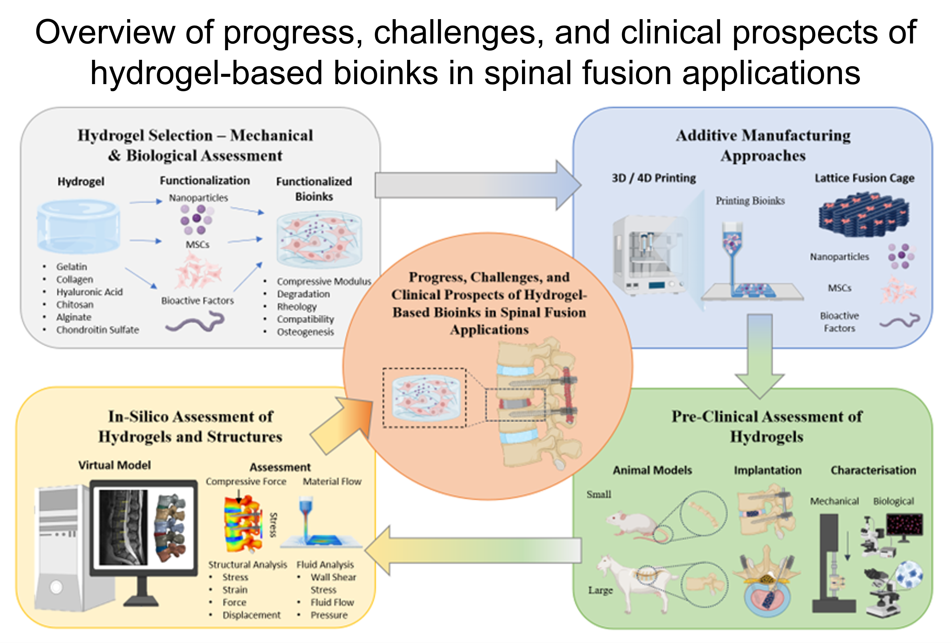
- Fehlings MG, Tetreault L, Nater A, et al. The Aging of the Global Population: The Changing Epidemiology of Disease and Spinal Disorders. Neurosurgery. 2015;77(suppl 4):S1-S5. doi: 10.1227/NEU.0000000000000953
- Epstein S, Sparer EH, Tran BN, et al. Prevalence of Work-Related Musculoskeletal Disorders Among Surgeons and Interventionalists: A Systematic Review and Meta-analysis. JAMA Surg. 2018;153(2):e174947. doi: 10.1001/jamasurg.2017.4947
- Rathbone J, Rackham M, Nielsen D, et al. A systematic review of anterior lumbar interbody fusion (ALIF) versus posterior lumbar interbody fusion (PLIF), transforaminal lumbar interbody fusion (TLIF), posterolateral lumbar fusion (PLF). Eur Spine J. 2023;32(6):1911-1926. doi: 10.1007/s00586-023-07567-x
- Epstein NE. A Review of Complication Rates for Anterior Cervical Diskectomy and Fusion (ACDF). Surg Neurol Int. 2019;10:100. doi: 10.25259/SNI-191-2019
- Taba HA, Williams SK. Lateral Lumbar Interbody Fusion. Neurosurg Clin N Am. 2020;31(1):33-42. doi: 10.1016/j.nec.2019.08.004
- Souslian FG, Patel PD. Review and analysis of modern lumbar spinal fusion techniques. Br J Neurosurg. 2024;38(1):61-67. doi: 10.1080/02688697.2021.1881041
- Battistelli M, Polli FM, D'Alessandris QG, et al. An Overview of Recent Advances in Anterior Cervical Decompression and Fusion Surgery. Surg Technol Int. 2022;43:309-315. doi: 10.52198/23.STI.43.NS1732
- Cheers GM, Weimer LP, Neuerburg C, et al. Advances in implants and bone graft types for lumbar spinal fusion surgery. Biomat Sci. 2024;12:4875–4902. doi: 10.1039/d4bm00848k
- Zhu Y, Qian J, Hu H, Zhou F, Yang H, Shi J. Sequential treatment of concomitant odontoid fracture and lower cervical fracture-dislocation: A case report. Int J Surg Case Rep. 2024;118:109636. doi: 10.1016/j.ijscr.2024.109636
- Fischer CR, Cassilly R, Cantor W, Edusei E, Hammouri Q, Errico T. A systematic review of comparative studies on bone graft alternatives for common spine fusion procedures. Eur Spine J. 2013;22(6):1423-1435. doi: 10.1007/s00586-013-2718-4
- Sandhu HS, Grewal HS, Parvataneni H. Bone grafting for spinal fusion. Orthop Clin North Am. 1999;30(4):685-698.
- Tavares WM, de França SA, Paiva WS, Teixeira MJ. A systematic review and meta-analysis of fusion rate enhancements and bone graft options for spine surgery. Sci Rep. 2022;12(1):7546. doi: 10.1038/s41598-022-11551-8
- Ganguly P, El-Jawhari JJ, Giannoudis PV, Burska AN, Ponchel F, Jones EA. Age-related Changes in Bone Marrow Mesenchymal Stromal Cells: A Potential Impact on Osteoporosis and Osteoarthritis Development. Cell Transplant. 2017;26(9):1520-1529. doi: 10.1177/0963689717721201
- Oliva F, Migliorini F, Cuozzo F, Torsiello E, Hildebrand F, Maffulli N. Outcomes and complications of the reamer irrigator aspirator versus traditional iliac crest bone graft harvesting: a systematic review and meta-analysis. J Orthop Traumatol. 2021;22(1):50. doi: 10.1186/s10195-021-00612-9
- Chun DS, Baker KC, Hsu WK. Lumbar pseudarthrosis: a review of current diagnosis and treatment. Neurosurg Focus. 2015;39(4):E10. doi: 10.3171/2015.7.FOCUS15292
- Amorim-Barbosa T, Pereira C, Catelas D, et al. Risk factors for cage subsidence and clinical outcomes after transforaminal and posterior lumbar interbody fusion. Eur J Orthop Surg Traumatol. 2022;32(7):1291-1299. doi: 10.1007/s00590-021-03103-z
- Umehara S, Zindrick MR, Patwardhan AG, et al. The biomechanical effect of postoperative hypolordosis in instrumented lumbar fusion on instrumented and adjacent spinal segments. Spine (Phila Pa 1976). 2000;25(13):1617-1624.
- Jiang S, Li W. Biomechanical study of proximal adjacent segment degeneration after posterior lumbar interbody fusion and fixation: a finite element analysis. J Orthop Surg Res. 2019;14(1):135. doi: 10.1186/s13018-019-1150-9
- Okuda S, Nagamoto Y, Matsumoto T, Sugiura T, Takahashi Y, Iwasaki M. Adjacent Segment Disease After Single Segment Posterior Lumbar Interbody Fusion for Degenerative Spondylolisthesis: Minimum 10 Years Follow-up. Spine (Phila Pa 1976). 2018;43(23):E1384-E1388. doi: 10.1097/BRS.0000000000002710
- Ansaripour H, Haeussler KL, Ferguson SJ, Flohr M. Prioritizing biomaterials for spinal disc implants by a fuzzy AHP and TOPSIS decision making method. Sci Rep. 2023;13(1):21531. doi: 10.1038/s41598-023-48735-9
- Teles AR, Yavin D, Zafeiris CP, et al. Fractures After Removal of Spinal Instrumentation: Revisiting the Stress-Shielding Effect of Instrumentation in Spine Fusion. World Neurosurg. 2018;116:e1137-e1143. doi: 10.1016/j.wneu.2018.05.187
- Reith G, Schmitz-Greven V, Hensel KO, et al. Metal implant removal: benefits and drawbacks--a patient survey. BMC Surg. 2015;15:96. doi: 10.1186/s12893-015-0081-6
- Farah S, Anderson DG, Langer R. Physical and mechanical properties of PLA, and their functions in widespread applications - A comprehensive review. Adv Drug Deliv Rev. 2016;107:367-392. doi: 10.1016/j.addr.2016.06.012
- Bose S, Banerjee D, Bandyopadhyay A. Chapter 1 - Introduction to Biomaterials and Devices for Bone Disorders. In: Bose S, Bandyopadhyay A, eds. Materials for Bone Disorders. Amsterdam, Netherlands: Elsevier; 2017:1-27. doi: 10.1016/b978-0-12-802792-9.00001-x
- Um S-H, Lee J, Chae M, et al. Biomedical Device Surface Treatment by Laser-Driven Hydroxyapatite Penetration-Synthesis Technique for Gapless PEEK-to-Bone Integration. Adv Healthc Mater. 2024;13(26):e2401260. doi: 10.1002/adhm.202401260
- Cheng TL, Valtchev P, Murphy CM, et al. A sugar-based phase-transitioning delivery system for bone tissue engineering. Eur Cell Mater. 2013;26:208-221. doi: 10.22203/ecm.v026a15
- Tobin EJ. Recent coating developments for combination devices in orthopedic and dental applications: A literature review. Adv Drug Deliv Rev. 2017;112:88-100. doi: 10.1016/j.addr.2017.01.007
- Cosgrave M, Kaur K, Simpson C, et al. Tuning Star Polymer Architecture to Tailor Secondary Structures and Mechanical Properties of Diblock Polypeptide Hydrogels for Direct Ink Writing. Biomacromolecules. 2024;26(1):670-678. doi: 10.1021/acs.biomac.4c01500
- Hölzl K, Lin S, Tytgat L, Van Vlierberghe S, Gu L, Ovsianikov A. Bioink properties before, during and after 3D bioprinting. Biofabrication. 2016;8(3):032002. doi: 10.1088/1758-5090/8/3/032002
- Li X, Liu B, Pei B, et al. Inkjet Bioprinting of Biomaterials. Chem Rev. 2020;120(19):10793-10833. doi: 10.1021/acs.chemrev.0c00008
- Ostrovidov S, Salehi S, Costantini M, et al. 3D Bioprinting in Skeletal Muscle Tissue Engineering. Small. 2019;15(24):e1805530. doi: 10.1002/smll.201805530
- Zhai X, Wu Y, Tan H. Gelatin-based Targeted Delivery Systems for Tissue Engineering. Curr Drug Targets. 2023;24(8):673-687. doi: 10.2174/1389450124666230605150303
- Van Nieuwenhove I, Salamon A, Adam S, Dubruel P, Van Vlierberghe S, Peters K. Gelatin- and starch-based hydrogels. Part B: In vitro mesenchymal stem cell behavior on the hydrogels. Carbohydr Polym. 2017;161:295-305. doi: 10.1016/j.carbpol.2017.01.010
- Entekhabi E, Haghbin Nazarpak M, Sedighi M, Kazemzadeh A. Predicting degradation rate of genipin cross-linked gelatin scaffolds with machine learning. Mater Sci Eng C Mater Biol Appl. 2020;107:110362. doi: 10.1016/j.msec.2019.110362
- Zhou Y, Lian X-J, Lu Y, et al. Harnessing oriented arrangement of collagen fibers by 3D printing for enhancing mechanical and osteogenic properties of mineralized collagen scaffolds. Biomed Mater. 2024;19(4):045020. doi: 10.1088/1748-605X/ad5244
- Buxton PG, Bitar M, Gellynck K, et al. Dense collagen matrix accelerates osteogenic differentiation and rescues the apoptotic response to MMP inhibition. Bone. 2008;43(2):377-385. doi: 10.1016/j.bone.2008.03.028
- Zhao N, Wang X, Qin L, et al. Effect of hyaluronic acid in bone formation and its applications in dentistry. J Biomed Mater Res A. 2016;104(6):1560-1569. doi: 10.1002/jbm.a.35681
- Cui X, Huang C, Chen Z, et al. Hyaluronic acid facilitates bone repair effects of calcium phosphate cement by accelerating osteogenic expression. Bioact Mater. 2021;6(11):3801-3811. doi: 10.1016/j.bioactmat.2021.03.028
- Hajam IA, Katiki M, McNally R, et al. Functional divergence of a bacterial enzyme promotes healthy or acneic skin. Nat Commun. 2023;14(1):8061. doi: 10.1038/s41467-023-43833-8
- Gholap AD, Rojekar S, Kapare HS, et al. Chitosan scaffolds: Expanding horizons in biomedical applications. Carbohydr Polym. 2023;323:121394. doi: 10.1016/j.carbpol.2023.121394.
- Cohen E, Poverenov E. Hydrophilic Chitosan Derivatives: Synthesis and Applications. Chemistry. 2022;28(67):e202202156. doi: 10.1002/chem.202202156
- Abourehab MAS, Baisakhiya S, Aggarwal A, et al. Chondroitin sulfate-based composites: a tour d'horizon of their biomedical applications. J Mater Chem B. 2022;10(44):9125-9178. doi: 10.1039/d2tb01514e
- Zhu D, Trinh P, Liu E, Yang F. Biochemical and Mechanical Gradients Synergize To Enhance Cartilage Zonal Organization in 3D. ACS Biomater Sci Eng. 2018;4(10):3561-3569. doi: 10.1021/acsbiomaterials.8b00775
- Bencherif SA, Srinivasan A, Horkay F, Hollinger JO, Matyjaszewski K, Washburn NR. Influence of the degree of methacrylation on hyaluronic acid hydrogels properties. Biomaterials. 2008;29(12):1739-1749. doi: 10.1016/j.biomaterials.2007.11.047
- Kurian AG, Singh RK, Patel KD, Lee J-H, Kim H-W. Multifunctional GelMA platforms with nanomaterials for advanced tissue therapeutics. Bioact Mater. 2022;8:267-295. doi: 10.1016/j.bioactmat.2021.06.027
- Pien N, Pezzoli D, Van Hoorick J, et al. Development of photo-crosslinkable collagen hydrogel building blocks for vascular tissue engineering applications: A superior alternative to methacrylated gelatin? Mater Sci Eng C Mater Biol Appl. 2021;130:112460. doi: 10.1016/j.msec.2021.112460
- Liu Y, Du J, Peng P, et al. Regulation of the inflammatory cycle by a controllable release hydrogel for eliminating postoperative inflammation after discectomy. Bioact Mater. 2021;6(1):146-157. doi: 10.1016/j.bioactmat.2020.07.008
- Ullah I, Hussain Z, Ullah S, et al. An osteogenic, antibacterial, and anti-inflammatory nanocomposite hydrogel platform to accelerate bone reconstruction. J Mater Chem B. 2023;11(25):5830-5845. doi: 10.1039/d3tb00641g
- Xing X, Su J, Liu Y, Lin H, Wang Y, Cheng H. A novel visible light-curing chitosan-based hydrogel membrane for guided tissue regeneration. Colloids Surf B Biointerfaces. 2022;218:112760. doi: 10.1016/j.colsurfb.2022.112760
- Santana MdV, Magulas MBS, Brito GC, et al. Cryogenic 3D Printing of GelMA/Graphene Bioinks: Improved Mechanical Strength and Structural Properties for Tissue Engineering. Int J Nanomedicine. 2024;19:10745-10765. doi: 10.2147/IJN.S486868
- Gao LT, Chen YM, Aziz Y, et al. Tough, self-healing and injectable dynamic nanocomposite hydrogel based on gelatin and sodium alginate. Carbohydr Polym. 2024;330:121812. doi: 10.1016/j.carbpol.2024.121812
- Kaur K, Paiva SS, Caffrey D, Cavanagh BL, Murphy CM. Injectable chitosan/collagen hydrogels nano-engineered with functionalized single wall carbon nanotubes for minimally invasive applications in bone. Mater Sci Eng C Mater Biol Appl. 2021;128:112340. doi: 10.1016/j.msec.2021.112340
- Montanari M, Korkeamäki JT, Campodoni E, et al. Effects of Magnesium-Doped Hydroxyapatite Nanoparticles on Bioink Formulation for Bone Tissue Engineering. ACS Appl Bio Mater. 2025;8(1):535-547. doi: 10.1021/acsabm.4c01418
- Zhu Y, Cheng Q, Liu C, et al. Integrated GelMA and liposome composite hydrogel with effective coupling of angiogenesis and osteogenesis for promoting bone regeneration. Int J Biol Macromol. 2025;297:139835. doi: 10.1016/j.ijbiomac.2025.139835
- Li T, Zhang X, Hu Y, Gao X, Yao X, Xu Z. Development of gelatin-methacryloyl composite carriers for bone morphogenetic Protein-2 delivery: A potential strategy for spinal fusion. J Biomater Appl. 2024;39(3):195-206. doi: 10.1177/08853282241258302
- Guo T, Lin S, Zou L, et al. Graphene Oxide Functionalized GelMA Platform Loaded With BFP-1 for Osteogenic Differentiation of BMSCs. J Biomed Mater Res A. 2025;113(1):e37829. doi: 10.1002/jbm.a.37829
- Yang F, Xue Y, Wang F, et al. Sustained release of magnesium and zinc ions synergistically accelerates wound healing. Bioact Mater. 2023;26:88-101. doi: 10.1016/j.bioactmat.2023.02.019
- Paiva SS, Ferreira A, Pakenham E, et al. Magnesium Ion-Mediated Regulation of Osteogenesis and Osteoclastogenesis in 2D Culture and 3D Collagen/Nano-Hydroxyapatite Scaffolds for Enhanced Bone Repair. J Funct Biomater. 2025;16(10):363. doi: 10.3390/jfb16100363
- Borciani G, Montalbano G, Perut F, Ciapetti G, Baldini N, Vitale-Brovarone C. Osteoblast and osteoclast activity on collagen-based 3D printed scaffolds enriched with strontium-doped bioactive glasses and hydroxyapatite nanorods for bone tissue engineering. Biomed Mater. 2024;19(6):065007. doi: 10.1088/1748-605X/ad72c3
- Hao L, Zhao S, Hao S, et al. Functionalized gelatin-alginate based bioink with enhanced manufacturability and biomimicry for accelerating wound healing. Int J Biol Macromol. 2023;240:124364. doi: 10.1016/j.ijbiomac.2023.124364
- Wang X, Tang S, Chai S, et al. Preparing printable bacterial cellulose based gelatin gel to promote in vivo bone regeneration. Carbohydr Polym. 2021;270:118342. doi: 10.1016/j.carbpol.2021.118342
- Cheng TL, Murphy CM, Ravarian R, Dehghani F, Little DG, Schindeler A. Bisphosphonate-adsorbed ceramic nanoparticles increase bone formation in an injectable carrier for bone tissue engineering. J Tissue Eng. 2015;6:2041731415609448. doi: 10.1177/2041731415609448
- Wang J, Wu Y, Li G, et al. Engineering Large-Scale Self-Mineralizing Bone Organoids with Bone Matrix-Inspired Hydroxyapatite Hybrid Bioinks. Adv Mater. 2024;36(30):e2309875. doi: 10.1002/adma.202309875
- Jonidi Shariatzadeh F, Solouk A, Bagheri Khoulenjani S, Bonakdar S, Mirzadeh H. Injectable and reversible preformed cryogels based on chemically crosslinked gelatin methacrylate (GelMA) and physically crosslinked hyaluronic acid (HA) for soft tissue engineering. Colloids Surf B Biointerfaces. 2021;203:111725. doi: 10.1016/j.colsurfb.2021.111725
- You J, Li Y, Wang C, et al. Mild Thermotherapy-Assisted GelMA/HA/MPDA@Roxadustat 3D-Printed Scaffolds with Combined Angiogenesis-Osteogenesis Functions for Bone Regeneration. Adv Healthc Mater. 2024;13(22):e2400545. doi: 10.1002/adhm.202400545
- Bharadwaj T, Chrungoo S, Verma D. Self-assembled chitosan/gelatin nanofibrous aggregates incorporated thermosensitive nanocomposite bioink for bone tissue engineering. Carbohydr Polym. 2024;324:121544. doi: 10.1016/j.carbpol.2023.121544
- Lu W, Zeng M, Liu W, et al. Human urine-derived stem cell exosomes delivered via injectable GelMA templated hydrogel accelerate bone regeneration. Mater Today Bio. 2023;19:100569. doi: 10.1016/j.mtbio.2023.100569
- Fan Y, Yue Z, Lucarelli E, Wallace GG. Hybrid Printing Using Cellulose Nanocrystals Reinforced GelMA/HAMA Hydrogels for Improved Structural Integration. Adv Healthc Mater. 2020;9(24):e2001410. doi: 10.1002/adhm.202001410
- Atturu P, Mudigonda S, Wang C-Z, et al. Adipose-derived stem cells loaded photocurable and bioprintable bioinks composed of GelMA, HAMA and PEGDA crosslinker to differentiate into smooth muscle phenotype. Int J Biol Macromol. 2024;265(Pt 2):130710. doi: 10.1016/j.ijbiomac.2024.130710
- Yue K, Trujillo-de Santiago G, Alvarez MM, Tamayol A, Annabi N, Khademhosseini A. Synthesis, properties, and biomedical applications of gelatin methacryloyl (GelMA) hydrogels. Biomaterials. 2015;73:254-271. doi: 10.1016/j.biomaterials.2015.08.045
- Ali SM, Patrawalla NY, Kajave NS, Brown AB, Kishore V. Species-Based Differences in Mechanical Properties, Cytocompatibility, and Printability of Methacrylated Collagen Hydrogels. Biomacromolecules. 2022;23(12):5137-5147. doi: 10.1021/acs.biomac.2c00985
- Yang Y, Zhang Y, Min Y, Chen J. Preparation of methacrylated hyaluronate/methacrylated collagen sponges with rapid shape recovery and orderly channel for fast blood absorption as hemostatic dressing. Int J Biol Macromol. 2022;222(Pt A):30-40. doi: 10.1016/j.ijbiomac.2022.09.054
- Bilici C, Tatar AG, Koc B. A UV-free and cytocompatible crosslinking strategy for methacrylated chitosan hydrogels via bisulfite-initiated polymerization for embedded 3D bioprinting. Int J Biol Macromol. 2025;328(Pt 2):147583. doi: 10.1016/j.ijbiomac.2025.147583
- Benito Alston C, Chadwick MG, Rupani SE, et al. Engineering Dynamic Hydrogels via GelMa-AlgMa Hybrids for Enhanced Swelling, Porosity, and Tissue Mimicry. ACS Biomater Sci Eng. 2026;12(3):1663-1674. doi: 10.1021/acsbiomaterials.5c01535
- Çelik E, Bayram C, Denkbaş EB. Zone-inspired hydrogel constructs promote spatially controlled chondrogenesis for osteochondral regeneration. Sci Rep. 2025;16(1):3370. doi: 10.1038/s41598-025-33222-0
- McCaffrey K, McCaffrey MH, Pelletier MH, Lovric V, Mobbs RJ, Walsh WR. Load Sharing and Endplate Pressure Distribution in Anterior Interbody Fusion Influenced by Graft Choice. World Neurosurg. 2020;146:e336-e340. doi: 10.1016/j.wneu.2020.10.084
- Cheng I, Oshtory R, Wildstein MS. The role of osteobiologics in spinal deformity. Neurosurg Clin N Am. 2007;18(2):393-401. doi: 10.1016/j.nec.2007.01.002
- Lee W, Debasitis JC, Lee VK, et al. Multi-layered culture of human skin fibroblasts and keratinocytes through three-dimensional freeform fabrication. Biomaterials. 2009;30(8):1587-1595. doi: 10.1016/j.biomaterials.2008.12.009
- Dababneh AB, Ozbolat IT. Bioprinting Technology: A Current State-of-the-Art Review. J Manuf Sci Eng. 2014;136(6). doi: 10.1115/1.4028512
- Alonzo M, AnilKumar S, Roman B, Tasnim N, Joddar B. 3D Bioprinting of cardiac tissue and cardiac stem cell therapy. Transl Res. 2019;211:64-83. doi: 10.1016/j.trsl.2019.04.004
- Wu L, Song Y. Recent innovations in interfacial strategies for DLP 3D printing process optimization. Mater Horiz. 2025;12(2):401-417. doi: 10.1039/d4mh01160k
- Xu N, Wei F, Liu X, et al. Reconstruction of the Upper Cervical Spine Using a Personalized 3D-Printed Vertebral Body in an Adolescent With Ewing Sarcoma. Spine (Phila Pa 1976). 2016;41(1):E50-E54. doi: 10.1097/BRS.0000000000001179
- Litak J, Szymoniuk M, Czyżewski W, et al. Metallic Implants Used in Lumbar Interbody Fusion. Materials (Basel). 2022;15(10):3650. doi: 10.3390/ma15103650
- Levy HA, Karamian BA, Yalla GR, Canseco JA, Vaccaro AR, Kepler CK. Impact of surface roughness and bulk porosity on spinal interbody implants. J Biomed Mater Res B Appl Biomater. 2023;111(2):478-489. doi: 10.1002/jbm.b.35161
- Li S, Huan Y, Zhu B, et al. Research progress on the biological modifications of implant materials in 3D printed intervertebral fusion cages. J Mater Sci Mater Med. 2021;33(1):2. doi: 10.1007/s10856-021-06609-4
- Ke D, Vu AA, Bandyopadhyay A, Bose S. Compositionally graded doped hydroxyapatite coating on titanium using laser and plasma spray deposition for bone implants. Acta Biomater. 2019;84:414-423. doi: 10.1016/j.actbio.2018.11.041
- Geng Z, Ji L, Li Z, et al. Nano-needle strontium-substituted apatite coating enhances osteoporotic osseointegration through promoting osteogenesis and inhibiting osteoclastogenesis. Bioact Mater. 2021;6(4):905-915. doi: 10.1016/j.bioactmat.2020.09.024
- Liu X, Astudillo Potes MD, Serdiuk V, et al. Injectable bioactive poly(propylene fumarate) and polycaprolactone based click chemistry bone cement for spinal fusion in rabbits. J Biomed Mater Res A. 2024;112(10):1803-1816. doi: 10.1002/jbm.a.37725
- Morimoto T, Hirata H, Eto S, et al. Development of Silver-Containing Hydroxyapatite-Coated Antimicrobial Implants for Orthopaedic and Spinal Surgery. Medicina (Kaunas). 2022;58(4):519. doi: 10.3390/medicina58040519
- Tibbits S. 4D Printing: Multi-Material Shape Change. Archit Des. 2014;84(1):116-121. doi: 10.1002/ad.1710
- Kuang X, Roach DJ, Wu J, et al. Advances in 4D Printing: Materials and Applications. Adv Funct Mater. 2019;29(2):1805290. doi: 10.1002/adfm.201805290
- Lin C, Zhang L, Liu Y, Liu L, Leng J. 4D printing of personalized shape memory polymer vascular stents with negative Poisson's ratio structure: A preliminary study. Sci China Technol Sci. 2020;63(4):578-588. doi: 10.1007/s11431-019-1468-2
- Ding A, Lee SJ, Ayyagari S, Tang R, Huynh CT, Alsberg E. 4D biofabrication via instantly generated graded hydrogel scaffolds. Bioact Mater. 2022;7:324-332. doi: 10.1016/j.bioactmat.2021.05.021
- Gugulothu SB, Chatterjee K. Visible Light-Based 4D-Bioprinted Tissue Scaffold. ACS Macro Lett. 2023;12(4):494-502. doi: 10.1021/acsmacrolett.3c00036
- Abolhassani S, Fattahi R, Safshekan F, Saremi J, Hasanzadeh E. Advances in 4D Bioprinting: The Next Frontier in Regenerative Medicine and Tissue Engineering Applications. Adv Healthc Mater. 2025;14(4):e2403065. doi: 10.1002/adhm.202403065
- Yu T, Zheng L, Chen G, et al. A Study to Compare the Efficacy of a Biodegradable Dynamic Fixation System With Titanium Devices in Posterior Spinal Fusion Between Articular Processes in a Canine Model. J Biomech Eng. 2020;143(3). doi: 10.1115/1.4049154
- Zhang F, Wang L, Zheng Z, Liu Y, Leng J. Magnetic programming of 4D printed shape memory composite structures. Compos Part A Appl Sci Manuf. 2019;125:105571. doi: 10.1016/j.compositesa.2019.105571
- Rana D, Rangel VR, Padmanaban P, et al. Bioprinting of Aptamer-Based Programmable Bioinks to Modulate Multiscale Microvascular Morphogenesis in 4D. Adv Healthc Mater. 2025;14(1):e2402302. doi: 10.1002/adhm.202402302
- Tok OE, Demirel G, Saatci Y, Akbulut Z, Kayalar O, Aktas RG. Culturing, Freezing, Processing, and Imaging of Entire Organoids and Spheroids While Still in a Hydrogel. J Vis Exp. 2022;(190). doi: 10.3791/64563
- Abe E, Nickel T, Buttermann GR, Lewis JL, Transfeldt EE. Lumbar intradiscal pressure after posterolateral fusion and pedicle screw fixation. Tohoku J Exp Med. 1998;186(4):243-253. doi: 10.1620/tjem.186.243
- Shen Y-W, Yang Y, Liu H, et al. Biomechanical Evaluation of Intervertebral Fusion Process After Anterior Cervical Discectomy and Fusion: A Finite Element Study. Front Bioeng Biotechnol. 2022;10:842382. doi: 10.3389/fbioe.2022.842382
- Chiellini E, Cinelli P, Grillo Fernandes E, Kenawy e-RS, Lazzeri A. Gelatin-based blends and composites. Morphological and thermal mechanical characterization. Biomacromolecules. 2001;2(3):806-811. doi: 10.1021/bm015519h
- Liao C, Li Y, Tjong SC. Polyetheretherketone and Its Composites for Bone Replacement and Regeneration. Polymers (Basel). 2020;12(12):2858. doi: 10.3390/polym12122858
- Su M, Ruan L, Dong X, et al. Current state of knowledge on intelligent-response biological and other macromolecular hydrogels in biomedical engineering: A review. Int J Biol Macromol. 2023;227:472-492. doi: 10.1016/j.ijbiomac.2022.12.148
- LeValley PJ, Neelarapu R, Sutherland BP, Dasgupta S, Kloxin CJ, Kloxin AM. Photolabile Linkers: Exploiting Labile Bond Chemistry to Control Mode and Rate of Hydrogel Degradation and Protein Release. J Am Chem Soc. 2020;142(10):4671-4679. doi: 10.1021/jacs.9b11564
- Mancha Sánchez E, Gómez-Blanco JC, López Nieto E, et al. Hydrogels for Bioprinting: A Systematic Review of Hydrogels Synthesis, Bioprinting Parameters, and Bioprinted Structures Behavior. Front Bioeng Biotechnol. 2020;8:776. doi: 10.3389/fbioe.2020.00776
- Meslier QA, Shefelbine SJ. Using Finite Element Modeling in Bone Mechanoadaptation. Curr Osteoporos Rep. 2023;21(2):105-116. doi: 10.1007/s11914-023-00776-9
- Thompson JMT, Kolston PJ. Finite-element modelling: a new tool for the biologist. Philos Trans R Soc A. 2000;358(1766):611-631. doi: 10.1098/rsta.2000.0548
- Viceconti M, Hunter P. The Virtual Physiological Human: Ten Years After. Annu Rev Biomed Eng. 2016;18:103-123. doi: 10.1146/annurev-bioeng-110915-114742
- Li J, Cao S, Zhao B. Biomechanical comparison of polyetheretherketone rods and titanium alloy rods in transforaminal lumbar interbody fusion: a finite element analysis. BMC Surg. 2024;24(1):169. doi: 10.1186/s12893-024-02462-8
- Wang MC, Kiapour A, Massaad E, Shin JH, Yoganandan N. A guide to finite element analysis models of the spine for clinicians. J Neurosurg Spine. 2024;40(1):38-44. doi: 10.3171/2023.7.SPINE23164
- Li H, Xiao C, Pan H, Lei Y, Wang H, Li S. Biomechanical study between percutaneous vertebroplasty combined with cement pedicle plasty improves vertebral biomechanical stability: A finite element analysis. BMC Musculoskelet Disord. 2024;25(1):597. doi: 10.1186/s12891-024-07689-3
- Eastlack RK, Nunley PD, Poelstra KA, et al. Finite element analysis comparing a PEEK posterior fixation device versus pedicle screws for lumbar fusion. J Orthop Surg Res. 2023;18(1):855. doi: 10.1186/s13018-023-04349-5
- Zou D, Yue L, Fan Z, et al. Biomechanical Analysis of Lumbar Interbody Fusion Cages With Various Elastic Moduli in Osteoporotic and Non-osteoporotic Lumbar Spine: A Finite Element Analysis. Global Spine J. 2024;14(7):2053-2061. doi: 10.1177/21925682231166612
- Li Z, Wang Y, Wu X, et al. Studying the Factors of Human Carotid Atherosclerotic Plaque Rupture, by Calculating Stress/Strain in the Plaque, Based on CEUS Images: A Numerical Study. Front Neuroinform. 2020;14:596340. doi: 10.3389/fninf.2020.596340
- Miranda P, Pajares A, Guiberteau F. Finite element modeling as a tool for predicting the fracture behavior of robocast scaffolds. Acta Biomater. 2008;4(6):1715-1724. doi: 10.1016/j.actbio.2008.05.020
- Hendrikson WJ, van Blitterswijk CA, Rouwkema J, Moroni L. The Use of Finite Element Analyses to Design and Fabricate Three-Dimensional Scaffolds for Skeletal Tissue Engineering. Front Bioeng Biotechnol. 2017;5:30. doi: 10.3389/fbioe.2017.00030
- Soufivand AA, Budday S. Predicting the hyperelastic properties of alginate-gelatin hydrogels and 3D bioprinted mesostructures. Sci Rep. 2023;13(1):21858. doi: 10.1038/s41598-023-48711-3
- Lv X, Wang S, Xu Z, et al. Structural Mechanical Properties of 3D Printing Biomimetic Bone Replacement Materials. Biomimetics (Basel). 2023;8(2):166. doi: 10.3390/biomimetics8020166
- Vila Pouca MCP, Cerqueira MRG, Ferreira JPS, et al. Simulating 3D printing on hydrogel inks: A finite element framework for predicting mechanical properties and scaffold deformation. Finite Elem Anal Des. 2024;230:104098. doi: 10.1016/j.finel.2023.104098
- Banerjee A, Datta S, Das A, Roy Chowdhury A, Datta P. A Micro-Scale Non-Linear Finite Element Model to Optimize the Mechanical Behavior of Bioprinted Constructs. 3D Print Addit Manuf. 2022;9(6):490-502. doi: 10.1089/3dp.2021.0238
- Vallejos Baier R, Contreras Raggio JI, Toro Arancibia C, et al. Structure-function assessment of 3D-printed porous scaffolds by a low-cost/open source fused filament fabrication printer. Mater Sci Eng C Mater Biol Appl. 2021;123:111945. doi: 10.1016/j.msec.2021.111945
- Ali D, Sen S. Finite element analysis of mechanical behavior, permeability and fluid induced wall shear stress of high porosity scaffolds with gyroid and lattice-based architectures. J Mech Behav Biomed Mater. 2017;75:262-270. doi: 10.1016/j.jmbbm.2017.07.035
- Gryko A, Prochor P, Sajewicz E. Finite element analysis of the influence of porosity and pore geometry on mechanical properties of orthopaedic scaffolds. J Mech Behav Biomed Mater. 2022;132:105275. doi: 10.1016/j.jmbbm.2022.105275
- Schipani R, Nolan DR, Lally C, Kelly DJ. Integrating finite element modelling and 3D printing to engineer biomimetic polymeric scaffolds for tissue engineering. Connect Tissue Res. 2020;61(2):174-189. doi: 10.1080/03008207.2019.1656720
- Castilho M, Hochleitner G, Wilson W, et al. Mechanical behavior of a soft hydrogel reinforced with three-dimensional printed microfibre scaffolds. Sci Rep. 2018;8(1):1245. doi: 10.1038/s41598-018-19502-y
- Ansari MAA, Makwana P, Dhimmar B, Vasita R, Jain PK, Nanda HS. Design and development of 3D printed shape memory triphasic polymer-ceramic bioactive scaffolds for bone tissue engineering. J Mater Chem B. 2024;12(28):6886-6904. doi: 10.1039/d4tb00785a
- Soufivand AA, Abolfathi N, Hashemi SA, Lee SJ. Prediction of mechanical behavior of 3D bioprinted tissue-engineered scaffolds using finite element method (FEM) analysis. Addit Manuf. 2020;33:101181. doi: 10.1016/j.addma.2020.101181
- Scocozza F, Di Gravina GM, Bari E, Auricchio F, Torre ML, Conti M. Prediction of the mechanical response of a 3D (bio)printed hybrid scaffold for improving bone tissue regeneration by structural finite element analysis. J Mech Behav Biomed Mater. 2023;142:105822. doi: 10.1016/j.jmbbm.2023.105822
- Saatchi AR, Seddiqi H, Amoabediny G, Helder MN, Zandieh-Doulabi B, Klein-Nulend J. Computational Fluid Dynamics in 3D-Printed Scaffolds with Different Strand-Orientation in Perfusion Bioreactors. Iran J Chem Chem Eng. 2020;39(5):307-320. doi: 10.30492/ijcce.2019.35867
- Emmermacher J, Spura D, Cziommer J, et al. Engineering considerations on extrusion-based bioprinting: interactions of material behavior, mechanical forces and cells in the printing needle. Biofabrication. 2020;12(2):025022. doi: 10.1088/1758-5090/ab7553
- Chand R, Muhire BS, Vijayavenkataraman S. Computational Fluid Dynamics Assessment of the Effect of Bioprinting Parameters in Extrusion Bioprinting. Int J Bioprint. 2022;8(2):545. doi: 10.18063/ijb.v8i2.545
- Lee S, Son M, Lee J, et al. Computational Fluid Dynamics Analysis and Empirical Evaluation of Carboxymethylcellulose/Alginate 3D Bioprinting Inks for Screw-Based Microextrusion. Polymers (Basel). 2024;16(8):1137. doi: 10.3390/polym16081137
- Drespe IH, Polzhofer GK, Turner AS, Grauer JN. Animal models for spinal fusion. Spine J. 2005;5(6):S209-S216. doi: 10.1016/j.spinee.2005.02.013
- Jaumard NV, Leung J, Gokhale AJ, Guarino BB, Welch WC, Winkelstein BA. Relevant Anatomic and Morphological Measurements of the Rat Spine: Considerations for Rodent Models of Human Spine Trauma. Spine (Phila Pa 1976). 2015;40(20):E1084-E1092. doi: 10.1097/BRS.0000000000001021
- Kronenberg D, Wieskoetter B, Soeger S, et al. Comparative Evaluation of Compression Testing Methods for Murine Lumbar Vertebral Bodies: Identifying Most Reliable and Reproducible Techniques for Assessing Compressive Strength. Bioengineering (Basel). 2025;12(3):273. doi: 10.3390/bioengineering12030273
- Hussein AI, Mason ZD, Morgan EF. Presence of intervertebral discs alters observed stiffness and failure mechanisms in the vertebra. J Biomech. 2013;46(10):1683-1688. doi: 10.1016/j.jbiomech.2013.04.004
- Likhitpanichkul M, Dreischarf M, Illien-Junger S, et al. Fibrin-genipin adhesive hydrogel for annulus fibrosus repair: performance evaluation with large animal organ culture, in situ biomechanics, and in vivo degradation tests. Eur Cell Mater. 2014;28:25-38. doi: 10.22203/ecm.v028a03
- Zimmerman MC, Vuono-Hawkins M, Parsons JR, et al. The mechanical properties of the canine lumbar disc and motion segment. Spine (Phila Pa 1976). 1992;17(2):213-220. doi: 10.1097/00007632-199202000-00016
- Yang M, Xiang D, Wang S, Liu W. In Vitro Studies for Investigating Creep of Intervertebral Discs under Axial Compression: A Review of Testing Environment and Results. Materials (Basel). 2022;15(7):2500. doi: 10.3390/ma15072500
- Showalter BL, Beckstein JC, Martin JT, et al. Comparison of animal discs used in disc research to human lumbar disc: torsion mechanics and collagen content. Spine (Phila Pa 1976). 2012;37(15):E900-E907. doi: 10.1097/BRS.0b013e31824d911c
- Holsgrove TP, Gill HS, Miles AW, Gheduzzi S. The dynamic, six-axis stiffness matrix testing of porcine spinal specimens. Spine J. 2015;15(1):176-184. doi: 10.1016/j.spinee.2014.09.001
- Hedlund J, Ekström L, Thoreson O. Porcine Functional Spine Unit in orthopedic research, a systematic scoping review of the methodology. J Exp Orthop. 2022;9(1):54. doi: 10.1186/s40634-022-00488-6
- Smit TH. The use of a quadruped as an in vivo model for the study of the spine - biomechanical considerations. Eur Spine J. 2002;11(2):137-144. doi: 10.1007/s005860100346
- Hou Y, Yuan W, Kang J, Liu Y. Influences of endplate removal and bone mineral density on the biomechanical properties of lumbar spine. PLoS One. 2013;8(11):e76843. doi: 10.1371/journal.pone.0076843
- Wang P, Qian L, Liang H, et al. A Polyvinyl Alcohol/Acrylamide Hydrogel with Enhanced Mechanical Properties Promotes Full-Thickness Skin Defect Healing by Regulating Immunomodulation and Angiogenesis Through Paracrine Secretion. Engineering. 2024;37(6):138-151. doi: 10.1016/j.eng.2024.02.005
- Yang Z, Huang R, Zheng B, et al. Highly Stretchable, Adhesive, Biocompatible, and Antibacterial Hydrogel Dressings for Wound Healing. Adv Sci (Weinh). 2021;8(8):2003627. doi: 10.1002/advs.202003627
- Moriarty TF, Schmid T, Post V, et al. A large animal model for a failed two-stage revision of intramedullary nail-related infection by methicillin-resistant Staphylococcus aureus. Eur Cell Mater. 2017;34:83-98. doi: 10.22203/eCM.v034a06
- Boot W, Foster AL, Guillaume O, et al. An Antibiotic-Loaded Hydrogel Demonstrates Efficacy as Prophylaxis and Treatment in a Large Animal Model of Orthopaedic Device-Related Infection. Front Cell Infect Microbiol. 2022;12:826392. doi: 10.3389/fcimb.2022.826392
- Liu T, Fang W, Wu G, Li Y, Pathak JL, Liu Y. Low Dose BMP2-Doped Calcium Phosphate Graft Promotes Bone Defect Healing in a Large Animal Model. Front Cell Dev Biol. 2020;8:613891. doi: 10.3389/fcell.2020.613891
- Anderson ML, Dhert WJ, de Bruijn JD, et al. Critical size defect in the goat's os ilium. A model to evaluate bone grafts and substitutes. Clin Orthop Relat Res. 1999;(364):231-239. doi: 10.1097/00003086-199907000-00030
- de Silva L, Longoni A, Staubli F, et al. Bone Regeneration in a Large Animal Model Featuring a Modular Off-the-Shelf Soft Callus Mimetic. Adv Healthc Mater. 2023;12(29):e2301717. doi: 10.1002/adhm.202301717
- Hu T, Naidu M, Yang Z, et al. Bone Regeneration by Controlled Release of Bone Morphogenetic Protein-2: A Rabbit Spinal Fusion Chamber Molecular Study. Tissue Eng Part A. 2019;25(19-20):1356-1368. doi: 10.1089/ten.TEA.2018.0281
- Lee HY, An SB, Hwang SY, et al. Synergistic enhancement of spinal fusion in preclinical models using low-dose rhBMP-2 and stromal vascular fraction in an injectable hydrogel composite. Mater Today Bio. 2025;30:101379. doi: 10.1016/j.mtbio.2024.101379
- Zhai W-J, Liu L, Gao Y-H, Qin S-L, Han P-F, Xu Y-F. Application of 3D‑printed porous titanium interbody fusion cage vs. polyether ether ketone interbody fusion cage in anterior cervical discectomy and fusion: A systematic review and meta‑analysis update. Exp Ther Med. 2024;28(1):290. doi: 10.3892/etm.2024.12579
- Mayer F, Heider F, Haasters F, Mehren C. Radiological and Clinical Outcomes after Anterior Cervical Discectomy and Fusion (ACDF) with an Innovative 3D Printed Cellular Titanium Cage Filled with Vertebral Bone Marrow. Biomed Res Int. 2022;2022:6339910. doi: 10.1155/2022/6339910
- Wang H, Li M, Yang J, et al. Hydrogel adhesives with a hydrodynamically induced liquid-solid transition for annular fissure sealing and inflammation modulation following microdiscectomy. J Orthop Translat. 2025;55:62-74. doi: 10.1016/j.jot.2025.08.004
- Yu Q, Zhu Z, Wang Y, et al. From disease model to therapeutic insight: An engineered hydrogel reveals the role of matrix viscous dissipation in intervertebral disc degeneration. Bioact Mater. 2026;61:408-428. doi: 10.1016/j.bioactmat.2026.02.021
- Jin J. JAMA patient page. FDA authorization of medical devices. JAMA. 2014;311(4):435. doi: 10.1001/jama.2013.286274
- Ricles LM, Coburn JC, Di Prima M, Oh SS. Regulating 3D-printed medical products. Sci Transl Med. 2018;10(461). doi: 10.1126/scitranslmed.aan6521
- Birkholz J, Roos J, Hasselbach H, et al. Mandatory Labelling of CMR Substances in Implants and Their Impact on Orthopedics and Trauma Surgery. Z Orthop Unfall. 2026. doi: 10.1055/a-2761-5480
- Mirza MA, Iqbal Z. 3D Printing in Pharmaceuticals: Regulatory Perspective. Curr Pharm Des. 2018;24(42):5081-5083. doi: 10.2174/1381612825666181130163027