

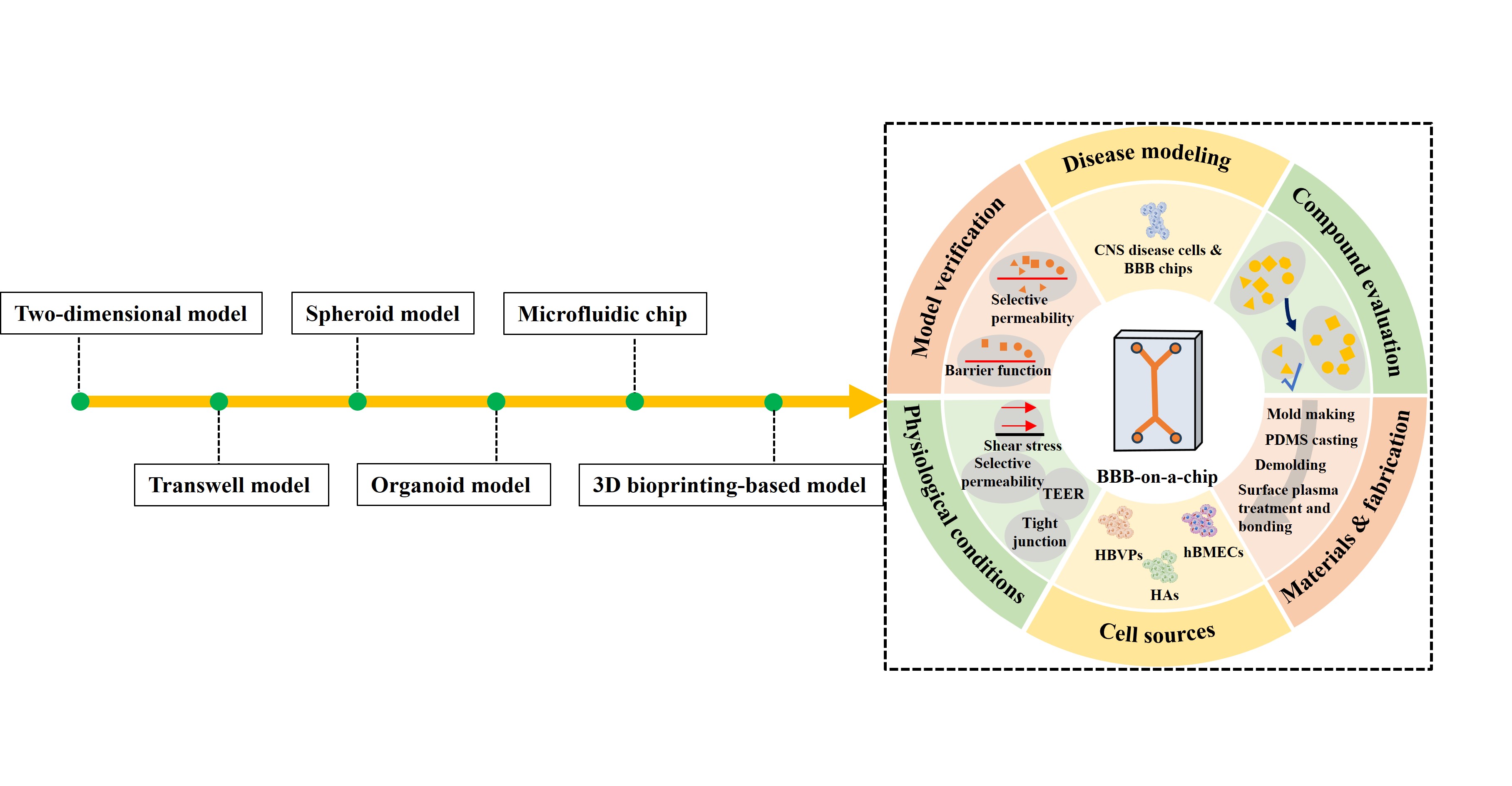
Advances in in vitro blood–brain barrier models: Integrating bioprinting with microfluidic chips for compound evaluation

The blood–brain barrier (BBB), a vital defense interface of the central nervous system, selectively regulates molecular transport into the brain and maintains brain homeostasis. Disruption of BBB integrity contributes to various neurological diseases, making the BBB a key target for therapeutic compounds. However, traditional in vitro models struggle to recreate the BBB’s complex structure and dynamic functions. Recent advances in microfluidics and three-dimensional bioprinting have enabled the construction of high-fidelity in vitro BBB models that recapitulate key aspects of the brain’s vascular microenvironment. By integrating principles from materials science, microfabrication, and cell biology, these “BBB‑on‑a‑chip” platforms support physiologically relevant shear stress, cell–cell interactions, and barrier properties, making them powerful tools for compound screening and mechanistic research. This review summarizes the advances in in vitro BBB models and the application of bioprinting and microfluidic technology for compound evaluation.
- Floryanzia SD, Nance E. Applications and considerations for microfluidic systems to model the blood–brain barrier. ACS Appl Bio Mater. 2023;6(9):3617-3632. doi: 10.1021/acsabm.3c00364
- Shamul JG, Wang Z, Gong H, et al. Meta-analysis of the make-up and properties of in vitro models of the healthy and diseased blood–brain barrier. Nat Biomed Eng. 2024;9(4):566-598. doi: 10.1038/s41551-024-01250-2
- Shi SM, Suh RJ, Shon DJ, et al. Glycocalyx dysregulation impairs blood–brain barrier in ageing and disease. Nature. 2025;639(8056):985-994. doi: 10.1038/s41586-025-08589-9
- Sumpio BE, Timothy Riley J, Dardik A, Cells in focus: endothelial cell. Int J Biochem Cell Biol. 2002;34(12):1508-1512. doi: 10.1016/S1357-2725(02)00075-4
- Bonkowski, D, Katyshev V, Balabanov RD, Borisov A, Dore- Duffy P. The CNS microvascular pericyte: pericyte-astrocyte crosstalk in the regulation of tissue survival. Fluids Barriers CNS. 2011;8(1):8. doi: 10.1186/2045-8118-8-8
- Candelario-Jalil E, Dijkhuizen RM, Magnus T. Neuroinflammation, stroke, blood-brain barrier dysfunction, and imaging modalities. Stroke. 2022;53(5):1473-1486. doi: 10.1161/strokeaha.122.036946
- Abbott NJ, Rönnbäck L, Hansson E. Astrocyte–endothelial interactions at the blood–brain barrier. Nat Rev Neurosci. 2006;7(1):41-53. doi: 10.1038/nrn1824
- Vetter J, Palagi I, Waisman A, Blaeser A. Recent advances in blood-brain barrier-on-a-chip models. Acta Biomater. 2025;197:1-28. doi: 10.1016/j.actbio.2025.03.041
- Katt ME, Shusta EV. In vitro models of the blood-brain barrier: building in physiological complexity. Curr Opin Chem Eng. 2020;30:42-52. doi: 10.1016/j.coche.2020.07.002
- Profaci CP, Munji RN, Pulido RS, Daneman R. The blood-brain barrier in health and disease: important unanswered questions. J Exp Med. 2020;217(4):e20190062. doi: 10.1084/jem.20190062
- Engelhardt B, Ransohoff RM. Capture, crawl, cross: the T cell code to breach the blood–brain barriers. Trends Immunol. 2012;33(12):579-589. doi: 10.1016/j.it.2012.07.004
- Gursoy-Ozdemir Y, Yemisci M, Dalkara T. Microvascular protection is essential for successful neuroprotection in stroke. J Neurochem. 2012;123(s2):2-11. doi: 10.1111/j.1471-4159.2012.07938.x
- Nance E, Pun SH, Saigal R, Sellers DL. Drug delivery to the central nervous system. Nat Rev Mater. 2022;7(4):314-331. doi: 10.1038/s41578-021-00394-w
- Gao Q, Hernandes MS. Sepsis-associated encephalopathy and blood-brain barrier dysfunction. Inflammation. 2021;44(6):2143-2150. doi: 10.1007/s10753-021-01501-3
- Yin P, Wang X. Progresses in the establishment, evaluation, and application of in vitro blood-brain barrier models. J Neurosci Res. 2024;102(6):e25359. doi: 10.1002/jnr.25359
- Pediaditakis I, Kodella KR, Manatakis DV, et al. Modeling alpha-synuclein pathology in a human brain-chip to assess blood-brain barrier disruption. Nat Commun. 2021;12(1):5907. doi: 10.1038/s41467-021-26066-5
- Liu X, Li J, Liu S, et al. Fabrication of a 3D bioprinting model for posterior capsule opacification using GelMA and PLMA hydrogel-coated resin. Regenerative Biomaterials. 2024;11:rbae020. doi: 10.1093/rb/rbae020
- Lee, G., Kim, S. J., Park, J. K., Fabrication of a self-assembled and vascularized tumor array via bioprinting on a microfluidic chip. Lab Chip. 2023;23(18):4079-4091. doi: 10.1039/d3lc00275f
- Dudman, J., Ferreira, A. M., Gentile, P., Wang, X., Dalgarno, K., Microvalve Bioprinting of MSC-Chondrocyte Co- Cultures. Cells. 2021;10(12):3329. doi: 10.3390/cells10123329
- Rueda-Gensini, L., Serna, J. A., Rubio, D., Orozco, J. C., Bolaños, N. I., Cruz, J. C., Muñoz-Camargo, C., Three-dimensional neuroimmune co-culture system for modeling Parkinson’s disease microenvironments in vitro. Biofabrication. 2023;15(4). doi: 10.1088/1758-5090/ace21b
- Li Y, Liu B, Zhao T, et al. Comparative study of extracellular vesicles derived from mesenchymal stem cells and brain endothelial cells attenuating blood-brain barrier permeability via regulating Caveolin-1-dependent ZO-1 and Claudin-5 endocytosis in acute ischemic stroke. J Nanobiotechnology. 2023;21(1):70. doi: 10.1186/s12951-023-01828-z
- Cho C-F, Wolfe JM, Fadzen CM, et al. Blood-brain-barrier spheroids as an in vitro screening platform for brain-penetrating agents. Nat Commun. 2017;8(1):15623. doi: 10.1038/ncomms15623
- Chai YC, To SK, Simorgh S, et al. Spatially self-organized three-dimensional neural concentroid as a novel reductionist humanized model to study neurovascular development. Adv Sci (Weinh). 2024;11(5):e2304421. doi: 10.1002/advs.202304421
- Middelkamp HHT, Verboven AHA, De Sá Vivas AG, et al. Cell type-specific changes in transcriptomic profiles of endothelial cells, iPSC-derived neurons and astrocytes cultured on microfluidic chips. Sci Rep. 2021;11(1):2281. doi: 10.1038/s41598-021-81933-x
- Galpayage Dona KNU, Ramirez SH, Andrews AM. A Next-generation 3D tissue-engineered model of the human brain microvasculature to study the blood-brain barrier. Bioengineering (Basel). 2023;10(7):817. doi: 10.3390/bioengineering10070817
- Mancuso S, Bhalerao A, Cucullo L. Advances and challenges of bioassembly strategies in neurovascular in vitro modeling: an overview of current technologies with a focus on three-dimensional bioprinting. Int J Mol Sci. 2024;25(20): 11000. doi: 10.3390/ijms252011000
- Audus KL, Borchardt RT. Characterization of an in vitro blood-brain barrier model system for studying drug transport and metabolism. Pharm Res. 1986;3(2):81-87. doi: 10.1023/a:1016337202335
- van Bree JB, de Boer AG, Danhof M, Ginsel LA, Breimer DD. Characterization of an “in vitro” blood-brain barrier: effects of molecular size and lipophilicity on cerebrovascular endothelial transport rates of drugs. J Pharmacol Exp Ther. 1988;247(3):1233-1239. doi: 10.1016/S0022-3565(25)13283-7
- Bernoud N, Fenart L, Bénistant C, et al. Astrocytes are mainly responsible for the polyunsaturated fatty acid enrichment in blood-brain barrier endothelial cells in vitro. J Lipid Res. 1998;39(9):1816-1824. doi: 10.1016/S0022-2275(20)32169-6
- Al-Ahmad AJ. Human-induced pluripotent stem cell-based model of the blood-brain at 10 years: a retrospective on past and current disease models. Handb Exp Pharmacol. 2023;281:141-156. doi: 10.1007/164_2023_645
- Stone NL, England TJ, O’Sullivan SE. A novel transwell blood brain barrier model using primary human cells. Front Cell Neurosci. 2019;13:230. doi: 10.3389/fncel.2019.00230
- Qi D, Lin H, Hu B, Wei Y. A review on in vitro model of the blood-brain barrier (BBB) based on hCMEC/D3 cells. J Control Release. 2023;358:78-97. doi: 10.1016/j.jconrel.2023.04.020
- Xie Y, Ye L, Zhang X, et al. Transport of nerve growth factor encapsulated into liposomes across the blood-brain barrier: in vitro and in vivo studies. J Control Release. 2005;105(1-2):106-119. doi: 10.1016/j.jconrel.2005.03.005
- Singh NR, Gromnicova R, Brachner A, et al. A hydrogel model of the human blood-brain barrier using differentiated stem cells. PLoS One. 2023;18(4):e0283954. doi: 10.1371/journal.pone.0283954
- Schofield C, Sarrigiannidis S, Moran-Horowich A, et al. An in vitro model of the blood-brain barrier for the investigation and isolation of the key drivers of barriergenesis. Adv Healthc Mater. 2024;13(32):e2303777. doi: 10.1002/adhm.202303777
- Chen S, Tang C, Ding H, et al. Maf1 ameliorates sepsis-associated encephalopathy by suppressing the NF-kB/ NLRP3 inflammasome signaling pathway. Front Immunol. 2020;11:594071. doi: 10.3389/fimmu.2020.594071
- Wang J, Tang W, Yang M, et al. Inflammatory tumor microenvironment responsive neutrophil exosomes-based drug delivery system for targeted glioma therapy. Biomaterials. 2021;273:120784. doi: 10.1016/j.biomaterials.2021.120784
- Maherally Z, Fillmore HL, Tan SL, et al. Real-time acquisition of transendothelial electrical resistance in an all-human, in vitro, 3-dimensional, blood-brain barrier model exemplifies tight-junction integrity. Faseb J. 2018;32(1):168-182. doi: 10.1096/fj.201700162R
- Jain MK, Chernomorsky A, Silver FH, Berg RA. Material properties of living soft tissue composites. J Biomed Mater Res. 1988;22(3 Suppl):311-326. doi: 10.1002/jbm.820221409
- Alpert, S. S., Banks, G., The concentration dependence of the hemoglobin mutual diffusion coefficient. Biophys Chem. 1976;4(3):287-296. doi: 10.1016/0301-4622(76)80077-4
- Schmitz KS, Shaw BR. Hydrodynamic evidence in support of spacer regions in chromatin. Science. 1977;197(4304):661-663. doi: 10.1126/science.877579
- Wang PY, Youson JH, Drakos TT. Mouse 3T3 cell filtrability correlating with concanavalin A agglutinability. Biochim Biophys Acta. 1984;802(3):467-476. doi: 10.1016/0304-4165(84)90366-0
- Oelze ML, O’Brien WD, Jr. Application of three scattering models to characterization of solid tumors in mice. Ultrason Imaging. 2006;28(2):83-96. doi: 10.1177/016173460602800202
- Pérez-López A, Torres-Suárez AI, Martín-Sabroso C, Aparicio-Blanco J. An overview of in vitro 3D models of the blood-brain barrier as a tool to predict the in vivo permeability of nanomedicines. Adv Drug Deliv Rev. 2023;196:114816. doi: 10.1016/j.addr.2023.114816
- Sokolova V, Nzou G, van der Meer SB, et al. Ultrasmall gold nanoparticles (2 nm) can penetrate and enter cell nuclei in an in vitro 3D brain spheroid model. Acta Biomater. 2020;111:349-362. doi: 10.1016/j.actbio.2020.04.023
- Urich E, Patsch C, Aigner S, Graf M, Iacone R, Freskgård PO. Multicellular self-assembled spheroidal model of the blood brain barrier. Sci Rep. 2013;3:1500. doi: 10.1038/srep01500
- Younis M, Faming W, Hongyan Z, Mengmeng T, Hang S, Liudi Y. Iguratimod encapsulated PLGA-NPs improves therapeutic outcome in glioma, glioma stem-like cells and temozolomide resistant glioma cells. Nanomedicine. 2019;22:102101. doi: 10.1016/j.nano.2019.102101
- Huang H-S, Chiang IT, Lawal B, et al. A novel isotope-labeled small molecule probe CC12 for anti-glioma via suppressing LYN-mediated progression and activating apoptosis pathways. Int J Biol Sci. 2023;19(10):3209-3225. doi: 10.7150/ijbs.82266
- Clevers H. Modeling development and disease with organoids. Cell. 2016;165(7):1586-1597. doi: 10.1016/j.cell.2016.05.082
- Ahn Y, An J-H, Yang H-J, et al. Human blood vessel organoids penetrate human cerebral organoids and form a vessel-like system. Cells. 2021;10(8):2036. doi: 10.3390/cells10082036
- Yan Y, Song L, Bejoy J, et al. Modeling neurodegenerative microenvironment using cortical organoids derived from human stem cells. Tissue Eng Part A. 2018;24(13-14):1125-1137. doi: 10.1089/ten.TEA.2017.0423
- Huang S, Zhang Z, Cao J, Yu Y, Pei G. Chimeric cerebral organoids reveal the essentials of neuronal and astrocytic APOE4 for Alzheimer’s tau pathology. Signal Transduct Target Ther. 2022;7(1):176. doi: 10.1038/s41392-022-01006-x
- Dao L, You Z, Lu L, et al. Modeling blood-brain barrier formation and cerebral cavernous malformations in human PSC-derived organoids. Cell Stem Cell. 2024;31(6):818-833.e11. doi: 10.1016/j.stem.2024.04.019
- Fang GC, Chen YC, Lu HX, Jin DY. Advances in spheroids and organoids on a chip. Adv Funct Mater. 2023;33(19):2215043. doi: 10.1002/adfm.202215043
- Chen X, Sun G, Tian E, et al. Modeling sporadic alzheimer’s disease in human brain organoids under serum exposure. Adv Sci. 2021;8(18):e2101462. doi: 10.1002/advs.202101462
- Sooriyaarachchi D, Maharubin S, Tan GZ. Fabrication of microtube-embedded chip to mimic blood–brain barrier capillary vessels. In: Stone N, ed. The Blood-Brain Barrier: Methods and Protocols. US, New York, NY: Springer; 2022: 241-249.
- Kawakita S, Mandal K, Mou L, et al. Organ-on-a-chip models of the blood-brain barrier: recent advances and future prospects. Small. 2022;18(39):e2201401. doi: 10.1002/smll.202201401
- Ponmozhi J, Dhinakaran S, Kocsis D, Iván K, Erdő F. Models for barrier understanding in health and disease in lab-on-a-chips. Tissue Barriers. 2024;12(2):2221632. doi: 10.1080/21688370.2023.2221632
- Nahon DM, Moerkens R, Aydogmus H, et al. Standardizing designed and emergent quantitative features in microphysiological systems. Nat Biomed Eng. 2024;8(8):941-962. doi: 10.1038/s41551-024-01236-0
- Sonninen TM, Peltonen S, Kalvala S, et al. From inserts to chips: microfluidic culture and 3D astrocyte co-culture drive functional and transcriptomic changes in hiPSC-derived endothelial cells. Fluids Barriers CNS. 2025;22(1):58. doi: 10.1186/s12987-025-00672-7
- Jeong S, Seo J-H, Garud KS, Park SW, Lee M-Y. Numerical approach-based simulation to predict cerebrovascular shear stress in a blood-brain barrier organ-on-a-chip. Biosens Bioelectron. 2021;183:113197. doi: 10.1016/j.bios.2021.113197
- Zakharova M, Palma do Carmo MA, van der Helm MW, et al. Multiplexed blood–brain barrier organ-on-chip. Lab Chip. 2020;20(17):3132-3143. doi: 10.1039/d0lc00399a
- Chang Y, Chen T, Geng S, et al. A scenario-adaptive microfluidic chip for constructing in vitro models of biological barriers. Anal Chem. 2025;97(7):3816-3821. doi: 10.1021/acs.analchem.4c06602
- Wu CA, Zhu Y, Woo YJ. Advances in 3D bioprinting: techniques, applications, and future directions for cardiac tissue engineering. Bioengineering (Basel). 2023;10(7):842. doi: 10.3390/bioengineering10070842
- Gungor-Ozkerim PS, Inci I, Zhang YS, Khademhosseini A, Dokmeci MR. Bioinks for 3D bioprinting: an overview. Biomater Sci. 2018;6(5):915-946. doi: 10.1039/c7bm00765e
- Shi W, Zhang Z, Wang X. The prospect of hepatic decellularized extracellular matrix as a bioink for liver 3D bioprinting. Biomolecules. 2024;14(8):1019. doi: 10.3390/biom14081019
- Tang M, Rich JN, Chen S. Biomaterials and 3D bioprinting strategies to model glioblastoma and the blood–brain barrier. Adv Mater. 2020;33(5):e2004776. doi: 10.1002/adma.202004776
- Galpayage Dona KNU, Hale JF, Salako T, et al. The use of tissue engineering to fabricate perfusable 3D brain microvessels in vitro. Front Physiol. 2021;12:715431. doi: 10.3389/fphys.2021.715431
- Oh H, Kang M, Bae E, et al. Fabrication of hydrogel microchannels using aqueous two-phase printing for 3D blood brain barrier. BioChip J. 2023;17(3):369-383. doi: 10.1007/s13206-023-00110-6
- Royse MK, Fowler M, Mai AK, et al. Development of a 3D printed perfusable in vitro blood-brain barrier model for use as a scalable screening tool. Biomater Sci. 2024;12(17):4363-4375. doi: 10.1039/d4bm00663a
- Choi, J., Mathew, S., Oerter, S., Appelt-Menzel, A., Hansmann, J., Schmitz, T., Online measurement system for dynamic flow bioreactors to study barrier integrity of hipsc-based blood– brain barrier in vitro models. Bioengineering. 2022;9(1):39. doi: 10.3390/bioengineering9010039
- Ohbuchi M, Shibuta M, Tetsuka K, et al. Modeling of blood-brain barrier (BBB) dysfunction and immune cell migration using human BBB-on-a-chip for drug discovery research. Int J Mol Sci. 2024;25(12):6496. doi: 10.3390/ijms25126496
- Sugiyama S, Sasaki T, Tanaka H, et al. The tight junction protein occludin modulates blood-brain barrier integrity and neurological function after ischemic stroke in mice. Sci Rep. 2023;13(1):2892. doi: 10.1038/s41598-023-29894-1
- Dudek KA, Dion-Albert L, Lebel M, et al. Molecular adaptations of the blood-brain barrier promote stress resilience vs. depression. Proc Natl Acad Sci USA. 2020;117(6):3326-3336. doi: 10.1073/pnas.1914655117
- Shu Y, Peng F, Zhao B, et al. Transfer of patient’s peripheral blood mononuclear cells (PBMCs) disrupts blood-brain barrier and induces anti-NMDAR encephalitis: a study of novel humanized PBMC mouse model. J Neuroinflammation. 2023;20(1):164. doi: 10.1186/s12974-023-02844-4
- Lin L, Bi H, Yang J, et al. Pasteurella multocida infection induces blood-brain barrier disruption by decreasing tight junctions and adherens junctions between neighbored brain microvascular endothelial cells. Vet Res. 2024;55(1):104. doi: 10.1186/s13567-024-01351-5
- Shi Y, He X, Wang H, et al. Construction of a novel blood brain barrier-glioma microfluidic chip model: applications in the evaluation of permeability and anti-glioma activity of traditional Chinese medicine components. Talanta. 2023;253:123971. doi: 10.1016/j.talanta.2022.123971
- Park JS, Choe K, Khan A, et al. Establishing co-culture blood-brain barrier models for different neurodegeneration conditions to understand its effect on BBB integrity. Int J Mol Sci. 2023;24(6):5283. doi: 10.3390/ijms24065283
- Zorkina YA, Volgina NE, Gorlachev GE, et al. Effect of γ-irradiation on expression of tight and adherens junction protein mRNA on in vitro blood-brain barrier model. Bull Exp Biol Med. 2014;158(1):127-136. doi: 10.1007/s10517-014-2708-5
- Petrovskaya AV, Barykin EP, Tverskoi AM, et al. Blood-brain barrier transwell modeling. Mol Biol (Mosk). 2022;56(6):1086-1094. doi: 10.31857/s0026898422060179
- Sun P, Hamblin MH, Yin KJ. Non-coding RNAs in the regulation of blood-brain barrier functions in central nervous system disorders. Fluids Barriers CNS. 2022;19(1):27. doi: 10.1186/s12987-022-00317-z
- Eigenmann DE, Xue G, Kim KS, Moses AV, Hamburger M, Oufir M. Comparative study of four immortalized human brain capillary endothelial cell lines, hCMEC/D3, hBMEC, TY10, and BB19, and optimization of culture conditions, for an in vitro blood-brain barrier model for drug permeability studies. Fluids Barriers CNS. 2013;10(1):33. doi: 10.1186/2045-8118-10-33
- Waithe OY, Peng X, Childs EW, Tharakan B. Measurement of transendothelial electrical resistance in blood-brain barrier endothelial cells. Methods Mol Biol. 2024;2711:199-203. doi: 10.1007/978-1-0716-3429-5_16
- Noorani B, Bhalerao A, Raut S, Nozohouri E, Bickel U, Cucullo L. A quasi-physiological microfluidic blood-brain barrier model for brain permeability studies. Pharmaceutics. 2021;13(9):1474. doi: 10.3390/pharmaceutics13091474
- Offeddu GS, Haase K, Gillrie MR, et al. An on-chip model of protein paracellular and transcellular permeability in the microcirculation. Biomaterials. 2019;212:115-125. doi: 10.1016/j.biomaterials.2019.05.022
- Kim Y, Lee S, Zhang H, et al. CLEC14A deficiency exacerbates neuronal loss by increasing blood-brain barrier permeability and inflammation. J Neuroinflammation. 2020;17(1):48. doi: 10.1186/s12974-020-1727-6
- Waithe OY, Peng X, Childs EW, Tharakan B. Measurement of blood-brain barrier hyperpermeability using evans blue extravasation assay. Methods Mol Biol. 2024;2711:177-184. doi: 10.1007/978-1-0716-3429-5_14
- Ahishali B, Kaya M. Evaluation of blood-brain barrier integrity using vascular permeability markers: evans blue, sodium fluorescein, albumin-alexa fluor conjugates, and horseradish peroxidase. Methods Mol Biol. 2021;2367:87-103. doi: 10.1007/7651_2020_316
- Ozgür B, Puris E, Brachner A, et al. Characterization of an iPSC-based barrier model for blood-brain barrier investigations using the SBAD0201 stem cell line. Fluids Barriers CNS. 2023;20(1):96. doi: 10.1186/s12987-023-00501-9
- Ito R, Morio H, Baba T, et al. In vitro-in vivo correlation of blood-brain barrier permeability of drugs: a feasibility study towards development of prediction methods for brain drug concentration in humans. Pharm Res. 2022;39(7):1575-1586. doi: 10.1007/s11095-022-03189-y
- Heidari H, Taylor H. Review article: capturing the physiological complexity of the brain’s neuro-vascular unit in vitro. Biomicrofluidics. 2018;12(5):051502. doi: 10.1063/1.5045126
- Koch EV, Ledwig V, Bendas S, Reichl S, Dietzel A. Tissue barrier-on-chip: a technology for reproducible practice in drug testing. Pharmaceutics. 2022;14(7):1451. doi: 10.3390/pharmaceutics14071451
- Guan M, Tang S, Chang H, et al. Development of alveolar-capillary-exchange (ACE) chip and its application for assessment of PM(2.5)-induced toxicity. Ecotoxicol Environ Saf. 2021;223:112601. doi: 10.1016/j.ecoenv.2021.112601
- Wang Z, Zhang Y, Li Z, Wang H, Li N, Deng Y. Microfluidic brain‐on‐a‐chip: from key technology to system integration and application. Small. 2023;19(52):e2304427. doi: 10.1002/smll.202304427
- Choi B, Choi JW, Jin H, et al. Condensed ECM-based nanofilms on highly permeable PET membranes for robust cell-to-cell communications with improved optical clarity. Biofabrication. 2021;13(4):045020. doi: 10.1088/1758-5090/ac23ad
- Alves ADH, Nucci MP, Ennes do Valle NM, et al. Current overview of induced pluripotent stem cell-based blood- brain barrier-on-a-chip. World J Stem Cells. 2023;15(6): 632-653. doi: 10.4252/wjsc.v15.i6.632
- Hosic S, Bindas AJ, Puzan ML, et al. Rapid prototyping of multilayer microphysiological systems. ACS Biomater Sci Eng. 2021;7(7):2949-2963. doi: 10.1021/acsbiomaterials.0c00190
- Pungjunun K, Yakoh A, Chaiyo S, et al. Laser engraved microapillary pump paper-based microfluidic device for colorimetric and electrochemical detection of salivary thiocyanate. Mikrochim Acta. 2021;188(4):140. doi: 10.1007/s00604-021-04793-2
- Wang YI, Abaci HE, Shuler ML. Microfluidic blood-brain barrier model provides in vivo-like barrier properties for drug permeability screening. Biotechnol Bioeng. 2017;114(1):184-194. doi: 10.1002/bit.26045
- Cenhrang K, Robart L, Castiaux AD, Martin RS. 3D printed devices with integrated collagen scaffolds for cell culture studies including transepithelial/transendothelial electrical resistance (TEER) measurements. Anal Chim Acta. 2022;1221:340166. doi: 10.1016/j.aca.2022.340166
- Matthiesen I, Jury M, Rasti Boroojeni F, et al. Astrocyte 3D culture and bioprinting using peptide functionalized hyaluronan hydrogels. Sci Technol Adv Mater. 2023;24(1):2165871. doi: 10.1080/14686996.2023.2165871
- Kim JA, Kim HN, Im S-K, Chung S, Kang JY, Choi N. Collagen-based brain microvasculature modelin vitrousing three-dimensional printed template. Biomicrofluidics. 2015;9(2):024115. doi: 10.1063/1.4917508
- Wang Z, Huang C, Shi Z, et al. Coaxial bioprinting of a three-layer vascular structure exhibiting blood-brain barrier function for neuroprotective drug screening. Colloids Surf B Biointerfaces. 2025;249:114494. doi: 10.1016/j.colsurfb.2025.114494
- Cucullo L, Hossain M, Puvenna V, Marchi N, Janigro D. The role of shear stress in Blood-Brain Barrier endothelial physiology. BMC Neurosci. 2011;12:40. doi: 10.1186/1471-2202-12-40
- Zhang YS, Davoudi F, Walch P, et al. Bioprinted thrombosis-on-a-chip. Lab Chip. 2016;16(21):4097-4105. doi: 10.1039/c6lc00380j
- Wang R, Zhang H, Li S, et al. Current progress ofin vitrovascular models on microfluidic chips. Biofabrication. 2025;17(2). doi: 10.1088/1758-5090/adb182
- Yang JY, Shin DS, Jeong M, et al. Evaluation of drug blood-brain-barrier permeability using a microfluidic chip. Pharmaceutics. 2024;16(5):574. doi: 10.3390/pharmaceutics16050574
- Ferreira L. What human blood-brain barrier models can tell us about BBB function and drug discovery? Expert Opin Drug Discov. 2019;14(11):1113-1123. doi: 10.1080/17460441.2019.1646722
- Nahon DM, Vila Cuenca M, van den Hil FE, et al. Self-assembling 3D vessel-on-chip model with hiPSC-derived astrocytes. Stem Cell Reports. 2024;19(7):946-956. doi: 10.1016/j.stemcr.2024.05.006
- Chim SM, Howell K, Kokkosis A, Zambrowicz B, Karalis K, Pavlopoulos E. A human brain-chip for modeling brain pathologies and screening blood-brain barrier crossing therapeutic strategies. Pharmaceutics. 2024;16(10):1314. doi: 10.3390/pharmaceutics16101314
- Choi JW, Kim K, Mukhambetiyar K, et al. Organ-on-a-chip approach for accelerating blood-brain barrier nanoshuttle discovery. ACS Nano. 2024;18(22):14388-14402. doi: 10.1021/acsnano.4c00994
- Stanness KA, Westrum LE, Fornaciari E, et al. Morphological and functional characterization of an in vitro blood-brain barrier model. Brain Res. 1997;771(2):329-42. doi: 10.1016/s0006-8993(97)00829-9
- Cucullo L, Couraud PO, Weksler B, et al. Immortalized human brain endothelial cells and flow-based vascular modeling: a marriage of convenience for rational neurovascular studies. J Cereb Blood Flow Metab. 2008;28(2):312-328. doi: 10.1038/sj.jcbfm.9600525
- Wang X, Xu B, Xiang M, et al. Advances on fluid shear stress regulating blood-brain barrier. Microvasc Res. 2020;128:103930. doi: 10.1016/j.mvr.2019.103930
- Griep LM, Wolbers F, de Wagenaar B, et al. BBB on chip: microfluidic platform to mechanically and biochemically modulate blood-brain barrier function. Biomed Microdevices. 2013;15(1):145-150. doi: 10.1007/s10544-012-9699-7
- Ceccarelli MC, Lefevre MC, Marino A, et al. Real-time monitoring of a 3D blood-brain barrier model maturation and integrity with a sensorized microfluidic device. Lab Chip. 2024;24(22):5085-5100. doi: 10.1039/d4lc00633j
- Pichler V, Schwingenschlögl-Maisetschläger V, Duman I, et al. Bioanalytic hybrid system merging 3-dimensional cell culture and chromatographic precision for unprecedented preclinical insights in molecular imaging. J Nucl Med. 2025;66(5):813-816. doi: 10.2967/jnumed.124.269133
- Mossburg KJ, Shepherd SJ, Barragan D, et al. Towards the clinical translation of a silver sulfide nanoparticle contrast agent: large scale production with a highly parallelized microfluidic chip. Eur J Nucl Med Mol Imaging. 2025;52(3):1177-1188. doi: 10.1007/s00259-024-06967-5
- Yan X, Liu X, Zhao C, Chen GQ. Applications of synthetic biology in medical and pharmaceutical fields. Signal Transduct Target Ther. 2023;8(1):199. doi: 10.1038/s41392-023-01440-5
- Lu YL, Lin CM, Huang JH. Triplicate dynamic cell culture platform for enhanced reproducibility in anti-cancer drug testing. ACS Biomater Sci Eng. 2025;11(2):1222-1231. doi: 10.1021/acsbiomaterials.4c02142
- Xu H, Li Z, Yu Y, et al. A dynamic in vivo-like organotypic blood-brain barrier model to probe metastatic brain tumors. Sci Rep. 2016;6(1):36670. doi: 10.1038/srep36670
- Maoz BM, Herland A, FitzGerald EA, et al. A linked organ-on-chip model of the human neurovascular unit reveals the metabolic coupling of endothelial and neuronal cells. Nat Biotechnol. 2018;36(9):865-874. doi: 10.1038/nbt.4226
- Liu L, Koo Y, Russell T, Yun Y. A three-dimensional brain-on-a-chip using human ipsc-derived gabaergic neurons and astrocytes. Methods Mol Biol. 2022;2492:117-128. doi: 10.1007/978-1-0716-2289-6_6
- Miller DR, McClain ES, Dodds JN, et al. Chlorpyrifos disrupts acetylcholine metabolism across model blood-brain barrier. Front Bioeng Biotechnol. 2021;9:622175. doi: 10.3389/fbioe.2021.622175
- Kim SS, Kim S, Kim Y, et al. Neurotoxic effects of citronellol induced by the conversion of kynurenine to 3-hydroxykynurenine. J Hazard Mater. 2025;486:136965. doi: 10.1016/j.jhazmat.2024.136965
- Arduino I, Di Fonte R, Sommonte F, et al. Fabrication of biomimetic hybrid liposomes via microfluidic technology: homotypic targeting and antitumor efficacy studies in glioma cells. Int J Nanomed. 2024;19:13217-13233. doi: 10.2147/ijn.S489872
- Shao X, Gao D, Chen Y, et al. Development of a blood-brain barrier model in a membrane-based microchip for characterization of drug permeability and cytotoxicity for drug screening. Anal Chim Acta. 2016;934:186-193. doi: 10.1016/j.aca.2016.06.028
- Yu F, Kumar NDOS, Foo LC, Ng SH, Hunziker W, Choudhury D. A pump‐free tricellular blood–brain barrier on‐a‐chip model to understand barrier property and evaluate drug response. Biotechnol Bioeng. 2020;117(4): 1127-1136. doi: 10.1002/bit.27260
- Mitchell MJ, Billingsley MM, Haley RM, Wechsler ME, Peppas NA, Langer R. Engineering precision nanoparticles for drug delivery. Nat Rev Drug Discov. 2020;20(2):101-124. doi: 10.1038/s41573-020-0090-8
- Palma-Florez S, López-Canosa A, Moralez-Zavala F, et al. BBB-on-a-chip with integrated micro-TEER for permeability evaluation of multi-functionalized gold nanorods against Alzheimer’s disease. Journal of Nanobiotechnology. 2023;21(1):115. doi: 10.1186/s12951-023-01798-2
- Garcia L, Palma-Florez S, Espinosa V, et al. Ferulic acid-loaded polymeric nanoparticles prepared from nano-emulsion templates facilitate internalisation across the blood–brain barrier in model membranes. Nanoscale. 2023;15(17):7929-7944. doi: 10.1039/d2nr07256d
- Cai X, Refaat A, Gan P-Y, et al. Angiopep-2-Functionalized Lipid Cubosomes for Blood–Brain Barrier Crossing and Glioblastoma Treatment. ACS Applied Materials & Interfaces. 2024;16(10):12161-12174. doi: 10.1021/acsami.3c14709
- Heard, S. C., Wu, G., Winter, J. M., Antifungal natural products. Curr Opin Biotechnol. 2021;69(232-241. doi: 10.1016/j.copbio.2021.02.001
- Shan L, Fan Y, Yuanchun C, et al. Sanhua Tang protects against ischemic stroke by preventing blood-brain barrier injury: a network pharmacology and experiments. J Tradit Chin Med. 2024;44(4):794-803. doi: 10.19852/j.cnki.jtcm.20240515.001
- Liu G, Liang Y, Xu M, et al. Protective mechanism of Erigeron breviscapus injection on blood-brain barrier injury induced by cerebral ischemia in rats. Sci Rep. 2021;11(1):18451. doi: 10.1038/s41598-021-97908-x
- Zhang Y, Shen L, Xie J, et al. Pushen capsule treatment promotes functional recovery after ischemic stroke. Phytomedicine. 2023;111(154664. doi: 10.1016/j.phymed.2023.154664
- Karthika C, Najda A, Klepacka J, et al. Involvement of resveratrol against brain cancer: a combination strategy with a pharmaceutical approach. Molecules. 2022;27(14):4663. doi: 10.3390/molecules27144663
- Liu Y, Yang G, Cui W, Zhang Y, Liang X. Regulatory mechanisms of tetramethylpyrazine on central nervous system diseases: a review. Front Pharmacol. 2022;13:948600. doi: 10.3389/fphar.2022.948600
- Ru J, Li P, Wang J, et al. TCMSP: a database of systems pharmacology for drug discovery from herbal medicines. J Cheminform. 2014;6:13. doi: 10.1186/1758-2946-6-13
- Meena M, Vandormael R, De Laere M, et al. A microfluidic in vitro three-dimensional dynamic model of the blood– brain barrier to study the transmigration of immune cells. Brain Sci. 2022;12(10):1293. doi: 10.3390/brainsci12101293
- Malekpour A, Chen X. Printability and cell viability in extrusion-based bioprinting from experimental, computational, and machine learning views. J Funct Biomater. 2022;13(2):40. doi: 10.3390/jfb13020040
- Billiet T, Gevaert E, De Schryver T, Cornelissen M, Dubruel P. The 3D printing of gelatin methacrylamide cell-laden tissue-engineered constructs with high cell viability. Biomaterials. 2014;35(1):49-62. doi: 10.1016/j.biomaterials.2013.09.078
- Naghieh S, Karamooz-Ravari MR, Sarker MD, Karki E, Chen X. Influence of crosslinking on the mechanical behavior of 3D printed alginate scaffolds: Experimental and numerical approaches. J Mech Behav Biomed Mater. 2018;80:111-118. doi: 10.1016/j.jmbbm.2018.01.034
- Xu Y, Kong D, Zhang T, Xiong Z, Sun W. A parallel multilayered neurovascular unit-on-a-chip for modeling neurovascular microenvironment and screening chemotherapeutic drugs. Int J Bioprint. 2024;10(3):1684. doi: 10.36922/ijb.1684
- Ahmad A, Zobaida Akter M, Kim SY, Choi YJ, Yi HG. High-throughput bioprinting method for modeling vascular permeability in standard six-well plates with size and pattern flexibility. J Vis Exp. 2024;210. doi: 10.3791/66676
- Mou L, Mandal K, Mecwan MM, et al. Integrated biosensors for monitoring microphysiological systems. Lab Chip. 2022;22(20):3801-3816. doi: 10.1039/d2lc00262k
- Gnatowski P, Ansariaghmiuni M, Piłat E, et al. Hydrogel membranes in organ-on-a-chip devices: a review. Colloids Surf B Biointerfaces. 2025;251:114591. doi: 10.1016/j.colsurfb.2025.114591
- Brown JA, Codreanu SG, Shi M, et al. Metabolic consequences of inflammatory disruption of the blood-brain barrier in an organ-on-chip model of the human neurovascular unit. J Neuroinflammation. 2016;13(1):306. doi: 10.1186/s12974-016-0760-y
- Su S-H, Song Y, Stephens A, et al. A tissue chip with integrated digital immunosensors: In situ brain endothelial barrier cytokine secretion monitoring. Biosens Bioelectron. 2023;224:115030. doi: 10.1016/j.bios.2022.115030
- Feng Y, Chen C-S, Ho J, et al. High-throughput chip assay for investigating Escherichia coli interaction with the blood–brain barrier using microbial and human proteome microarrays (dual-microarray technology). Anal Chem. 2018;90(18):10958-10966. doi: 10.1021/acs.analchem.8b02513
- Koenig L, Ramme AP, Faust D, et al. A human stem cell-derived brain-liver chip for assessing blood-brain-barrier permeation of pharmaceutical drugs. Cells. 2022;11(20):3295. doi: 10.3390/cells11203295
- Ahn SI, Kim Y. Human blood–brain barrier on a chip: featuring unique multicellular cooperation in pathophysiology. Trends Biotechnol. 2021;39(8):749-752. doi: 10.1016/j.tibtech.2021.01.010
- Oddo A, Peng B, Tong Z, et al. Advances in microfluidic blood-brain barrier (BBB) models. Trends Biotechnol. 2019;37(12):1295-1314. doi: 10.1016/j.tibtech.2019.04.006