

Bioprinting strategies for skeletal muscle regeneration: Advances in bioinks, technologies, and functional reconstruction

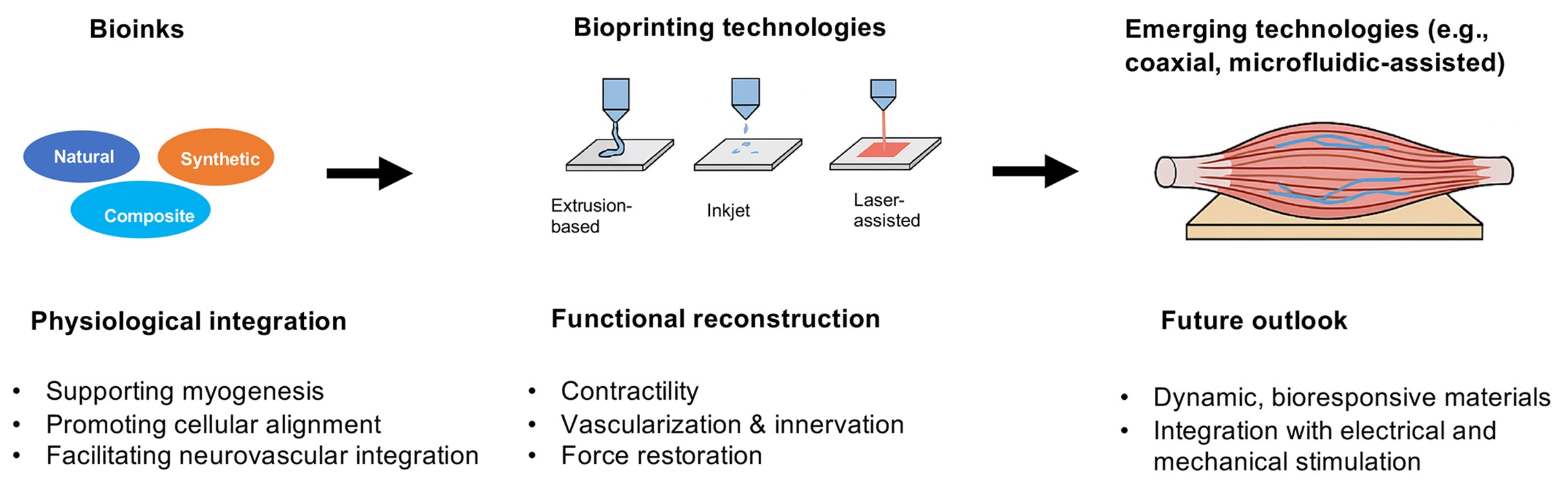
Volumetric muscle loss (VML) presents a significant clinical challenge because the intrinsic regenerative capacity of skeletal muscle is insufficient to repair extensive defects, and current therapeutic strategies remain inadequate. Bioprinting has emerged as a transformative approach, enabling the spatially controlled deposition of cells, biomaterials, and biochemical cues to create functional, biomimetic muscle tissues. This review offers a comprehensive overview of recent advancements in bioink development, bioprinting technologies, and functional reconstruction strategies for skeletal muscle regeneration. Bioinks derived from natural, synthetic, and composite materials are examined in terms of their effectiveness in supporting myogenesis, promoting cellular alignment, and facilitating neurovascular integration. We compare key bioprinting techniques—including extrusion-based, inkjet, and laser-assisted printing—highlighting their respective strengths and limitations in achieving structural fidelity and multicellular complexity. Emerging technologies such as coaxial and microfluidic-assisted printing are also discussed for their potential to fabricate aligned, anisotropic muscle constructs with hierarchical architectures. Functional outcomes are synthesized from in vitro assays (e.g., contractility, gene expression) and in vivo studies using VML models, with a focus on vascularization, innervation, and force restoration. Despite significant progress, substantial challenges remain in achieving complete neurovascular integration, long-term functionality, and clinical scalability. Moving forward, future efforts should emphasize the development of dynamic, bioresponsive materials, integration with electrical and mechanical stimulation, and the establishment of standardized preclinical protocols. By bridging material innovation, structural design, and biological functionality, bioprinting holds great promise for next-generation, clinically relevant skeletal muscle regeneration.
- Relaix F, Bencze M, Borok MJ, et al. Perspectives on skeletal muscle stem cells. Nat Commun. 2021;12(1):692. doi: 10.1038/s41467-020-20760-6
- Judson RN, Rossi FMV. Towards stem cell therapies for skeletal muscle repair. NJP Regen Med. 2020;5(1):10. doi: 10.1038/s41536-020-0094-3
- Sousa-Victor P, García-Prat L, Muñoz-Cánoves P. Control of satellite cell function in muscle regeneration and its disruption in ageing. Nat Rev Mol Cell Bio. 2022;23(3):204-226. doi: 10.1038/s41580-021-00421-2
- Qi L, Zhang F, Wang K, et al. Advancements in skeletal muscle tissue engineering: Strategies for repair and regeneration of skeletal muscle beyond self-repair. Regen Biomater. 2025;12:rbaf050. doi: 10.1093/rb/rbaf050
- Maimaiti D, Ge X, Wang C, et al. Extracellular matrix-mimicking cryogels composed of methacrylated fucoidan enhance vascularized skeletal muscle regeneration following volumetric muscle loss. Int J Biol Macromol. 2024;283:137122. doi: 10.1016/j.ijbiomac.2024.137122
- Sonaye SY, Sikder P. Bioengineered constructs as a tissue engineering-based therapy for volumetric muscle loss. Tissue Eng Part B Rev. 2025. doi: 10.1089/ten.teb.2025.0017
- Cai CW, Grey JA, Hubmacher D, Han WM. Biomaterial-based regenerative strategies for volumetric muscle loss: challenges and solutions. Adv Wound Care. 2025;14(3):159-175. doi: 10.1089/wound.2024.0079
- Edouard P, Reurink G, Mackey AL, et al. Traumatic muscle injury. Nat Rev Dis Primers. 2023;9(1):56. doi: 10.1038/s41572-023-00469-8
- SantAnna JPC, Pedrinelli A, Hernandez AJ, Fernandes TL. Muscle injury: pathophysiology, diagnosis, and treatment. Rev Bras Ortop. 2022;57(1):1-13. doi: 10.1055/s-0041-1731417
- Topaloğlu H, Poorshiri B. The congenital muscular dystrophies. Ann Child Neurol Soc. 2024;2(1):27-39. doi: 10.1002/cns3.20050
- Ostojic M, Indelli PF, Lovrekovic B, et al. Graft selection in anterior cruciate ligament reconstruction: a comprehensive review of current trends. Medicina. 2024;60(12):2090. doi: 10.3390/medicina60122090
- Gahlawat S, Oruc D, Paul N, et al. Tissue engineered 3D constructs for volumetric muscle loss. Ann Biomed Eng. 2024;52(9):2325-2347. doi: 10.1007/s10439-024-03541-w
- Boyer O, Butler-Browne G, Chinoy H, et al. Myogenic cell transplantation in genetic and acquired diseases of skeletal muscle. Front Genet. 2021;12. doi: 10.3389/fgene.2021.702547
- Liu H, Cheema U, Player DJ. Photobiomodulation therapy (PBMT) in skeletal muscle regeneration: a comprehensive review of mechanisms, clinical applications, and future directions. Photodiagn Photodyn. 2025;53:104634. doi: 10.1016/j.pdpdt.2025.104634
- de Sire A, Marotta N, Lippi L, et al. Pharmacological treatment for acute traumatic musculoskeletal pain in athletes. Medicina. 2021;57(11):1208. doi: 10.3390/medicina57111208
- Wei Q, An Y, Zhao X, Li M, Zhang J. Three-dimensional bioprinting of tissue-engineered skin: biomaterials, fabrication techniques, challenging difficulties, and future directions: a review. Int J Biol Macromol. 2024;266:131281. doi: 10.1016/j.ijbiomac.2024.131281
- Saini G, Segaran N, Mayer JL, Saini A, Albadawi H, Oklu R. Applications of 3D bioprinting in tissue engineering and regenerative medicine. J Clin Med. 2021;10(21):4966. doi: 10.3390/jcm10214966
- Jain P, Kathuria H, Dubey N. Advances in 3D bioprinting of tissues/organs for regenerative medicine and in-vitro models. Biomaterials. 2022;287:121639. doi: 10.1016/j.biomaterials.2022.121639
- Samandari M, Quint J, Rodríguez-delaRosa A, Sinha I, Pourquié O, Tamayol A. Bioinks and bioprinting strategies for skeletal muscle tissue engineering. Adv Mater. 2022;34(12):2105883. doi: 10.1002/adma.202105883
- Sabetkish S, Currie P, Meagher L. Recent trends in 3D bioprinting technology for skeletal muscle regeneration. Acta Biomater. 2024;181:46-66. doi: 10.1016/j.actbio.2024.04.038
- Lee H, Kim W, Lee J, et al. Self-aligned myofibers in 3D bioprinted extracellular matrix-based construct accelerate skeletal muscle function restoration. Appl Phys Rev. 2021;8(2):021405. doi: 10.1063/5.0039639
- Sicherer ST, Haque N, Parikh Y, Grasman JM. Current methodologies for inducing aligned myofibers in tissue constructs for skeletal muscle tissue regeneration. Adv Wound Care. 2025;14(2):114-131. doi: 10.1089/wound.2024.0111
- Zhang H, Wu C. 3D printing of biomaterials for vascularized and innervated tissue regeneration. Int J Bioprinting. 2023;9(3):706. doi: 10.18063/ijb.706
- Thangadurai M, Ajith A, Budharaju H, Sethuraman S, Sundaramurthi D. Advances in electrospinning and 3D bioprinting strategies to enhance functional regeneration of skeletal muscle tissue. Biomater Adv. 2022;142:213135. doi: 10.1016/j.bioadv.2022.213135
- Miramini S, Fegan KL, Green NC, Espino DM, Zhang L, Thomas-Seale LEJ. The status and challenges of replicating the mechanical properties of connective tissues using additive manufacturing. J Mech Behav Biomed. 2020;103:103544. doi: 10.1016/j.jmbbm.2019.103544
- García-Lizarribar A, Villasante A, Lopez-Martin JA, et al. 3D bioprinted functional skeletal muscle models have potential applications for studies of muscle wasting in cancer cachexia. Biomater Adv. 2023;150:213426. doi: 10.1016/j.bioadv.2023.213426
- Alave Reyes-Furrer A, De Andrade S, Bachmann D, et al. Matrigel 3D bioprinting of contractile human skeletal muscle models recapitulating exercise and pharmacological responses. Commun Biol. 2021;4(1):1183. doi: 10.1038/s42003-021-02691-0
- Mancuso S, Bhalerao A, Cucullo L. Advances and challenges of bioassembly strategies in neurovascular in vitro modeling: an overview of current technologies with a focus on three-dimensional bioprinting. Int J Mol Sci. 2024;25(20):11000. doi: 10.3390/ijms252011000
- Lee SJ, Jeong W, Atala A. 3D bioprinting for engineered tissue constructs and patient‐specific models: current progress and prospects in clinical applications. Adv Mater. 2024;36(49):e2408032. doi: 10.1002/adma.202408032
- Mathur V, Agarwal P, Kasturi M, Srinivasan V, Seetharam RN, Vasanthan KS. Innovative bioinks for 3D bioprinting: exploring technological potential and regulatory challenges. J Tissue Eng. 2025;16:20417314241308022. doi: 10.1177/20417314241308022
- Liu H, Xing F, Yu P, et al. Biomimetic fabrication bioprinting strategies based on decellularized extracellular matrix for musculoskeletal tissue regeneration: current status and future perspectives. Mater Des. 2024;243:113072. doi: 10.1016/j.matdes.2024.113072
- Chu J, Lu M, Pfeifer CG, Alt V, Docheva D. Rebuilding tendons: a concise review on the potential of dermal fibroblasts. Cells-basel. 2020;9(9):2047. doi: 10.3390/cells9092047
- Stocco TD, Zhang T, Dimitrov E, et al. Carbon nanomaterial-based hydrogels as scaffolds in tissue engineering: a comprehensive review. Int J Nanomed. 2023;18:6153-6183. doi: 10.2147/IJN.S436867
- Brooks SV, Guzman SD, Ruiz LP. Skeletal muscle structure, physiology, and function. Handb Clin Neurol. 2023;195:3-16. doi: 10.1016/b978-0-323-98818-6.00013-3
- Feng LT, Chen ZN, Bian H. Skeletal muscle: molecular structure, myogenesis, biological functions, and diseases. MedComm. 2024;5(7):e649. doi: 10.1002/mco2.649
- Roy BC, Bruce HL. Contribution of intramuscular connective tissue and its structural components on meat tenderness-revisited: a review. Crit Rev Food Sci. 2024;64(25):9280-9310. doi: 10.1080/10408398.2023.2211671
- Sunadome K, Erickson AG, Kah D, et al. Directionality of developing skeletal muscles is set by mechanical forces. Nat Commun. 2023;14(1):3060. doi: 10.1038/s41467-023-38647-7
- Wohlgemuth RP, Brashear SE, Smith LR. Alignment, cross linking, and beyond: a collagen architect’s guide to the skeletal muscle extracellular matrix. Am J Physiol Cell Physiol. 2023;325(4):C1017-C1030. doi: 10.1152/ajpcell.00287.2023
- Kim W, Kim G. Bioprinting 3D muscle tissue supplemented with endothelial-spheroids for neuromuscular junction model. Appl Phys Rev. 2023;10(3):031410. doi: 10.1063/5.0152924
- Rudolf R, Kettelhut IC, Navegantes LCC. Sympathetic innervation in skeletal muscle and its role at the neuromuscular junction. J Muscle Res Cell Motil. 2024;45(2):79-86. doi: 10.1007/s10974-024-09665-9
- Poole DC, Kano Y, Koga S, Musch TI. August krogh: muscle capillary function and oxygen delivery. Comp Biochem Physiol A Mol Integr Physiol. 2021;253:110852. doi: 10.1016/j.cbpa.2020.110852
- Mondrinos MJ, Alisafaei F, Yi AY, et al. Surface-directed engineering of tissue anisotropy in microphysiological models of musculoskeletal tissue. Sci Adv. 2021;7(11):eabe9446. doi: 10.1126/sciadv.abe9446
- Ferrara PJ, Yee EM, Petrocelli JJ, et al. Macrophage immunomodulation accelerates skeletal muscle functional recovery in aged mice following disuse atrophy. J Appl Physiol. 2022;133(4):919-931. doi: 10.1152/japplphysiol.00374.2022
- Wirth G, Juusola G, Tarvainen S, Laakkonen JP, Korpisalo P, Ylä-Herttuala S. Capillary dynamics regulate post-ischemic muscle damage and regeneration in experimental hindlimb ischemia. Cells Basel. 2023;12(16):2060. doi: 10.3390/cells12162060
- Pajalunga D, Crescenzi M. Restoring the cell cycle and proliferation competence in terminally differentiated skeletal muscle myotubes. Cells Basel. 2021;10(10):2753. doi: 10.3390/cells10102753
- Fukada S ichiro, Higashimoto T, Kaneshige A. Differences in muscle satellite cell dynamics during muscle hypertrophy and regeneration. Skelet Muscle. 2022;12(1):17. doi: 10.1186/s13395-022-00300-0
- Andre AB, Rees KP, O’Connor S, et al. Single cell analysis reveals satellite cell heterogeneity for proinflammatory chemokine expression. Front Cell Dev Biol. 2023;11. doi: 10.3389/fcell.2023.1084068
- Zhong T, Gao N, Niu H, et al. Targeted delivery of engineered extracellular vesicles to simultaneously promote vascularization and muscle regeneration in ischemic limbs. J Control Release. 2025;384:113938. doi: 10.1016/j.jconrel.2025.113938
- Wang X, Zhou L. The many roles of macrophages in skeletal muscle injury and repair. Front Cell Dev Biol. 2022;10. doi: 10.3389/fcell.2022.952249
- Wang X, Zhou L. The multifaceted role of macrophages in homeostatic and injured skeletal muscle. Front Immunol. 2023;14. doi: 10.3389/fimmu.2023.1274816
- Zelada D, Bermedo-García F, Collao N, Henríquez JP. Motor function recovery: Deciphering a regenerative niche at the neuromuscular synapse. Biol Rev. 2021;96(2):752-766. doi: 10.1111/brv.12675
- Guzman SD, Brooks SV. Skeletal muscle innervation: reactive oxygen species as regulators of neuromuscular junction dynamics and motor unit remodeling. Free Radical Bio Med. 2025;230:58-65. doi: 10.1016/j.freeradbiomed.2025.01.053
- Cordelle MZ, Snelling SJB, Mouthuy PA. Skeletal muscle tissue engineering: from tissue regeneration to biorobotics. Cyborg Bionic Syst. 2025;6:0279. doi: 10.34133/cbsystems.0279
- Langridge B, Griffin M, Butler PE. Regenerative medicine for skeletal muscle loss: a review of current tissue engineering approaches. J Mater Sci-mater M. 2021;32(1):15. doi: 10.1007/s10856-020-06476-5
- Xing J, Liu N, Xu N, Chen W, Xing D. Engineering complex anisotropic scaffolds beyond simply uniaxial alignment for tissue engineering. Adv Funct Mater. 2022;32(15):2110676. doi: 10.1002/adfm.202110676
- Filippi M, Yasa O, Giachino J, et al. Perfusable biohybrid designs for bioprinted skeletal muscle tissue. Adv Healthc Mater. 2023;12(18):2300151. doi: 10.1002/adhm.202300151
- Shiwarski DJ, Hudson AR, Tashman JW, et al. 3D bioprinting of collagen-based high-resolution internally perfusable scaffolds for engineering fully biologic tissue systems. Sci Adv. 2025;11(17):eadu5905. doi: 10.1126/sciadv.adu5905
- Gordon T. Peripheral nerve regeneration and muscle reinnervation. Int J Mol Sci. 2020;21(22):8652. doi: 10.3390/ijms21228652
- Kim JH, Kim I, Seol YJ, et al. Neural cell integration into 3D bioprinted skeletal muscle constructs accelerates restoration of muscle function. Nat Commun. 2020;11(1):1025. doi: 10.1038/s41467-020-14930-9
- Christensen KW, Turner J, Coughenour K, et al. Assembled cell-decorated collagen (AC-DC) fiber bioprinted implants with musculoskeletal tissue properties promote functional recovery in volumetric muscle loss. Adv Healthc Mater. 2022;11(3):2101357. doi: 10.1002/adhm.202101357
- Park W, Gao G, Cho DW. Tissue-specific decellularized extracellular matrix bioinks for musculoskeletal tissue regeneration and modeling using 3D bioprinting technology. Int J Mol Sci. 2021;22(15):7837. doi: 10.3390/ijms22157837
- Cakal SD, Radeke C, Alcala JF, et al. A simple and scalable 3D printing methodology for generating aligned and extended human and murine skeletal muscle tissues. Biomed Mater. 2022;17(4):045013. doi: 10.1088/1748-605X/ac6b71
- Volpi M, Paradiso A, Costantini M, Świȩszkowski W. Hydrogel-based fiber biofabrication techniques for skeletal muscle tissue engineering. ACS Biomater Sci Eng. 2022;8(2):379-405. doi: 10.1021/acsbiomaterials.1c01145
- Baiguera S, Del Gaudio C, Di Nardo P, Manzari V, Carotenuto F, Teodori L. 3D printing decellularized extracellular matrix to design biomimetic scaffolds for skeletal muscle tissue engineering. BioMed Res Int. 2020;2020(1):2689701. doi: 10.1155/2020/2689701
- Bertassoni LE. Bioprinting of complex multicellular organs with advanced functionality—recent progress and challenges ahead. Adv Mater. 2022;34(3):2101321. doi: 10.1002/adma.202101321
- Zhuang P, An J, Chua CK, Tan LP. Bioprinting of 3D in vitro skeletal muscle models: a review. Mater Des. 2020;193:108794. doi: 10.1016/j.matdes.2020.108794
- Ronzoni FL, Aliberti F, Scocozza F, et al. Myoblast 3D bioprinting to burst in vitro skeletal muscle differentiation. J Tissue Eng Regen Med. 2022;16(5):484-495. doi: 10.1002/term.3293
- Kim WJ, Kim GH. 3D bioprinting of functional cell-laden bioinks and its application for cell-alignment and maturation. Appl Mater Today. 2020;19:100588. doi: 10.1016/j.apmt.2020.100588
- Gilbert-Honick J, Grayson W. Vascularized and innervated skeletal muscle tissue engineering. Adv Healthc Mater. 2020;9(1):1900626. doi: 10.1002/adhm.201900626
- Blake C, Massey O, Boyd-Moss M, et al. Replace and repair: biomimetic bioprinting for effective muscle engineering. APL Bioeng. 2021;5(3):031502. doi: 10.1063/5.0040764
- Davoodi E, Sarikhani E, Montazerian H, et al. Extrusion and microfluidic-based bioprinting to fabricate biomimetic tissues and organs. Adv Mater Technol. 2020;5(8):1901044. doi: 10.1002/admt.201901044
- Abrishamkar A, Nilghaz A, Saadatmand M, Naeimirad M, deMello AJ. Microfluidic-assisted fiber production: potentials, limitations, and prospects. Biomicrofluidics. 2022;16(6):061504. doi: 10.1063/5.0129108
- Adhikari J, Roy A, Das A, et al. Effects of processing parameters of 3D bioprinting on the cellular activity of bioinks. Macromol Biosci. 2021;21(1):2000179. doi: 10.1002/mabi.202000179
- Ngan CGY, Quigley A, Williams RJ, et al. Matured myofibers in bioprinted constructs with in vivo vascularization and innervation. Gels Basel. 2021;7(4):171. doi: 10.3390/gels7040171
- Kang MS, Lee SH, Park WJ, Lee JE, Kim B, Han DW. Advanced techniques for skeletal muscle tissue engineering and regeneration. Bioengineering. 2020;7(3):99. doi: 10.3390/bioengineering7030099
- Lee S, Kim W, Kim G. Efficient myogenic activities achieved through blade-casting-assisted bioprinting of aligned myoblasts laden in collagen bioink. Biomacromolecules. 2023;24(11):5219-5229. doi: 10.1021/acs.biomac.3c00749
- Xiaorui L, Fuyin Z, Xudong W, et al. Biomaterial inks for extrusion-based 3D bioprinting: Property, classification, modification, and selection. Int J Bioprinting. 2022;9(2):649. doi: 10.18063/ijb.v9i2.649
- Smoak MM, Hogan KJ, Grande-Allen KJ, Mikos AG. Bioinspired electrospun dECM scaffolds guide cell growth and control the formation of myotubes. Sci Adv. 2021;7(20):eabg4123. doi: 10.1126/sciadv.abg4123
- Hwangbo H, Lee H, Jin EJ, et al. Bio-printing of aligned GelMa-based cell-laden structure for muscle tissue regeneration. Bioact Mater. 2022;8(1):57-70. doi: 10.1016/j.bioactmat.2021.06.031
- Yang GH, Kim W, Kim J, Kim G. A skeleton muscle model using GelMA-based cell-aligned bioink processed with an electric-field assisted 3D/4D bioprinting. Theranostics. 2021;11(1):48-63. doi: 10.7150/thno.50794
- Kim D, Hwangbo H, Kim G. Engineered myoblast-laden collagen filaments fabricated using a submerged bioprinting process to obtain efficient myogenic activities. Biomacromolecules. 2021;22(12):5042-5051. doi: 10.1021/acs.biomac.1c01006
- Li S, Dan X, Chen H, et al. Developing fibrin-based biomaterials/scaffolds in tissue engineering. Bioact Mater. 2024;40:597-623. doi: 10.1016/j.bioactmat.2024.08.006
- Xuan Z, Peng Q, Larsen T, et al. Tailoring hydrogel composition and stiffness to control smooth muscle cell differentiation in bioprinted constructs. Tissue Eng Regen Med. 2023;20(2):199-212. doi: 10.1007/s13770-022-00500-1
- Lou H, Lu H, Zhang S, et al. Highly aligned myotubes formation of piscine satellite cells in 3D fibrin hydrogels of cultured meat. Int J Biol Macromol. 2024;282:136879. doi: 10.1016/j.ijbiomac.2024.136879
- Liu J, Song Q, Yin W, et al. Bioactive scaffolds for tissue engineering: a review of decellularized extracellular matrix applications and innovations. Exploration. 2025;5(1):20230078. doi: 10.1002/EXP.20230078
- Askari M, Naniz MA, Kouhi M, Saberi A, Zolfagharian A, Bodaghi M. Recent progress in extrusion 3D bioprinting of hydrogel biomaterials for tissue regeneration: a comprehensive review with focus on advanced fabrication techniques. Biomater Sci. 2021;9(3):535-573. doi: 10.1039/D0BM00973C
- Saldin LT, Cramer MC, Velankar SS, White LJ, Badylak SF. Extracellular matrix hydrogels from decellularized tissues: structure and function. Acta Biomater. 2017;49(1):1-15. doi: 10.1016/j.actbio.2016.11.068
- Zhang H, Wang Y, Zheng Z, et al. Strategies for improving the 3D printability of decellularized extracellular matrix bioink. Theranostics. 2023;13(8):2562-2587. doi: 10.7150/thno.81785
- Marzi J, Fuhrmann E, Brauchle E, et al. Non-invasive three-dimensional cell analysis in bioinks by Raman imaging. ACS Appl Mater Interfaces. 2022;14(27):30455-30465. doi: 10.1021/acsami.1c24463
- Kalva SN, Zakaria Y, Velasquez CA, Koç M. Tailoring the mechanical and degradation properties of 3DP PLA/PCL scaffolds for biomedical applications. Rev Adv Mater Sci. 2025;64(1):12. doi: 10.1515/rams-2025-0098
- Dias JR, Sousa A, Augusto A, Bártolo PJ, Granja PL. Electrospun polycaprolactone (PCL) degradation: an in vitro and in vivo study. Polymers Basel. 2022;14(16):3397. doi: 10.3390/polym14163397
- Hakim Khalili M, Zhang R, Wilson S, Goel S, Impey SA, Aria AI. Additive manufacturing and physicomechanical characteristics of PEGDA hydrogels: recent advances and perspective for tissue engineering. Polymers Basel. 2023;15(10):2341. doi: 10.3390/polym15102341
- Yuan Z, Bai X, Li S, et al. Multimaterial and multidimensional bioprinting in regenerative medicine: advances, limitations, and future directions. Adv Healthc Mater. 2025 14(18):e2500475. doi: 10.1002/adhm.202500475
- Sung TC, Wang T, Liu Q, et al. Cell-binding peptides on the material surface guide stem cell fate of adhesion, proliferation and differentiation. J Mater Chem B. 2023; 11(7):1389-1415. doi: 10.1039/D2TB02601E
- Cámara-Torres M, Sinha R, Scopece P, et al. Tuning cell behavior on 3D scaffolds fabricated by atmospheric plasma-assisted additive manufacturing. ACS Appl Mater Interfaces. 2021;13(3):3631-3644. doi: 10.1021/acsami.0c19687
- Péter B, Boldizsár I, Kovács GM, et al. Natural compounds as target biomolecules in cellular adhesion and migration: from biomolecular stimulation to label-free discovery and bioactivity-based isolation. Biomedicines. 2021;9(12):1781. doi: 10.3390/biomedicines9121781
- Bramhe P, Rarokar N, Kumbhalkar R, Saoji S, Khedekar P. Natural and synthetic polymeric hydrogel: a bioink for 3D bioprinting of tissue models. J Drug Deliv Sci Technol. 2024;101:106204. doi: 10.1016/j.jddst.2024.106204
- Khoeini R, Nosrati H, Akbarzadeh A, et al. Natural and synthetic bioinks for 3D bioprinting. Adv NanoBiomed Res. 2021;1(8):2000097. doi: 10.1002/anbr.202000097
- Kumar S, Tharayil A, Thomas S. 3D bioprinting of nature-inspired hydrogel inks based on synthetic polymers. ACS Appl Polym Mater. 2021;3(8):3685-3701. doi: 10.1021/acsapm.1c00567
- Fatimi A, Okoro OV, Podstawczyk D, Siminska-Stanny J, Shavandi A. Natural hydrogel-based bio-inks for 3D bioprinting in tissue engineering: a review. Gels Basel. 2022;8(3):179. doi: 10.3390/gels8030179
- Kumar S. Synthetic polymer-derived single-network inks/bioinks for extrusion-based 3D printing towards bioapplications. Mater Adv. 2021;2(21):6928-6941. doi: 10.1039/D1MA00525A
- Satchanska G, Davidova S, Petrov PD. Natural and synthetic polymers for biomedical and environmental applications. Polymers Basel. 2024;16(8):1159. doi: 10.3390/polym16081159
- Reddy MSB, Ponnamma D, Choudhary R, Sadasivuni KK. A comparative review of natural and synthetic biopolymer composite scaffolds. Polymers Basel. 2021;13(7):1105. doi: 10.3390/polym13071105
- Serna JA, Rueda-Gensini L, Céspedes-Valenzuela DN, Cifuentes J, Cruz JC, Muñoz-Camargo C. Recent advances on stimuli-responsive hydrogels based on tissue-derived ECMs and their components: towards improving functionality for tissue engineering and controlled drug delivery. Polymers Basel. 2021;13(19):3263. doi: 10.3390/polym13193263
- Tripathi S, Dash M, Chakraborty R, et al. Engineering considerations in the design of tissue specific bioink for 3D bioprinting applications. Biomater Sci. 2025;13(1):93-129. doi: 10.1039/D4BM01192A
- Lee G, Kim YH, Kim D, Lee DH, Bhang SH, Lee K. PCL-fibrin-alginate hydrogel based cell co-culture system for improving angiogenesis and immune modulation in limb ischemia. Colloid Surface B. 2025;250:114553. doi: 10.1016/j.colsurfb.2025.114553
- Grzelak A, Hnydka A, Higuchi J, et al. Recent achievements in the development of biomaterials improved with platelet concentrates for soft and hard tissue engineering applications. Int J Mol Sci. 2024;25(3):1525. doi: 10.3390/ijms25031525
- Xiang Y, Gao Y, Cheng Q, et al. Recombinant collagen coating 3D printed PEGDA hydrogel tube loading with differentiable BMSCs to repair bile duct injury. Colloid Surface B. 2024;241:114064. doi: 10.1016/j.colsurfb.2024.114064
- Bartholomew K, Mahapatro A. Rheological characterization and print quality studies of gelatin/collagen I/ PEGDA hydrogels. Int J Polym Mater Polym Biomater. 2025;74(11):975-986. doi: 10.1080/00914037.2024.2391964
- Lee J, Lee H, Jin EJ, Ryu D, Kim GH. 3D bioprinting using a new photo-crosslinking method for muscle tissue restoration. NPJ Regen Med. 2023;8(1):18. doi: 10.1038/s41536-023-00292-5
- Altunbek M, Afghah F, Caliskan OS, Yoo JJ, Koc B. Design and bioprinting for tissue interfaces. Biofabrication. 2023;15(2):022002. doi: 10.1088/1758-5090/acb73d
- Heo G, Kim W, Kim G. Comb-assisted 3D bioprinting for highly aligned 3D muscle bioconstructs with enhanced cellular mechanotransduction. Virtual Phys Prototyp. 2025;20(1):e2499440. doi: 10.1080/17452759.2025.2499440
- Zhu Y, Yu X, Liu H, et al. Strategies of functionalized GelMA-based bioinks for bone regeneration: recent advances and future perspectives. Bioact Mater. 2024;38:346-373. doi: 10.1016/j.bioactmat.2024.04.032
- Luo W, Zhang H, Wan R, et al. Biomaterials-based technologies in skeletal muscle tissue engineering. Adv Healthc Mater. 2024;13(18):2304196. doi: 10.1002/adhm.202304196
- Poerio A, Mashanov V, Lai D, et al. Towards innervation of bioengineered muscle constructs: development of a sustained neurotrophic factor delivery and release system. Bioprinting. 2022;27:e00220. doi: 10.1016/j.bprint.2022.e00220
- Sueters J, van Heiningen R, de Vries R, Guler Z, Huirne J, Smit T. Advances in tissue engineering of peripheral nerve and tissue innervation – a systematic review. J Tissue Eng. 2025;16:20417314251316918. doi: 10.1177/20417314251316918
- P. Quint J, Samandari M, Abbasi L, et al. Nanoengineered myogenic scaffolds for skeletal muscle tissue engineering. Nanoscale. 2022;14(3):797-814. doi: 10.1039/D1NR06143G
- Quint JP, Mostafavi A, Endo Y, et al. In vivo printing of nanoenabled scaffolds for the treatment of skeletal muscle injuries. Adv Healthc Mater. 2021;10(10):e2002152. doi: 10.1002/adhm.202002152
- Huang B. Carbon nanotubes and their polymeric composites: the applications in tissue engineering. Biomanuf Rev. 2020;5(1):3. doi: 10.1007/s40898-020-00009-x
- Kandhola G, Park S, Lim JW, et al. Nanomaterial-based scaffolds for tissue engineering applications: a review on graphene, carbon nanotubes and nanocellulose. Tissue Eng Regen Med. 2023;20(3):411-433. doi: 10.1007/s13770-023-00530-3
- Boularaoui S, Shanti A, Lanotte M, et al. Nanocomposite conductive bioinks based on low-concentration GelMA and MXene nanosheets/gold nanoparticles providing enhanced printability of functional skeletal muscle tissues. ACS Biomater Sci Eng. 2021;7(12):5810-5822. doi: 10.1021/acsbiomaterials.1c01193
- Ghaziof S, Shojaei S, Mehdikhani M, Khodaei M, Jafari Nodoushan M. Electro-conductive 3D printed polycaprolactone/gold nanoparticles nanocomposite scaffolds for myocardial tissue engineering. J Mech Behav Biomed. 2022;132:105271. doi: 10.1016/j.jmbbm.2022.105271
- Aparicio-Collado JL, García-San-Martín N, Molina-Mateo J, et al. Electroactive calcium-alginate/polycaprolactone/ reduced graphene oxide nanohybrid hydrogels for skeletal muscle tissue engineering. Colloid Surface B. 2022;214:112455. doi: 10.1016/j.colsurfb.2022.112455
- Jo SB, Erdenebileg U, Dashnyam K, et al. Nano-graphene oxide/polyurethane nanofibers: mechanically flexible and myogenic stimulating matrix for skeletal tissue engineering. J Tissue Eng. 2020;11: 2041731419900424. doi: 10.1177/2041731419900424
- Han S, Cruz SH, Park S, Shin SR. Nano-biomaterials and advanced fabrication techniques for engineering skeletal muscle tissue constructs in regenerative medicine. Nano Converg. 2023;10(1):48. doi: 10.1186/s40580-023-00398-y
- Jo HJ, Kang MS, Heo HJ, et al. Skeletal muscle regeneration with 3D bioprinted hyaluronate/gelatin hydrogels incorporating MXene nanoparticles. Int J Biol Macromol. 2024;265:130696. doi: 10.1016/j.ijbiomac.2024.130696
- Liu L, Wang C, Wu Z, Xing Y. Ultralow-voltage-drivable artificial muscles based on a 3D structure MXene- PEDOT:PSS/AgNWs electrode. ACS Appl Mater Interfaces. 2022;14(16):18150-18158. doi: 10.1021/acsami.2c00760
- Oldroyd P, Oldroyd S, Meng M, et al. Stretchable device for simultaneous measurements of contractility and electrophysiology of neuromuscular tissue in the gastrointestinal tract. Adv Mater. 2024;36(19):2312735. doi: 10.1002/adma.202312735
- Nain A, Chakraborty S, Jain N, et al. 4D hydrogels: fabrication strategies, stimulation mechanisms, and biomedical applications. Biomater Sci. 2024;12(13):3249-3272. doi: 10.1039/D3BM02044D
- Arif ZU, Khalid MY, Zolfagharian A, Bodaghi M. 4D bioprinting of smart polymers for biomedical applications: Recent progress, challenges, and future perspectives. React Funct Polym. 2022;179:105374. doi: 10.1016/j.reactfunctpolym.2022.105374
- Wang Y, Cui H, Esworthy T, Mei D, Wang Y, Zhang LG. Emerging 4D printing strategies for next-generation tissue regeneration and medical devices. Adv Mater. 2022;34(20):2109198. doi: 10.1002/adma.202109198
- Fuentes J, Guix M, Cenev ZM, et al. Ferrofluid-based bioink for 3D printed skeletal muscle tissues with enhanced force and magnetic response. Adv Mater Interfaces. 2025;12(13):2400824. doi: 10.1002/admi.202400824
- Tuftee C, Alsberg E, Ozbolat IT, Rizwan M. Emerging granular hydrogel bioinks to improve biological function in bioprinted constructs. Trends Biotechnol. 2024;42(3):339-352. doi: 10.1016/j.tibtech.2023.09.007
- Zhang S, Li G, Man J, et al. Fabrication of microspheres from high-viscosity bioink using a novel microfluidic-based 3D bioprinting nozzle. Micromachines Basel. 2020;11(7):681. doi: 10.3390/mi11070681
- Yang J, Zhang X, Xie J, et al. Porous mullite ceramics with hierarchical pores constructed via vat photopolymerization of multi-phase pickering emulsion. Addit Manuf. 2024;79:103943. doi: 10.1016/j.addma.2023.103943
- Tuladhar S, Clark S, Habib A. Tuning shear thinning factors of 3D bio-printable hydrogels using short fiber. Materials. 2023;16(2):572. doi: 10.3390/ma16020572
- Sánchez-Sánchez R, Rodríguez-Rego JM, Macías-García A, Mendoza-Cerezo L, Díaz-Parralejo A. Relationship between shear-thinning rheological properties of bioinks and bioprinting parameters. Int J Bioprinting. 2023;9(2):687. doi: 10.18063/ijb.687
- de Barros NR, Darabi MA, Ma X, et al. Enhanced maturation of 3D bioprinted skeletal muscle tissue constructs encapsulating soluble factor-releasing microparticles. Macromol Biosci. 2023;23(12):e2300276. doi: 10.1002/mabi.202300276
- Cho S, Jang J. Recent trends in biofabrication technologies for studying skeletal muscle tissue-related diseases. Front Bioeng Biotechnol. 2021;9. doi: 10.3389/fbioe.2021.782333
- Fornetti E, De Paolis F, Fuoco C, et al. A novel extrusion-based 3D bioprinting system for skeletal muscle tissue engineering. Biofabrication. 2023;15(2):025009. doi: 10.1088/1758-5090/acb573
- Zhan Y, Jiang W, Liu Z, Wang Z, Guo K, Sun J. Utilizing bioprinting to engineer spatially organized tissues from the bottom-up. Stem Cell Res Ther. 2024;15(1):101. doi: 10.1186/s13287-024-03712-5
- Zoghi S. Advancements in tissue engineering: a review of bioprinting techniques, scaffolds, and bioinks. Biomed Eng Comput Biol. 2024;15:11795972241288099. doi: 10.1177/11795972241288099
- Sonaye SY, Ertugral EG, Kothapalli CR, Sikder P. Extrusion 3D (bio)printing of alginate-gelatin-based composite scaffolds for skeletal muscle tissue engineering. Materials. 2022;15(22):7945. doi: 10.3390/ma15227945
- Malekpour A, Chen X. Printability and cell viability in extrusion-based bioprinting from experimental, computational, and machine learning views. J Funct Biomater. 2022;13(2):40. doi: 10.3390/jfb13020040
- McCauley PJ, Fromen CA, Bayles AV. Cell viability in extrusion bioprinting: The impact of process parameters, bioink rheology, and cell mechanics. Rheol Acta. 2025. doi: 10.1007/s00397-025-01504-z
- Betancourt N, Chen X. Review of extrusion-based multi-material bioprinting processes. Bioprinting. 2022;25:e00189. doi: 10.1016/j.bprint.2021.e00189
- Rossi A, Pescara T, Gambelli AM, et al. Biomaterials for extrusion-based bioprinting and biomedical applications. Front Bioeng Biotechnol. 2024;12:1393641. doi: 10.3389/fbioe.2024.1393641
- Müller SJ, Fabry B, Gekle S. Predicting cell stress and strain during extrusion bioprinting. Phys Rev Appl. 2023;19(6):064061. doi: 10.1103/PhysRevApplied.19.064061
- Lombardi L, Scalzone A, Ausilio C, Gentile P, Tammaro D. Optimizing nozzle design in extrusion-based 3D bioprinting to minimize mechanical stress and enhance cell viability. Int J Bioprinting. 2025:e025190182. doi: 10.36922/IJB025190182
- Reina-Romo E, Mandal S, Amorim P, Bloemen V, Ferraris E, Geris L. Towards the experimentally-informed in silico nozzle design optimization for extrusion-based bioprinting of shear-thinning hydrogels. Front Bioeng Biotechnol. 2021;9:701778. doi: 10.3389/fbioe.2021.701778
- Rasouli R, Sweeney C, Frampton JP. Heterogeneous and composite bioinks for 3D-bioprinting of complex tissue. Biomed Mater Devices. 2025;3(1):108-126. doi: 10.1007/s44174-024-00171-7
- Budharaju H, Sundaramurthi D, Sethuraman S. Embedded 3D bioprinting – an emerging strategy to fabricate biomimetic & large vascularized tissue constructs. Bioact Mater. 2024;32:356-384. doi: 10.1016/j.bioactmat.2023.10.012
- Kumar P, Ebbens S, Zhao X. Inkjet printing of mammalian cells – theory and applications. Bioprinting. 2021;23:e00157. doi: 10.1016/j.bprint.2021.e00157
- Guida L, Cavallaro M, Levi M. Advancements in high-resolution 3D bioprinting: Exploring technological trends, bioinks and achieved resolutions. Bioprinting. 2024;44:e00376. doi: 10.1016/j.bprint.2024.e00376
- Huang J, Zhou G, Jiang Q, Li L. In situ 3D bioprinting: the future of regenerative medicine. Fundam Res. 2025. doi: 10.1016/j.fmre.2025.06.004
- Ng WL, Shkolnikov V. Optimizing cell deposition for inkjet-based bioprinting. Int J Bioprinting. 2024;10(2):2135. doi: 10.36922/ijb.2135
- Cheng C, Williamson EJ, Chiu GTC, Han B. Engineering biomaterials by inkjet printing of hydrogels with functional particulates. Med-X. 2024;2(1):9. doi: 10.1007/s44258-024-00024-4
- Xu HQ, Liu JC, Zhang ZY, Xu CX. A review on cell damage, viability, and functionality during 3D bioprinting. Mil Med Res 2022;9(1):70. doi: 10.1186/s40779-022-00429-5
- Ghosh E, Rego GP, Ghosh RN, et al. Advances in in situ bioprinting: a focus on extrusion and inkjet-based bioprinting techniques. Regen Eng Transl Med. 2025. doi: 10.1007/s40883-025-00420-1
- Chang J, Sun X. Laser-induced forward transfer based laser bioprinting in biomedical applications. Front Bioeng Biotechnol. 2023;11:1255782. doi: 10.3389/fbioe.2023.1255782
- Kryou C, Zergioti I. Laser-induced forward transfer on regenerative medicine applications. Biomed Mater Devices. 2023;1(1):5-20. doi: 10.1007/s44174-022-00040-1
- Leberfinger AN, Dinda S, Wu Y, et al. Bioprinting functional tissues. Acta Biomater. 2019;95(1):32-49. doi: 10.1016/j.actbio.2019.01.009
- Bosmans C, Ginés Rodriguez N, Karperien M, et al. Towards single-cell bioprinting: micropatterning tools for organ-on-chip development. Trends Biotechnol. 2024;42(6):739-759. doi: 10.1016/j.tibtech.2023.11.014
- Sun J, Gong Y, He Y, et al. Process optimization for coaxial extrusion-based bioprinting: a comprehensive analysis of material behavior, structural precision, and cell viability. Addit Manuf. 2025;100:104682. doi: 10.1016/j.addma.2025.104682
- Mohan TS, Datta P, Nesaei S, Ozbolat V, Ozbolat IT. 3D coaxial bioprinting: process mechanisms, bioinks and applications. Prog Biomed Eng (Bristol). 2022;4(2):022003. doi: 10.1088/2516-1091/ac631c
- Eghosasere E, Osasumwen E, Emmanuella O. 3D bioprinting in tissue engineering: Advancements, challenges, and pathways to clinical translation. JSM Regen Med Bioeng. 2025;7(1):1-15. doi: 10.47739/2379-0490/1023
- Volpi M, Paradiso A, Walejewska E, Gargioli C, Costantini M, Swieszkowski W. Automated microfluidics-assisted hydrogel-based wet-spinning for the biofabrication of biomimetic engineered myotendinous junction. Adv Healthc Mater. 2024;13(32):e2402075. doi: 10.1002/adhm.202402075
- Mohammadi S, Cidonio G. Unravelling hierarchical patterning of biomaterial inks with 3D microfluidic-assisted spinning: a paradigm shift in bioprinting technologies. Front Biomater Sci. 2023;2:1279061. doi: 10.3389/fbiom.2023.1279061
- Serpe F, Casciola CM, Ruocco G, Cidonio G, Scognamiglio C. Microfluidic fiber spinning for 3D bioprinting: harnessing microchannels to build macrotissues. Int J Bioprinting. 2024;10(1):1404. doi: 10.36922/ijb.1404
- Hosseinabadi HG, Dogan E, Miri AK, Ionov L. Digital light processing bioprinting advances for micro-tissue models. ACS Biomater Sci Eng. 2022;8(4):1381-1395. doi: 10.1021/acsbiomaterials.1c01509
- Jeong YG, Yoo JJ, Lee SJ, Kim MS. 3D digital light process bioprinting: cutting-edge platforms for resolution of organ fabrication. Mater Today Bio. 2024;29:101284. doi: 10.1016/j.mtbio.2024.101284
- Seo JW, Kim GM, Choi Y, Cha JM, Bae H. Improving printability of digital-light-processing 3D bioprinting via photoabsorber pigment adjustment. Int J Mol Sci. 2022;23(10):5428. doi: 10.3390/ijms23105428
- Karakaidos P, Kryou C, Simigdala N, Klinakis A, Zergioti I. Laser bioprinting of cells using UV and visible wavelengths: a comparative DNA damage study. Bioengineering. 2022;9(8):378. doi: 10.3390/bioengineering9080378
- Silva C, Cortés-Rodriguez CJ, Hazur J, Reakasame S, Boccaccini AR. Rational design of a triple-layered coaxial extruder system: in silico and in vitro evaluations directed toward optimizing cell viability. Int J Bioprinting. 2020;6(4):282. doi: 10.18063/ijb.v6i4.282
- Zhang P, Liu C, Modavi C, Abate A, Chen H. Printhead on a chip: empowering droplet-based bioprinting with microfluidics. Trends Biotechnol. 2024;42(3):353-368. doi: 10.1016/j.tibtech.2023.09.001
- Duong VT, Lin CC. Digital light processing 3D bioprinting of gelatin-norbornene hydrogel for enhanced vascularization. Macromol Biosci. 2023;23(12):e2300213. doi: 10.1002/mabi.202300213
- Cao T, Warren CR. From 2D myotube cultures to 3D engineered skeletal muscle constructs: a comprehensive review of in vitro skeletal muscle models and disease modeling applications. Cells Basel. 2025;14(12):882. doi: 10.3390/cells14120882
- Kim J, Lee H, Lee G, Ryu D, Kim G. Fabrication of fully aligned self-assembled cell-laden collagen filaments for tissue engineering via a hybrid bioprinting process. Bioact Mater. 2024;36:14-29. doi: 10.1016/j.bioactmat.2024.02.020
- Hwangbo H, Koo Y, Nacionales F, Kim J, Chae S, Kim GH. Stimulus-assisted in situ bioprinting: advancing direct bench-to-bedside delivery. Trends Biotechnol. 2025;43(5):1015-1030. doi: 10.1016/j.tibtech.2024.11.001
- Hwangbo H, Chae S, Ryu D, Kim G. In situ magnetic-field-assisted bioprinting process using magnetorheological bioink to obtain engineered muscle constructs. Bioact Mater. 2025;45:417-433. doi: 10.1016/j.bioactmat.2024.11.035
- Gao H, Xiao J, Wei Y, Wang H, Wan H, Liu S. Regulation of myogenic differentiation by topologically microgrooved surfaces for skeletal muscle tissue engineering. ACS Omega. 2021;6(32):20931-20940. doi: 10.1021/acsomega.1c02347
- Kamal KY, Othman MA, Kim JH, Lawler JM. Bioreactor development for skeletal muscle hypertrophy and atrophy by manipulating uniaxial cyclic strain: proof of concept. NPJ Microgravity. 2024;10(1):62. doi: 10.1038/s41526-023-00320-0
- Borisov V, Gili Sole L, Reid G, et al. Upscaled skeletal muscle engineered tissue with in vivo vascularization and innervation potential. Bioengineering. 2023;10(7):800. doi: 10.3390/bioengineering10070800
- Wang X, Dong W, Dong H, et al. Bioprinting of wearable sensors, brain-machine interfaces, and exoskeleton robots. Int J Bioprinting. 2024;10(6):3590. doi: 10.36922/ijb.3590
- Singh S, Choudhury D, Yu F, Mironov V, Naing MW. In situ bioprinting - bioprinting from benchside to bedside? Acta Biomater. 2020;101(1):14-25. doi: 10.1016/j.actbio.2019.08.045
- Gorbenko N, Vaccaro JC, Fagan R, et al. Perfusion bioreactor conditioning of small-diameter plant-based vascular grafts. Tissue Eng Regen Med. 2024;21(8):1189-1201. doi: 10.1007/s13770-024-00670-0
- Ng WL, An J, Chua CK. Process, material, and regulatory considerations for 3D printed medical devices and tissue constructs. Engineering. 2024;36(5):146-166. doi: 10.1016/j.eng.2024.01.028
- Mladenovska T, Choong PF, Wallace GG, O’Connell CD. The regulatory challenge of 3D bioprinting. Regen Med. 2023;18(8):659-674. doi: 10.2217/rme-2022-0194
- Yoshida A, Baba K, Takahashi H, Nagase K, Shimizu T. One-step fabrication of 3D-aligned human skeletal muscle tissue and measurement of contractile force for preclinical drug testing. Mater Today Bio. 2025;31:101456. doi: 10.1016/j.mtbio.2025.101456
- Gao L, Li L, Wu W, et al. 3D-bioprinted in vitro skeletal muscle with pennate fiber architecture to enhance contractile function. Int J Bioprinting. 2024;10(6):4371. doi: 10.36922/ijb.4371
- Carraro E, Rossi L, Maghin E, Canton M, Piccoli M. 3D in vitro models of pathological skeletal muscle: which cells and scaffolds to elect? Front Bioeng Biotechnol. 2022;10:941623. doi: 10.3389/fbioe.2022.941623
- Mazzoldi EL, Gaudenzi G, Ginestra PS, Ceretti E, Giliani SC. Evaluating cells metabolic activity of bioinks for bioprinting: the role of cell-laden hydrogels and 3D printing on cell survival. Front Bioeng Biotechnol. 2024;12:1450838. doi: 10.3389/fbioe.2024.1450838
- Pellegrini E, Desando G, Petretta M, et al. A 3D collagen-based bioprinted model to study osteosarcoma invasiveness and drug response. Polymers. 2022;14(19):4070. doi: 10.3390/polym14194070
- Avnet S, Pompo GD, Borciani G, Fischetti T, Graziani G, Baldini N. Advantages and limitations of using cell viability assays for 3D bioprinted constructs. Biomed Mater. 2024;19(2):025033. doi: 10.1088/1748-605X/ad2556
- VanGenderen CA, Granet JA, Filippelli RL, Liu Y, Chang NC. Modulating myogenesis: an optimized in vitro assay to pharmacologically influence primary myoblast differentiation. Curr Protoc. 2022;2(9):e565. doi: 10.1002/cpz1.565
- Mueller C, Trujillo-Miranda M, Maier M, Heath DE, O’Connor AJ, Salehi S. Effects of external stimulators on engineered skeletal muscle tissue maturation. Adv Mater Interfaces. 2021;8(1):2001167. doi: 10.1002/admi.202001167
- Vesga-Castro C, Aldazabal J, Vallejo-Illarramendi A, Paredes J. Contractile force assessment methods for in vitro skeletal muscle tissues. Huang CLH, Zaidi M, eds. Elife. 2022;11:e77204. doi: 10.7554/eLife.77204
- Khodabukus A. Tissue-engineered skeletal muscle models to study muscle function, plasticity, and disease. Front Physiol. 2021;12:619710. doi: 10.3389/fphys.2021.619710
- Ebrahimi M, Lad H, Fusto A, et al. De novo revertant fiber formation and therapy testing in a 3D culture model of duchenne muscular dystrophy skeletal muscle. Acta Biomater. 2021;132(1):227-244. doi: 10.1016/j.actbio.2021.05.020
- Tejedera-Villafranca A, Montolio M, Ramón-Azcón J, Fernández-Costa JM. Mimicking sarcolemmal damagein vitro: a contractile 3D model of skeletal muscle for drug testing in duchenne muscular dystrophy. Biofabrication. 2023;15(4). doi: 10.1088/1758-5090/acfb3d
- Torres MJ, Zhang X, Slentz DH, et al. Chemotherapeutic drug screening in 3D-bioengineered human myobundles provides insight into taxane-induced myotoxicities. Iscience. 2022;25(10):105189. doi: 10.1016/j.isci.2022.105189
- Madden L, Juhas M, Kraus WE, Truskey GA, Bursac N. Bioengineered human myobundles mimic clinical responses of skeletal muscle to drugs. Wagers AJ, ed. Elife. 2015;4:e04885. doi: 10.7554/eLife.04885
- Raman R, Cvetkovic C, Uzel SGM, et al. Optogenetic skeletal muscle-powered adaptive biological machines. Proc Natl Acad Sci U S A. 2016;113(13):3497-3502. doi: 10.1073/pnas.1516139113
- Filippi M, Badolato A, Georgopoulou A, et al. Bioprinting of piezoresistive organohydrogel networks for advanced real-time mechanosensing in engineered tissue models. Trends Biotechnol. 2025:S0167-7799(25)00212-4. doi: 10.1016/j.tibtech.2025.05.026
- Loi G, Scocozza F, Benedetti L, et al. Design, development, and benchmarking of a bioreactor integrated with 3D bioprinting: application to skeletal muscle regeneration. Bioprinting. 2024;42:e00352. doi: 10.1016/j.bprint.2024.e00352
- Rodriguez Ayala A, Christ G, Griffin D. Cell-scale porosity minimizes foreign body reaction and promotes innervated myofiber formation after volumetric muscle loss. NPJ Regen Med. 2025;10(1):12. doi: 10.1038/s41536-025-00395-1
- Niknezhad SV, Mehrali M, Khorasgani FR, et al. Enhancing volumetric muscle loss (VML) recovery in a rat model using super durable hydrogels derived from bacteria. Bioact Mater. 2024;38:540-558. doi: 10.1016/j.bioactmat.2024.04.006
- Zhao N, Huang Y, Cheng X, et al. A critical size volumetric muscle loss model in mouse masseter with impaired mastication on nutrition. Cell Proliferat. 2024; 57(6):e13610. doi: 10.1111/cpr.13610
- Whitaker R, Sung S, Tylek T, et al. Effects of injury size on local and systemic immune cell dynamics in volumetric muscle loss. NPJ Regen Med. 2025;10(1):9. doi: 10.1038/s41536-025-00397-z
- Fischer EO, Tsukerman A, Machour M, et al. Bioprinting perfusable and vascularized skeletal muscle flaps for the treatment of volumetric muscle loss. Adv Healthc Mater. 2025;14(13):e2404542. doi: 10.1002/adhm.202404542
- Yamaoka Y, Chan WI, Seno S, Iwamori K, Fukada SI, Matsuda H. Quantifying the recovery process of skeletal muscle on hematoxylin and eosin stained images via learning from label proportion. Sci Rep. 2024; 14(1):27044. doi: 10.1038/s41598-024-78433-z
- Rana SP, Dey M. Detrended fluctuation analysis of gait cycles: a study of neuromuscular and ground force dynamics. Sensors Basel. 2025;25(13):4122. doi: 10.3390/s25134122
- Hoffman DB, Schifino AG, Cooley MA, et al. Low intensity, high frequency vibration training to improve musculoskeletal function in a mouse model of volumetric muscle loss. J Orthop Res. 2025;43(3):622-631. doi: 10.1002/jor.26023
- Johnson D, Tobo C, Au J, et al. Combined regenerative rehabilitation improves recovery following volumetric muscle loss injury in a rat model. J Biomed Mater Res B. 2024;112(7):e35438. doi: 10.1002/jbm.b.35438
- Mehrotra P, Jablonski J, Toftegaard J, et al. Skeletal muscle reprogramming enhances reinnervation after peripheral nerve injury. Nat Commun. 2024;15(1):9218. doi: 10.1038/s41467-024-53276-4
- Hoffman DB, Raymond-Pope CJ, Pritchard EE, et al. Differential evaluation of neuromuscular injuries to understand re-innervation at the neuromuscular junction. Exp Neurol. 2024;382(1):114996. doi: 10.1016/j.expneurol.2024.114996
- Fabre P, Molina T, Larose J, et al. Bioactive lipid mediator class switching regulates myogenic cell progression and muscle regeneration. Nat Commun. 2025;16(1):5578. doi: 10.1038/s41467-025-60586-8
- Zhu C, Sklyar K, Karvar M, Endo Y, Sinha I. Scaffold tissue engineering strategies for volumetric muscle loss. PAR. 2023;10:58. doi: 10.20517/2347-9264.2022.89
- Bülow A, Schäfer B, Beier JP. Three-dimensional bioprinting in soft tissue engineering for plastic and reconstructive surgery. Bioengineering. 2023;10(10):1232. doi: 10.3390/bioengineering10101232
- Lee MC, Jodat YA, Endo Y, et al. Engineering large-scale hiPSC-derived vessel-integrated muscle-like lattices for enhanced volumetric muscle regeneration. Trends Biotechnol. 2024;42(12):1715-1744. doi: 10.1016/j.tibtech.2024.08.001
- Zhou Z, Liu J, Xiong T, Liu Y, Tuan RS, Li ZA. Engineering innervated musculoskeletal tissues for regenerative orthopedics and disease modeling. Small. 2024;20(23):2310614. doi: 10.1002/smll.202310614
- Sicherer ST, Venkatarama RS, Grasman JM. Recent trends in injury models to study skeletal muscle regeneration and repair. Bioengineering. 2020;7(3):76. doi: 10.3390/bioengineering7030076
- Fakhr MJ, Kavakebian F, Ababzadeh S, Rezapour A. Challenges and advances in peripheral nerve tissue engineering critical factors affecting nerve regeneration. J Tissue Eng Regen Med. 2024;1:8868411. doi: 10.1155/2024/8868411
- Garg K, Brockhouse J, McAndrew CM, et al. Regenerative rehabilitation: navigating the gap between preclinical promises and clinical realities for treating trauma‐induced volumetric muscle loss. J Physiol. 2025. doi: 10.1113/JP286551
- Jo B, Motoi K, Morimoto Y, Takeuchi S. Dynamic and static workout of in vitro skeletal muscle tissue through a weight training device. Adv Healthc Mater. 2024. doi: 10.1002/adhm.202401844
- Vesga-Castro C, Mosqueira-Martín L, Ubiria-Urkola P, et al. Development of an in vitro platform for the analysis of contractile and calcium dynamics in single human myotubes. Lab Chip. 2024;24(20):4741-4754. doi: 10.1039/D3LC00442B
- Rousseau E, Raman R, Tamir T, et al. Actuated tissue engineered muscle grafts restore functional mobility after volumetric muscle loss. Biomaterials. 2023;302(1): 122317. doi: 10.1016/j.biomaterials.2023.122317
- De Paolis F, Testa S, Guarnaccia G, et al. Long-term longitudinal study on swine VML model. Biol Direct. 2023;18(1):42. doi: 10.1186/s13062-023-00399-1
- Costantini M, Testa S, Fornetti E, et al. Biofabricating murine and human myo-substitutes for rapid volumetric muscle loss restoration. EMBO Mol Med. 2021;13(3): e12778. doi: 10.15252/emmm.202012778
- Humphrey JD. Constrained mixture models of soft tissue growth and remodeling – twenty years after. J Elasticity. 2021;145(1):49-75. doi: 10.1007/s10659-020-09809-1
- Roche CD, Sharma P, Ashton AW, Jackson C, Xue M, Gentile C. Printability, durability, contractility and vascular network formation in 3D bioprinted cardiac endothelial cells using alginate–gelatin hydrogels. Front Bioeng Biotechnol. 2021;9. doi: 10.3389/fbioe.2021.636257
- Kamaraj M, Moghimi N, Joshi A, et al. Recent advances in handheld and robotic bioprinting approach for tissue engineering. Adv Mater Technol. 2025;10(15):2500206. doi: 10.1002/admt.202500206
- Freeman S, Calabro S, Williams R, Jin S, Ye K. Bioink formulation and machine learning-empowered bioprinting optimization. Front Bioeng Biotechnol. 2022;10. doi: 10.3389/fbioe.2022.913579
- Chen H, Liu Y, Balabani S, Hirayama R, Huang J. Machine learning in predicting printable biomaterial formulations for direct ink writing. Research. 2023;0197. doi: 10.34133/research.0197
- Zhang Z, Zhou X, Fang Y, Xiong Z, Zhang T. AI-driven 3D bioprinting for regenerative medicine: from bench to bedside. Bioact Mater. 2025;45:201-230. doi: 10.1016/j.bioactmat.2024.11.021
- Kim MH, Singh YP, Celik N, et al. High-throughput bioprinting of spheroids for scalable tissue fabrication. Nat Commun. 2024;15(1):10083. doi: 10.1038/s41467-024-54504-7
- Jung H, Lee H, Shin M, Son D. Adhesive bioelectronics for closed-loop therapy. Med-X. 2025;3(1):11. doi: 10.1007/s44258-025-00055-5
- Boufidis D, Garg R, Angelopoulos E, Cullen DK, Vitale F. Bio-inspired electronics: Soft, biohybrid, and “living” neural interfaces. Nat Commun. 2025;16(1):1861. doi: 10.1038/s41467-025-57016-0
- Ploeger H, Gonzalez-Molina J. Designing biomimetic protocells and prototissues as smart biomaterials. Cell Biomater. 2025;(in press) doi: 10.1016/j.celbio.2025.100097
- Briones Y, Pascua B, Tiangco N, Crisostomo I, Casiguran S, Remenyi R. Assessing the landscape of clinical and observational trials involving bioprinting: a scoping review. 3D Print Med. 2025;11(1):5. doi: 10.1186/s41205-025-00253-2