

3D printing of the keloid scar using tunable GelMA-based bioinks for skin fibrosis modeling

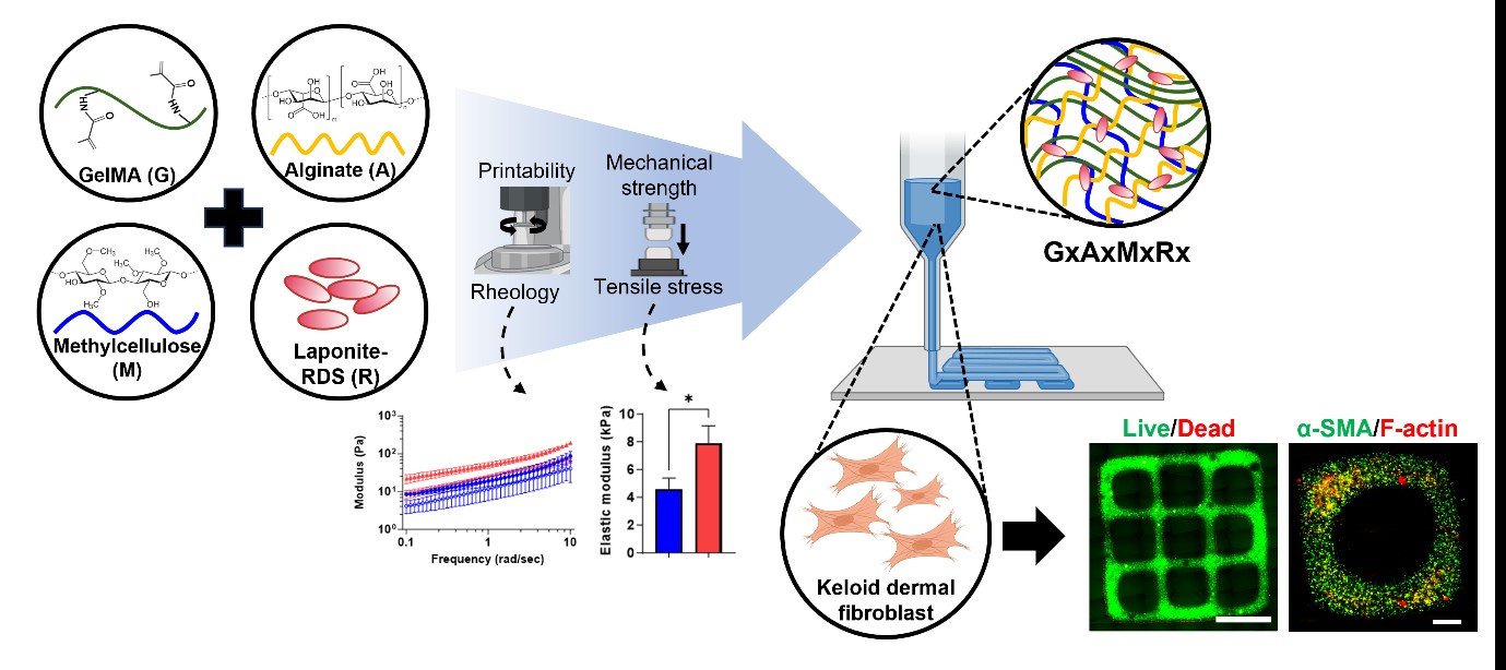
The development of mechanically tunable and cytocompatible hydrogels is critical for advancing three-dimensional (3D) bioprinting in tissue engineering. Here, we report a composite bioink composed of gelatin methacrylate (GelMA), methylcellulose, sodium alginate, and laponite-RDS. This formulation supports extrusion-based printing without ionic crosslinkers, mimics the extracellular matrix (ECM), and maintains stable viscoelasticity under physiological conditions (37°C, pH 7.4). Electrostatic and hydrogen bonding interactions among the charged polymers enhance pre-gel viscosity, shear-thinning behavior, and print fidelity. To evaluate its potential in disease modeling, patient-derived keloid fibroblasts were encapsulated in 3D-bioprinted constructs using two GelMA-based formulations with different stiffness levels, such as soft (G4A1M1R1, 2.1 kPa) and stiff (G5A1M1R1, 7.9 kPa), chosen to replicate the mechanical properties of normal dermis and keloid tissue, respectively. Both constructs exhibited excellent cell viability after three days, confirming cytocompatibility. Furthermore, matrix stiffness significantly regulated fibrotic gene expression. The stiffer hydrogel induced higher expression of COL1, MMP2, and IL6, suggesting enhanced myofibroblast activation and ECM remodeling. Immunofluorescence staining further confirmed elevated protein levels of α-SMA, FSP1, and actin stress fibers (F-actin) in the stiff construct, consistent with keloid pathology. Taken together, these results demonstrate that the GelMA-based bioink enables stiffness-dependent modulation of fibrotic responses, offering a simplified yet relevant 3D model of fibrotic skin. This platform may provide a useful basis for future studies on keloid progression and preliminary antifibrotic drug screening.
- Gu BK, Choi DJ, Park SJ, Kim MS, Kang CM, Kim C-H. 3-Dimensional bioprinting for tissue engineering applications. Biomater Res. 2016;20(1):12. doi: 10.1186/s40824-016-0058-2
- Choi K, Park CY, Choi JS, et al. The effect of the mechanical properties of the 3D printed gelatin/hyaluronic acid scaffolds on hMSCs differentiation towards chondrogenesis. Tissue Eng Regen Med. 2023;20(4):593-605. doi: 10.1007/s13770-023-00545-w
- Rossi A, Pescara T, Gambelli AM, et al. Biomaterials for extrusion-based bioprinting and biomedical applications. Front Bioeng Biotechnol. 2024;12:1393641. doi: 10.3389/fbioe.2024.1393641
- Chen XB, Fazel Anvari-Yazdi A, Duan X, et al. Biomaterials/ bioinks and extrusion bioprinting. Bioact Mater. 2023;28:511-536. doi: 10.1016/j.bioactmat.2023.06.006
- Yoon J, Han H, Jang J. Nanomaterials-incorporated hydrogels for 3D bioprinting technology. Nano Converg. 2023;10(1):52. doi: 10.1186/s40580-023-00402-5
- Nath R, Thomas J, Janardanan A, et al. An insight into synthesis, properties and applications of gelatin methacryloyl hydrogel for 3D bioprinting. Mater Adv. 2023;22:5496-5529. doi: 10.1039/D3MA00715D
- Wu Y, Xiang Y, Fang J, et al. The influence of the stiffness of GelMA substrate on the outgrowth of PC12 cells. Biosci Rep. 2019;39(1):BSR20181748. doi: 10.1042/BSR20181748
- Piao Y, You H, Xu T, et al. Biomedical applications of gelatin methacryloyl hydrogels. Eng Regen. 2021;2:47-56. doi: 10.1016/j.engreg.2021.03.002
- Axpe E, Oyen ML. Applications of alginate-based bioinks in 3D bioprinting. Int J Mol Sci. 2016;17(12):1976. doi: 10.3390/ijms17121976
- Aldana AA, Valente F, Dilley R, Doyle B. Development of 3D bioprinted GelMA-alginate hydrogels with tunable mechanical properties. Bioprinting. 2021;21:e00105. doi: 10.1016/j.bprint.2020.e00105
- Contessi N, Altomare L, Filipponi A, Farè S. Thermo-responsive properties of methylcellulose hydrogels for cell sheet engineering. Mater Lett. 2017;207:157-160. doi: 10.1016/j.matlet.2017.07.023
- Chang C, Zhang L. Cellulose-based hydrogels: present status and application prospects. Carbohydr Polym. 2011;84(1):40-53. doi: 10.1016/j.carbpol.2010.12.023
- Contessi Negrini N, Bonetti L, Contili L, Farè S. 3D printing of methylcellulose-based hydrogels. Bioprinting. 2018;10:e00024. doi: 10.1016/j.bprint.2018.e00024
- Ma Z, He H, Deng C, et al. 3D bioprinting of proangiogenic constructs with induced immunomodulatory microenvironments through a dual cross-linking procedure using laponite incorporated bioink. Compos B Eng. 2022;229:109399. doi: 10.1016/j.compositesb.2021.109399
- Choi D, Heo J, Aviles Milan J, et al. Structured nanofilms comprising Laponite® and bone extracellular matrix for osteogenic differentiation of skeletal progenitor cells. Mater Sci Eng C Mater Biol Appl. 2021;118:111440. doi: 10.1016/j.msec.2020.111440
- Li C, Hou Y, He M, et al. Laponite lights calcium flickers by reprogramming lysosomes to steer DC migration for an effective antiviral CD8(+) T-cell response. Adv Sci (Weinh). 2023;10(30):e230300. doi: 10.1002/advs.202303006.
- Wang Y, Zhao T, Jiao Y, et al. Silicate nanoplatelets promotes neuronal differentiation of neural stem cells and restoration of spinal cord injury. Adv Healthc Mater. 2023;12(19):e2203051. doi: 10.1002/adhm.202203051.
- Kim H, Anggradita LD, Lee SJ, et al. Ameliorating fibrotic phenotypes of keloid dermal fibroblasts through an epidermal growth factor-mediated extracellular matrix remodeling. Int J Mol Sci. 2021;22(4):2198. doi: 10.3390/ijms22042198
- Olejnik A, Semba JA, Kulpa A, Dańczak-Pazdrowska A, Rybka JD, Gornowicz-Porowska J. 3D bioprinting in skin related research: recent achievements and application perspectives. ACS Synth Biol. 2022;11(1):26-38. doi: 10.1021/acssynbio.1c00547
- Kwon SH, Lee J, Yoo JA-O, Jung YA-O. Artificial keloid skin models: understanding the pathophysiological mechanisms and application in therapeutic studies. Biomater Sci. 2024;25(13):3321-3334. doi: 10.1039/d4bm00005
- Kang H, Shih Y-RV, Hwang Y, et al. Mineralized gelatin methacrylate-based matrices induce osteogenic differentiation of human induced pluripotent stem cells. Acta Biomater. 2014;10(12):4961-4970. doi: 10.1016/j.actbio.2014.08.010.
- Temirel M, Dabbagh SR, Tasoglu S. Shape fidelity evaluation of alginate-based hydrogels through extrusion-based bioprinting. J Funct Biomater. 2022;13(4):225. doi: 10.3390/jfb13040225
- Livak KJ, Schmittgen TD. Analysis of relative gene expression data using real-time quantitative PCR and the 2(-Delta Delta C(T)) method. Methods. 2001;25(4):402-8. doi: 10.1006/meth.2001.1262. PMID: 11846609.
- Wang K-Y, Jin X-Y, Ma Y-H, et al. Injectable stress relaxation gelatin-based hydrogels with positive surface charge for adsorption of aggrecan and facile cartilage tissue regeneration. J Nanobiotechnol. 2021;19(1):214. doi: 10.1186/s12951-021-00950-0
- Vigata M, Meinert C, Bock N, Dargaville BL, Hutmacher DW. Deciphering the molecular mechanism of water interaction with gelatin methacryloyl hydrogels: role of ionic strength, pH, drug loading and hydrogel network characteristics. Biomedicines. 2021;9(5):574. doi: 10.3390/biomedicines9050574
- Xiao S, Zhao T, Wang J, et al. Gelatin methacrylate (GelMA)-based hydrogels for cell transplantation: an effective strategy for tissue engineering. Stem Cell Rev Rep. 2019;15(5):664-679. doi: 10.1007/s12015-019-09893-4.
- Fang W, Yang M, Wang L, et al. Hydrogels for 3D bioprinting in tissue engineering and regenerative medicine: current progress and challenges. Int J Bioprint. 2023;9(5):759. doi: 10.18063/ijb.759. PMID: 37457925
- Stealey ST, Gaharwar AK, Zustiak SP. Laponite-based nanocomposite hydrogels for drug delivery applications. Pharmaceuticals. 2023;16(6):821. doi: 10.3390/ph16060821
- de Barros NR, Gomez A, Ermis M, et al. Gelatin methacryloyl and Laponite bioink for 3D bioprinted organotypic tumor modeling. Biofabrication. 2023;15(4):045005. doi: 10.1088/1758-5090/ace0db
- Ashammakhi N, Ahadian S, Xu C, et al. Bioinks and bioprinting technologies to make heterogeneous and biomimetic tissue constructs. Mater Today Bio. 2019;1:100008. doi: 10.1016/j.mtbio.2019.100008
- Muthuramalingam K, Lee HA-O. Effect of GelMA hydrogel properties on long-term encapsulation and myogenic differentiation of C(2)C(12) spheroids. Gels. 2023;9(12):925. doi: 10.3390/gels9120925.
- Gao T, Gillispie GJ, Copus JS, et al. Optimization of gelatin-alginate composite bioink printability using rheological parameters: a systematic approach. Biofabrication. 2018;10(3):034106. doi: 10.1088/1758-5090/aacdc7.
- Moreira R, Chenlo F, Silva C, Torres MD. Rheological behaviour of aqueous methylcellulose systems: effect of concentration, temperature and presence of tragacanth. LWT. 2017;84:764-770. doi: 10.1016/j.lwt.2017.06.050
- Schütz K, Placht A-M, Paul B, Brüggemeier S, Gelinsky M, Lode A. Three-dimensional plotting of a cell-laden alginate/ methylcellulose blend: towards biofabrication of tissue engineering constructs with clinically relevant dimensions. J Tissue Eng Regen Med. 2017;11(5):1574-1587. doi: 10.1002/term.2058
- Rastin H, Ormsby RT, Atkins GJ, Losic DA-OX. 3D bioprinting of methylcellulose/gelatin-methacryloyl (MC/ GelMA) bioink with high shape integrity. ACS Appl Bio Mater. 2020;3(3):1815-1826. doi: 10.1021/acsabm.0c00169
- Afghah F, Altunbek M, Dikyol C, Koc B. Preparation and characterization of nanoclay-hydrogel composite support-bath for bioprinting of complex structures. Sci Rep. 2020;10(1):5257. doi: 10.1038/s41598-020-61606-x
- Mignon A, Pezzoli D, Prouvé E, et al. Combined effect of laponite and polymer molecular weight on the cell-interactive properties of synthetic PEO-based hydrogels. React Funct Polym. 2019;136:95-106. doi: 10.1016/j.reactfunctpolym.2018.12.017
- Aazmi A, Zhang D, Mazzaglia C, et al. Biofabrication methods for reconstructing extracellular matrix mimetics. Bioact Mater. 2023;31:475-496. doi: 10.1016/j.bioactmat.2023.08.018.
- GhavamiNejad A, Ashammakhi N, Wu XY, Khademhosseini A. Crosslinking strategies for 3D bioprinting of polymeric hydrogels. Small. 2020;16(35):2002931. doi: 10.1002/smll.202002931
- Karaca MA, Khalili V, Ege D. Highly flexible methyl cellulose/gelatin hydrogels for potential cartilage tissue engineering applications. Biopolymers. 2025;116(1):e23641. doi: 10.1002/bip.23641
- Šebenik U, Lapasin R, Krajnc M. Rheology of aqueous dispersions of laponite and TEMPO-oxidized nanofibrillated cellulose. Carbohydr Polym. 2020;240:116330. doi: 10.1016/j.carbpol.2020.116330
- Yue K, Trujillo-de Santiago G, Alvarez MM, Tamayol A, Annabi N, Khademhosseini A. Synthesis, properties, and biomedical applications of gelatin methacryloyl (GelMA) hydrogels. Biomaterials. 2015;73:254-271. doi: 10.1016/j.biomaterials.2015.08.045
- Li H, Chen S, Dissanayaka WL, Wang M. Gelatin methacryloyl/sodium alginate/cellulose nanocrystal inks and 3D printing for dental tissue engineering applications. ACS Omega. 2024;9(49):48361-48373. doi: 10.1021/acsomega.4c06458
- Xu L, Zhang Z, Jorgensen AM, et al. Bioprinting a skin patch with dual-crosslinked gelatin (GelMA) and silk fibroin (SilMA): an approach to accelerating cutaneous wound healing. Mater Today Bio. 2023;18:100550. doi: 10.1016/j.mtbio.2023.100550
- Zeimaran E, Pourshahrestani S, Röder J, Detsch R, Boccaccini AR. 3D printing of photocrosslinked alginate dialdehyde-gelatin hydrogels reinforced with cobalt-containing mesoporous bioactive glass nanoparticles for developing skin wound dressings. Adv Mater Interfaces. 2025;12(11):2400913. doi: 10.1002/admi.202400913
- Limandjaja GC, Niessen FB, Scheper RJ, Gibbs S. The Keloid disorder: heterogeneity, histopathology, mechanisms and models. Front Cell Dev Biol. 2020;8:360. doi: 10.3389/fcell.2020.00360.
- Hsu CK, Lin HH, Harn HI, et al. Caveolin-1 controls hyperresponsiveness to mechanical stimuli and fibrogenesis-associated RUNX2 activation in keloid fibroblasts. J Invest Dermatol. 2018;138(1):208-218. doi: 10.1016/j.jid.2017.05.041
- Saraswati S, Marrow SMW, Watch LA, Young PP. Identification of a pro-angiogenic functional role for FSP1- positive fibroblast subtype in wound healing. Nat Commun. 2019;10(1):3027. doi: 10.1038/s41467-019-10965-9
- Camman M, Nieswic N, Joanne P, et al. Fibrotic-like collagen matrices as innovative 3D in vitro models for investigating the impact of pathological ECM on muscle regeneration in muscular dystrophies. bioRxiv. 2024. doi: 10.1101/2024.12.23.630059
- Tai Y, Woods EL, Dally J, et al. Myofibroblasts: function, formation, and scope of molecular therapies for skin fibrosis. Biomolecules. 2021;11(8):1095. doi: 10.3390/biom11081095
- Lee W, Debasitis JC, Lee VK, et al. Multi-layered culture of human skin fibroblasts and keratinocytes through three-dimensional freeform fabrication. Biomaterials. 2009;30(8):1587-1595. doi: 10.1016/j.biomaterials.2008.12.009
- Jang Y, Jang J, Kim BY, Song YS, Lee DY. Effect of gelatin content on degradation behavior of PLLA/gelatin hybrid membranes. Tissue Eng Regen Med. 2024;21(4):557-569. doi: 10.1007/s13770-024-00626-4
- Lee YJ, Oh JH, Park S, et al. The application of L-serine-incorporated gelatin sponge into the calvarial defect of the ovariectomized rats. Tissue Eng Regen Med. 2025;22(1):91-104. doi: 10.1007/s13770-024-00686-6
- Vigata M, Meinert C, Pahoff S, Bock N, Hutmacher DW. Gelatin methacryloyl hydrogels control the localized delivery of albumin-bound paclitaxel. Polymers. 2020;12(2):501. doi: 10.3390/polym12020501
- Zhu M, Wang Y, Ferracci G, Zheng J, Cho NJ, Lee BH. Gelatin methacryloyl and its hydrogels with an exceptional degree of controllability and batch-to-batch consistency. Sci Rep. 2019;9(1):6863. doi: 10.1038/s41598-019-42186-x