

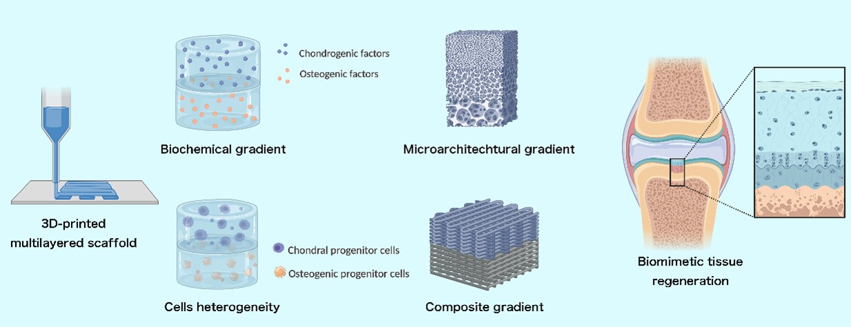
Hierarchical 3D-printed scaffolds for osteochondral regeneration: From biomimetic design to functional integration

Osteochondral defects, characterized by the structural and functional disruption of articular cartilage and subchondral bone, present significant clinical challenges due to the tissue’s limited intrinsic regenerative capacity. Scaffold-based tissue engineering has paved the way for osteochondral defect treatment; however, fully restoring the complex structure and composition of native osteochondral tissue remains challenging. Recent advances in three-dimensional (3D) printing have enabled the fabrication of layered, anisotropic scaffolds designed to biomimetically recapitulate the native tissue’s zonal properties through precise hierarchical design. High-resolution fabrication techniques facilitate the construction of delicate microarchitectures, while advanced bioprinting methods allow for the incorporation of bioactive factors and cells into the scaffold matrix. This review emphasizes the following four scaffold design paradigms: composite gradients, microarchitectural patterning, biochemical gradients, and cellular heterogeneity. Moreover, key properties of multilayered scaffolds are discussed, including mechanical performance, interfacial strength, and degradation behavior. In addition, several obstacles associated with the in vivo scaffold application are discussed, providing insights to guide future clinical translation in osteochondral defects treatment.
- Mahmoudian A, Lohmander LS, Mobasheri A, Englund M, Luyten FP. Early-stage symptomatic osteoarthritis of the knee - time for action. Nat Rev Rheumatol. 2021;17(10):621-632. doi: 10.1038/s41584-021-00673-4
- He B, Wu JP, Kirk TB, Carrino JA, Xiang C, Xu J. High-resolution measurements of the multilayer ultra-structure of articular cartilage and their translational potential. Arthritis Res Ther. 2014;16(2):205. doi: 10.1186/ar4506
- Soderquist M, Barnes L. Osteochondral allograft transplantation for articular humeral head defect from ballistic trauma. JSES Rev Rep Tech. 2024;4(3):540-546. doi: 10.1016/j.xrrt.2024.04.008
- GBD 2021 Osteoarthritis Collaborators. Global, regional, and national burden of osteoarthritis, 1990-2020 and projections to 2050: a systematic analysis for the Global Burden of Disease Study 2021. Lancet Rheumatol. 2023;5(9):e508-e522. doi: 10.1016/s2665-9913(23)00163-7
- Weng Q, Chen Q, Jiang T, et al. Global burden of early-onset osteoarthritis, 1990-2019: results from the Global Burden of Disease Study 2019. Ann Rheum Dis. 2024;83(7):915-925. doi: 10.1136/ard-2023-225324
- Abdulghani S, Morouço PG. Biofabrication for osteochondral tissue regeneration: bioink printability requirements. J Mater Sci Mater Med. 2019;30(2):20. doi: 10.1007/s10856-019-6218-x
- Brophy RH, Fillingham YA. AAOS clinical practice guideline summary: management of osteoarthritis of the knee (nonarthroplasty), third edition. J Am Acad Orthop Surg. 2022;30(9):e721-e729. doi: 10.5435/jaaos-d-21-01233
- Medina J, Garcia-Mansilla I, Fabricant PD, Kremen TJ, Sherman SL, Jones K. Microfracture for the treatment of symptomatic cartilage lesions of the knee: a survey of International Cartilage Regeneration & Joint Preservation Society. Cartilage. 2021;13(1_suppl):1148s-1155s. doi: 10.1177/1947603520954503
- Patel S, Caldwell JM, Doty SB, et al. Integrating soft and hard tissues via interface tissue engineering. J Orthop Res. 2018;36(4):1069-1077. doi: 10.1002/jor.23810
- Lee CH, Cook JL, Mendelson A, Moioli EK, Yao H, Mao JJ. Regeneration of the articular surface of the rabbit synovial joint by cell homing: a proof of concept study. Lancet. 2010;376(9739):440-448. doi: 10.1016/s0140-6736(10)60668-x
- Patel JM, Merriam AR, Culp BM, Gatt CJ, Jr., Dunn MG. One-year outcomes of total meniscus reconstruction using a novel fiber-reinforced scaffold in an ovine model. Am J Sports Med. 2016;44(4):898-907. doi: 10.1177/0363546515624913
- Ayers DC. How common is revision for adverse reaction to metal debris after total hip replacement with a metal-on-polyethylene bearing surface?: commentary on an article by Anders Persson, MD, et al.: “Revision for Symptomatic Pseudotumor Following Primary Total Hip Arthroplasty with a Standard Femoral Stem”. J Bone Joint Surg Am. 2018;100(11):e82. doi: 10.2106/jbjs.18.00193
- Ansari S, Khorshidi S, Karkhaneh A. Engineering of gradient osteochondral tissue: from nature to lab. Acta Biomater. 2019;87:41-54. doi: 10.1016/j.actbio.2019.01.071
- Bedell ML, Wang Z, Hogan KJ, et al. The effect of multi-material architecture on the ex vivo osteochondral integration of bioprinted constructs. Acta Biomater. 2023;155:99-112. doi: 10.1016/j.actbio.2022.11.014
- McKee TJ, Perlman G, Morris M, Komarova SV. Extracellular matrix composition of connective tissues: a systematic review and meta-analysis. Sci Rep. 2019;9(1):10542. doi: 10.1038/s41598-019-46896-0
- Di Luca A, Szlazak K, Lorenzo-Moldero I, et al. Influencing chondrogenic differentiation of human mesenchymal stromal cells in scaffolds displaying a structural gradient in pore size. Acta Biomater. 2016;36:210-219. doi: 10.1016/j.actbio.2016.03.014
- Shen G. The role of type X collagen in facilitating and regulating endochondral ossification of articular cartilage. Orthod Craniofac Res. 2005;8(1):11-17. doi: 10.1111/j.1601-6343.2004.00308.x
- Horkay F, Basser PJ, Geissler E. Cartilage extracellular matrix polymers: hierarchical structure, osmotic properties, and function. Soft Matter. 2024;20(30):6033-6043. doi: 10.1039/d4sm00617h
- Rose FR, Oreffo RO. Bone tissue engineering: hope vs hype. Biochem Biophys Res Commun. 2002;292(1):1-7. doi: 10.1006/bbrc.2002.6519
- Gonzalez-Fernandez T, Rathan S, Hobbs C, et al. Pore-forming bioinks to enable spatio-temporally defined gene delivery in bioprinted tissues. J Control Release. 2019;301:13-27. doi: 10.1016/j.jconrel.2019.03.006
- Rowland CR, Glass KA, Ettyreddy AR, et al. Regulation of decellularized tissue remodeling via scaffold-mediated lentiviral delivery in anatomically-shaped osteochondral constructs. Biomaterials. 2018;177:161-175. doi: 10.1016/j.biomaterials.2018.04.049
- Liu Y, Huang C, Bai M, Pi C, Zhang D, Xie J. The roles of Runx1 in skeletal development and osteoarthritis: a concise review. Heliyon. 2022;8(12):e12656. doi: 10.1016/j.heliyon.2022.e12656
- Li W, Zhang S, Liu J, Liu Y, Liang Q. Vitamin K2 stimulates MC3T3‑E1 osteoblast differentiation and mineralization through autophagy induction. Mol Med Rep. 2019;19(5):3676-3684. doi: 10.3892/mmr.2019.10040
- Grogan SP, Duffy SF, Pauli C, et al. Zone-specific gene expression patterns in articular cartilage. Arthritis Rheum. 2013;65(2):418-428. doi: 10.1002/art.37760
- Zuscik MJ, Hilton MJ, Zhang X, Chen D, O’Keefe RJ. Regulation of chondrogenesis and chondrocyte differentiation by stress. J Clin Invest. 2008;118(2):429-438. doi: 10.1172/jci34174
- Ma J, Wang Z, Zhao J, Miao W, Ye T, Chen A. Resveratrol attenuates lipopolysaccharides (LPS)-induced inhibition of osteoblast differentiation in MC3T3-E1 cells. Med Sci Monit. 2018;24:2045-2052. doi: 10.12659/msm.905703
- Kitaura H, Marahleh A, Ohori F, et al. Osteocyte-related cytokines regulate osteoclast formation and bone resorption. Int J Mol Sci. 2020;21(14):5169. doi: 10.3390/ijms21145169
- Zhang D, Deng X, Liu Y, et al. MMP-10 deficiency effects differentiation and death of chondrocytes associated with endochondral osteogenesis in an endemic osteoarthritis. Cartilage. 2022;13(3):19476035221109226. doi: 10.1177/19476035221109226
- Boyce BF, Xing L. Functions of RANKL/RANK/OPG in bone modeling and remodeling. Arch Biochem Biophys. 2008;473(2):139-146. doi: 10.1016/j.abb.2008.03.018
- Quinn TM, Häuselmann HJ, Shintani N, Hunziker EB. Cell and matrix morphology in articular cartilage from adult human knee and ankle joints suggests depth-associated adaptations to biomechanical and anatomical roles. Osteoarthritis Cartilage. 2013;21(12):1904-1912.
- Niu H, Liu C, Li A, et al. Relationship between triphasic mechanical properties of articular cartilage and osteoarthritic grade. Sci China Life Sci. 2012;55(5):444-451.
doi: 10.1007/s11427-012-4326-7
- Sun Y, Zhang K, Dong H, et al. Layered mechanical and electrical properties of porcine articular cartilage. Med Biol Eng Comput. 2022;60(10):3019-3028. doi: 10.1007/s11517-022-02653-6
- Burr DB, Gallant MA. Bone remodelling in osteoarthritis. Nat Rev Rheumatol. 2012;8(11):665-673. doi: 10.1038/nrrheum.2012.130
- Flachsmann ER, Broom ND, Oloyede A. A biomechanical investigation of unconstrained shear failure of the osteochondral region under impact loading. Clin Biomech (Bristol). 1995;10(3):156-165. doi: 10.1016/0268-0033(95)93706-y
- Madry H, van Dijk CN, Mueller-Gerbl M. The basic science of the subchondral bone. Knee Surg Sports Traumatol Arthrosc. 2010;18(4):419-433. doi: 10.1007/s00167-010-1054-z
- Broom ND, Oloyede A, Flachsmann R, Hows M. Dynamic fracture characteristics of the osteochondral junction undergoing shear deformation. Med Eng Phys. 1996;18(5):396-404. doi: 10.1016/1350-4533(95)00067-4
- Mieloch AA, Richter M, Trzeciak T, Giersig M, Rybka JD. Osteoarthritis severely decreases the elasticity and hardness of knee joint cartilage: a nanoindentation study. J Clin Med. 2019;8(11):1865. doi: 10.3390/jcm8111865
- Davis S, Zekonyte J, Karali A, Roldo M, Blunn G. Early degenerative changes in a spontaneous osteoarthritis model assessed by nanoindentation. Bioengineering (Basel). 2023;10(9):995. doi: 10.3390/bioengineering10090995
- Peters AE, Akhtar R, Comerford EJ, Bates KT. The effect of ageing and osteoarthritis on the mechanical properties of cartilage and bone in the human knee joint. Sci Rep. 2018;8(1):5931. doi: 10.1038/s41598-018-24258-6
- Hu YJ, Yu YE, Cooper HJ, et al. Mechanical and structural properties of articular cartilage and subchondral bone in human osteoarthritic knees. J Bone Miner Res. 2024;39(8):1120-1131. doi: 10.1093/jbmr/zjae094
- Guo J, Li Q, Zhang R, et al. Loose pre-cross-linking mediating cellulose self-assembly for 3D printing strong and tough biomimetic scaffolds. Biomacromolecules. 2022;23(3):877-888. doi: 10.1021/acs.biomac.1c01330
- Zhang X, Liu Y, Zuo Q, et al. 3D bioprinting of biomimetic bilayered scaffold consisting of decellularized extracellular matrix and silk fibroin for osteochondral repair. Int J Bioprint. 2021;7(4):401. doi: 10.18063/ijb.v7i4.401
- Nedrelow DS, Rassi A, Ajeeb B, et al. Regenerative engineering of a biphasic patient-fitted temporomandibular joint condylar prosthesis. Tissue Eng Part C Methods. 2023;29(7):307-320. doi: 10.1089/ten.TEC.2023.0093
- Wu Z, Yao H, Sun H, et al. Enhanced hyaline cartilage formation and continuous osteochondral regeneration via 3D-Printed heterogeneous hydrogel with multi-crosslinking inks. Mater Today Bio. 2024;26:101080. doi: 10.1016/j.mtbio.2024.101080
- Diloksumpan P, de Ruijter M, Castilho M, et al. Combining multi-scale 3D printing technologies to engineer reinforced hydrogel-ceramic interfaces. Biofabrication. 2020;12(2):025014. doi: 10.1088/1758-5090/ab69d9
- Liu Y, Peng L, Li L, et al. 3D-bioprinted BMSC-laden biomimetic multiphasic scaffolds for efficient repair of osteochondral defects in an osteoarthritic rat model. Biomaterials. 2021;279:121216. doi: 10.1016/j.biomaterials.2021.121216
- Gao J, Ding X, Yu X, et al. Cell-free bilayered porous scaffolds for osteochondral regeneration fabricated by continuous 3d-printing using nascent physical hydrogel as ink. Adv Healthc Mater. 2021;10(3):e2001404. doi: 10.1002/adhm.202001404
- Wang Z, Cao W, Wu F, et al. A triphasic biomimetic BMSC-loaded scaffold for osteochondral integrated regeneration in rabbits and pigs. Biomater Sci. 2023;11(8):2924-2934. doi: 10.1039/d2bm02148j
- Braxton T, Lim K, Alcala-Orozco C, et al. Mechanical and physical characterization of a biphasic 3D printed silk-infilled scaffold for osteochondral tissue engineering. ACS Biomater Sci Eng. 2024;10(12):7606-7618. doi: 10.1021/acsbiomaterials.4c01865
- Wang S, Luo B, Bai B, et al. 3D printed chondrogenic functionalized PGS bioactive scaffold for cartilage regeneration. Adv Healthc Mater. 2023;12(27): e2301006. doi: 10.1002/adhm.202301006
- Wang H, Zhang J, Bai H, et al. 3D printed cell-free bilayer porous scaffold based on alginate with biomimetic microenvironment for osteochondral defect repair. Biomater Adv. 2025;167:214092. doi: 10.1016/j.bioadv.2024.214092
- Ding X, Gao J, Yu X, et al. 3D-printed porous scaffolds of hydrogels modified with TGF-β1 binding peptides to promote in vivo cartilage regeneration and animal gait restoration. ACS Appl Mater Interfaces. 2022;14(14):15982-15995. doi: 10.1021/acsami.2c00761
- Li Q, Yu H, Zhao F, et al. 3D printing of microenvironment-specific bioinspired and exosome-reinforced hydrogel scaffolds for efficient cartilage and subchondral bone regeneration. Adv Sci (Weinh). 2023;10(26):e2303650. doi: 10.1002/advs.202303650
- Cui X, Alcala-Orozco CR, Baer K, et al. 3D bioassembly of cell-instructive chondrogenic and osteogenic hydrogel microspheres containing allogeneic stem cells for hybrid biofabrication of osteochondral constructs. Biofabrication. 2022;14(3):034101. doi: 10.1088/1758-5090/ac61a3
- Wei W, Liu W, Kang H, et al. A one-stone-two-birds strategy for osteochondral regeneration based on a 3D printable biomimetic scaffold with kartogenin biochemical stimuli gradient. Adv Healthc Mater. 2023;12(15):e2300108. doi: 10.1002/adhm.202300108
- Coyle A, Chakraborty A, Huang J, Shamiya Y, Luo W, Paul A. In vitro engineered ECM-incorporated hydrogels for osteochondral tissue repair: a cell-free approach. Adv Healthc Mater. 2025;14(4):e2402701. doi: 10.1002/adhm.202402701
- Sun T, Feng Z, He W, et al. Novel 3D-printing bilayer GelMA-based hydrogel containing BP,β-TCP and exosomes for cartilage-bone integrated repair. Biofabrication. 2023;16(1):015008. doi: 10.1088/1758-5090/ad04fe
- Eckstein KN, Hergert JE, Uzcategui AC, et al. Controlled mechanical property gradients within a digital light processing printed hydrogel-composite osteochondral scaffold. Ann Biomed Eng. 2024;52(8):2162-2177. doi: 10.1007/s10439-024-03516-x
- Golebiowska A, Nukavarapu SP. Bio-inspired zonal-structured matrices for bone-cartilage interface engineering. Biofabrication. 2022;14(2):025016. doi: 10.1088/1758-5090/ac52e1
- Sun Y, Wu Q, Zhang Y, Dai K, Wei Y. 3D-bioprinted gradient-structured scaffold generates anisotropic cartilage with vascularization by pore-size-dependent activation of HIF1α/FAK signaling axis. Nanomedicine. 2021; 37:102426. doi: 10.1016/j.nano.2021.102426
- Gu Y, Zou Y, Huang Y, et al. 3D-printed biomimetic scaffolds with precisely controlled and tunable structures guide cell migration and promote regeneration of osteochondral defect. Biofabrication. 2023;16(1):015003. doi: 10.1088/1758-5090/ad0071
- Kilian D, Cometta S, Bernhardt A, et al. Core-shell bioprinting as a strategy to apply differentiation factors in a spatially defined manner inside osteochondral tissue substitutes. Biofabrication. 2022;14(1):014108. doi: 10.1088/1758-5090/ac457b
- Zhang Y, Li D, Liu Y, et al. 3D-bioprinted anisotropic bicellular living hydrogels boost osteochondral regeneration via reconstruction of cartilage-bone interface. Innovation (Camb). 2024;5(1):100542. doi: 10.1016/j.xinn.2023.100542
- Maherani M, Eslami H, Poursamar SA, Ansari M. A modular approach to 3D-printed bilayer composite scaffolds for osteochondral tissue engineering. J Mater Sci Mater Med. 2024;35(1):62. doi: 10.1007/s10856-024-06824-9
- O’Shea DG, Hodgkinson T, Curtin CM, O’Brien FJ. An injectable and 3D printable pro-chondrogenic hyaluronic acid and collagen type II composite hydrogel for the repair of articular cartilage defects. Biofabrication. 2023;16(1):015007. doi: 10.1088/1758-5090/ad047a
- Zineh BR, Roshangar L, Meshgi S, Shabgard M. 3D printing of alginate/thymoquinone/halloysite nanotube bio-scaffolds for cartilage repairs: experimental and numerical study. Med Biol Eng Comput. 2022;60(11):3069-3080. doi: 10.1007/s11517-022-02654-5
- Naranda J, Bračič M, Vogrin M, Maver U. Recent advancements in 3d printing of polysaccharide hydrogels in cartilage tissue engineering. Materials (Basel). 2021;14(14):3977. doi: 10.3390/ma14143977
- Majumder N, Roy C, Doenges L, Martin I, Barbero A, Ghosh S. Covalent conjugation of small molecule inhibitors and growth factors to a silk fibroin-derived bioink to develop phenotypically stable 3D bioprinted cartilage. ACS Appl Mater Interfaces. 2024;16(8):9925-9943. doi: 10.1021/acsami.3c18903
- Zhang W, Lian Q, Li D, et al. Cartilage repair and subchondral bone migration using 3D printing osteochondral composites: a one-year-period study in rabbit trochlea. Biomed Res Int. 2014;2014:746138. doi: 10.1155/2014/746138
- Golebiowska AA, Nukavarapu SP. Bio-inspired zonal-structured matrices for bone-cartilage interface engineering. Biofabrication. 2022;14(2):025016. doi: 10.1088/1758-5090/ac5413
- Jiang G, Li S, Yu K, et al. A 3D-printed PRP-GelMA hydrogel promotes osteochondral regeneration through M2 macrophage polarization in a rabbit model. Acta Biomater. 2021;128:150-162. doi: 10.1016/j.actbio.2021.04.010
- Parisi C, Salvatore L, Veschini L, et al. Biomimetic gradient scaffold of collagen-hydroxyapatite for osteochondral regeneration. J Tissue Eng. 2020;11:2041731419896068. doi: 10.1177/2041731419896068
- Sa MW, Nguyen BB, Moriarty RA, Kamalitdinov T, Fisher JP, Kim JY. Fabrication and evaluation of 3D printed BCP scaffolds reinforced with ZrO(2) for bone tissue applications. Biotechnol Bioeng. 2018;115(4):989-999. doi: 10.1002/bit.26514
- Liu L, Yu F, Chen L, Xia L, Wu C, Fang B. Lithium-containing biomaterials stimulate cartilage repair through bone marrow stromal cells-derived exosomal miR-455-3p and Histone H3 acetylation. Adv Healthc Mater. 2023;12(11):e2202390. doi: 10.1002/adhm.202202390
- Zheng J, Mao X, Ling J, Chen C, Zhang W. Role of magnesium transporter subtype 1 (MagT1) in the osteogenic differentiation of rat bone marrow stem cells. Biol Trace Elem Res. 2016;171(1):131-137. doi: 10.1007/s12011-015-0459-4
- Wang X, Gao L, Han Y, et al. Silicon-enhanced adipogenesis and angiogenesis for vascularized adipose tissue engineering. Adv Sci (Weinh). 2018;5(11):1800776. doi: 10.1002/advs.201800776
- Rahaman MN, Day DE, Bal BS, et al. Bioactive glass in tissue engineering. Acta Biomater. 2011;7(6):2355-2373. doi: 10.1016/j.actbio.2011.03.016
- Morgan EF, Salisbury Palomares KT, Gleason RE, et al. Correlations between local strains and tissue phenotypes in an experimental model of skeletal healing. J Biomech. 2010;43(12):2418-2424. doi: 10.1016/j.jbiomech.2010.04.019
- Claes L, Eckert-Hübner K, Augat P. The effect of mechanical stability on local vascularization and tissue differentiation in callus healing. J Orthop Res. 2002;20(5):1099-1105. doi: 10.1016/s0736-0266(02)00044-x
- Chen C, Xie J, Deng L, Yang L. Substrate stiffness together with soluble factors affects chondrocyte mechanoresponses. ACS Appl Mater Interfaces. 2014;6(18):16106-16116. doi: 10.1021/am504135b
- Yang Y, Feng Y, Qu R, et al. Synthesis of aligned porous polyethylene glycol/silk fibroin/hydroxyapatite scaffolds for osteoinduction in bone tissue engineering. Stem Cell Res Ther. 2020;11(1):522. doi: 10.1186/s13287-020-02024-8
- Cao B, Li J, Wang X, et al. Mechanosensitive miR-99b mediates the regulatory effect of matrix stiffness on bone marrow mesenchymal stem cell fate both in vitro and in vivo. APL Bioeng. 2023;7(1):016106. doi: 10.1063/5.0131125
- Lai Q, Li B, Chen L, Zhou Y, Bao H, Li H. Substrate stiffness regulates the proliferation and inflammation of chondrocytes and macrophages through exosomes. Acta Biomater. 2025;192:77-89. doi: 10.1016/j.actbio.2024.12.021
- Tortorici M, Petersen A, Ehrhart K, Duda GN, Checa S. Scaffold-dependent mechanical and architectural cues guide osteochondral defect healing in silico. Front Bioeng Biotechnol. 2021;9:642217. doi: 10.3389/fbioe.2021.642217
- Radhakrishnan J, Manigandan A, Chinnaswamy P, Subramanian A, Sethuraman S. Gradient nano-engineered in situ forming composite hydrogel for osteochondral regeneration. Biomaterials. 2018;162:82-98. doi: 10.1016/j.biomaterials.2018.01.056
- Pattnaik A, Sanket AS, Pradhan S, et al. Designing of gradient scaffolds and their applications in tissue regeneration. Biomaterials. 2023;296:122078. doi: doi: 10.1016/j.biomaterials.2023.122078
- Zhang W, Chen J, Tao J, et al. The use of type 1 collagen scaffold containing stromal cell-derived factor-1 to create a matrix environment conducive to partial-thickness cartilage defects repair. Biomaterials. 2013;34(3):713-723. doi: 10.1016/j.biomaterials.2012.10.027
- Hayashi K, Shimabukuro M, Kishida R, Tsuchiya A, Ishikawa K. Honeycomb scaffolds capable of achieving barrier membrane-free guided bone regeneration. Mater Adv. 2021;2(23):7638-7649. doi: 10.1039/D1MA00698C
- Zhao Y, Liang Y, Ding S, Zhang K, Mao HQ, Yang Y. Application of conductive PPy/SF composite scaffold and electrical stimulation for neural tissue engineering. Biomaterials. 2020;255:120164. doi: 10.1016/j.biomaterials.2020.120164
- He J, Sun C, Gu Z, et al. Morphology, migration, and transcriptome analysis of schwann cell culture on butterfly wings with different surface architectures. ACS Nano. 2018;12(10):9660-9668. doi: 10.1021/acsnano.8b00552
- Zhang J, Wu Y, Thote T, Lee EH, Ge Z, Yang Z. The influence of scaffold microstructure on chondrogenic differentiation of mesenchymal stem cells. Biomed Mater. 2014;9(3):035011. doi: 10.1088/1748-6041/9/3/035011
- DeLise AM, Fischer L, Tuan RS. Cellular interactions and signaling in cartilage development. Osteoarthritis Cartilage. 2000;8(5):309-334. doi: 10.1053/joca.1999.0306
- Li S, Tallia F, Mohammed AA, Stevens MM, Jones JR. Scaffold channel size influences stem cell differentiation pathway in 3-D printed silica hybrid scaffolds for cartilage regeneration. Biomater Sci. 2020;8(16):4458-4466. doi: 10.1039/c9bm01829h
- Zanetti NC, Solursh M. Induction of chondrogenesis in limb mesenchymal cultures by disruption of the actin cytoskeleton. J Cell Biol. 1984;99(1 Pt 1):115-123. doi: 10.1083/jcb.99.1.115
- Vanden Berg-Foels WS. In situ tissue regeneration: chemoattractants for endogenous stem cell recruitment. Tissue Eng Part B Rev. 2014;20(1):28-39.doi: 10.1089/ten.TEB.2013.0100
- Salgado AJ, Coutinho OP, Reis RL. Bone tissue engineering: state of the art and future trends. Macromol Biosci. 2004;4(8):743-765. doi: 10.1002/mabi.200400026
- Zhang X, Chen X, Hong H, Hu R, Liu J, Liu C. Decellularized extracellular matrix scaffolds: recent trends and emerging strategies in tissue engineering. Bioact Mater. 2022;10:15-31. doi: 10.1016/j.bioactmat.2021.09.014
- Jang J, Kim TG, Kim BS, Kim SW, Kwon SM, Cho DW. Tailoring mechanical properties of decellularized extracellular matrix bioink by vitamin B2-induced photo-crosslinking. Acta Biomater. 2016;33:88-95. doi: 10.1016/j.actbio.2016.01.013
- Zhang X, Liu Y, Luo C, et al. Crosslinker-free silk/ decellularized extracellular matrix porous bioink for 3D bioprinting-based cartilage tissue engineering. Mater Sci Eng C Mater Biol Appl. 2021;118:111388. doi: 10.1016/j.msec.2020.111388
- Joyce M, Hodgkinson T, Lemoine M, González-Vázquez A, Kelly DJ, O’Brien FJ. Development of a 3D-printed bioabsorbable composite scaffold with mechanical properties suitable for treating large, load-bearingarticular cartilage defects. Eur Cell Mater. 2023;45:158-172. doi: 10.22203/eCM.v045a11
- Johnson K, Zhu S, Tremblay MS, et al. A stem cell-based approach to cartilage repair. Science. 2012;336(6082):717-721. doi: 10.1126/science.1215157
- Stefani RM, Lee AJ, Tan AR, et al. Sustained low-dose dexamethasone delivery via a PLGA microsphere-embedded agarose implant for enhanced osteochondral repair. Acta Biomaterialia. 2020;102:326-340. doi: doi: 10.1016/j.actbio.2019.11.052
- Brito Barrera YA, Husteden C, Alherz J, Fuhrmann B, Wölk C, Groth T. Extracellular matrix-inspired surface coatings functionalized with dexamethasone-loaded liposomes to induce osteo- and chondrogenic differentiation of multipotent stem cells. Mater Sci Eng C Mater Biol Appl. 2021;131:112516. doi: 10.1016/j.msec.2021.112516
- Yan X, Chen YR, Song YF, et al. Scaffold-based gene therapeutics for osteochondral tissue engineering. Front Pharmacol. 2019;10:1534. doi: 10.3389/fphar.2019.01534
- An C, Cheng Y, Yuan Q, Li J. IGF-1 and BMP-2 induces differentiation of adipose-derived mesenchymal stem cells into chondrocytes-like cells. Ann Biomed Eng. 2010;38(4):1647-1654. doi: 10.1007/s10439-009-9892-x
- Tao K, Frisch J, Rey-Rico A, et al. Co-overexpression of TGF-β and SOX9 via rAAV gene transfer modulates the metabolic and chondrogenic activities of human bone marrow-derived mesenchymal stem cells. Stem Cell Res Ther. 2016;7:20. doi: 10.1186/s13287-016-0280-9
- Lee JM, Im GI. SOX trio-co-transduced adipose stem cells in fibrin gel to enhance cartilage repair and delay the progression of osteoarthritis in the rat. Biomaterials. 2012;33(7):2016-2024. doi: 10.1016/j.biomaterials.2011.11.050
- Vermeij EA, Broeren MG, Bennink MB, et al. Disease-regulated local IL-10 gene therapy diminishes synovitis and cartilage proteoglycan depletion in experimental arthritis. Ann Rheum Dis. 2015;74(11):2084-2091. doi: 10.1136/annrheumdis-2014-205223
- Kay JD, Gouze E, Oligino TJ, et al. Intra-articular gene delivery and expression of interleukin-1Ra mediated by self-complementary adeno-associated virus. J Gene Med. 2009;11(7):605-614. doi: 10.1002/jgm.1334
- Bellavia D, Veronesi F, Carina V, et al. Gene therapy for chondral and osteochondral regeneration: is the future now? Cell Mol Life Sci. 2018;75(4):649-667. doi: 10.1007/s00018-017-2637-3
- Lu H, Wei J, Liu K, et al. Radical-scavenging and subchondral bone-regenerating nanomedicine for osteoarthritis treatment. ACS Nano. 2023;17(6):6131-6146. doi: 10.1021/acsnano.3c01789
- Chen Y, Huang H, Zhong W, Li L, Lu Y, Si HB. miR- 140-5p protects cartilage progenitor/stem cells from fate changes in knee osteoarthritis. Int Immunopharmacol. 2023; 114:109576. doi: 10.1016/j.intimp.2022.109576
- Xing H, Zhang Z, Mao Q, et al. Injectable exosome-functionalized extracellular matrix hydrogel for metabolism balance and pyroptosis regulation in intervertebral disc degeneration. J Nanobiotechnol. 2021;19(1):264. doi: 10.1186/s12951-021-00991-5
- Zhu W, Wang H, Feng B, et al. Self-healing hyaluronic acid-based hydrogel with miRNA140-5p loaded MON-PEI nanoparticles for chondrocyte regeneration: Schiff base self-assembly approach. Adv Sci (Weinh). 2025;12(1):e2406479. doi: 10.1002/advs.202406479
- Tuan RS, Chen AF, Klatt BA. Cartilage regeneration. J Am Acad Orthop Surg. 2013;21(5):303-311. doi: 10.5435/jaaos-21-05-303
- Wang LT, Ting CH, Yen ML, et al. Human mesenchymal stem cells (MSCs) for treatment towards immune- and inflammation-mediated diseases: review of current clinical trials. J Biomed Sci. 2016;23(1):76. doi: 10.1186/s12929-016-0289-5
- Bush CJ, Grant JA, Krych AJ, Bedi A. The role of mesenchymal stromal cells in the management of knee chondral defects. J Bone Joint Surg Am. 2022;104(3):284-292.doi: 10.2106/jbjs.20.01800
- Armiento AR, Alini M, Stoddart MJ. Articular fibrocartilage - why does hyaline cartilage fail to repair? Adv Drug Deliv Rev. 2019;146:289-305. doi: 10.1016/j.addr.2018.12.015
- Knutsen G, Engebretsen L, Ludvigsen TC, et al. Autologous chondrocyte implantation compared with microfracture in the knee. A randomized trial. J Bone Joint Surg Am. 2004;86(3):455-464. doi: 10.2106/00004623-200403000-00001
- Zhen G, Wen C, Jia X, et al. Inhibition of TGF-β signaling in mesenchymal stem cells of subchondral bone attenuates osteoarthritis. Nat Med. 2013;19(6):704-712. doi: 10.1038/nm.3143
- Lam J, Lu S, Kasper FK, Mikos AG. Strategies for controlled delivery of biologics for cartilage repair. Adv Drug Deliv Rev. 2015;84:123-134. doi: doi: 10.1016/j.addr.2014.06.006
- Möller T, Amoroso M, Hägg D, et al. In vivo chondrogenesis in 3d bioprinted human cell-laden hydrogel constructs. Plast Reconstr Surg Glob Open. 2017;5(2):e1227. doi: 10.1097/gox.0000000000001227
- Wu H, Wang X, Wang G, et al. Advancing scaffold-assisted modality for in situ osteochondral regeneration: a shift from biodegradable to bioadaptable. Adv Mater. 2024;36(47):e2407040. doi: 10.1002/adma.202407040
- Karageorgiou V, Kaplan D. Porosity of 3D biomaterial scaffolds and osteogenesis. Biomaterials. 2005;26(27): 5474-5491. doi: 10.1016/j.biomaterials.2005.02.002
- Brauer DS. Bioactive glasses—structure and properties. Angew Chem Int Ed Engl. 2015;54(14):4160-4181. doi: 10.1002/anie.201405310
- Bejarano J, Boccaccini AR, Covarrubias C, Palza H. Effect of Cu- and Zn-doped bioactive glasses on the in vitro bioactivity, mechanical and degradation behavior of biodegradable PDLLA scaffolds. Materials (Basel). 2020;13(13):2908. doi: 10.3390/ma13132908
- Heiden M, Nauman E, Stanciu L. Bioresorbable Fe–Mn and Fe–Mn–HA materials for orthopedic implantation: enhancing degradation through porosity control. Adv Healthc Mater. 2017;6(13):1700120. doi: doi: 10.1002/adhm.201700120
- Kim JA, Lim J, Naren R, Yun HS, Park EK. Effect of the biodegradation rate controlled by pore structures in magnesium phosphate ceramic scaffolds on bone tissue regeneration in vivo. Acta Biomater. 2016;44:155-167. doi: 10.1016/j.actbio.2016.08.039
- Qu Z, Liu L, Deng Y, et al. Relationship between biodegradation rate and grain size itself excluding other structural factors caused by alloying additions and deformation processing for pure Mg. Materials (Basel). 2022;15(15):5295. doi: 10.3390/ma15155295
- Hu Q, Ecker M. Overview of MMP-13 as a promising target for the treatment of osteoarthritis. Int J Mol Sci. 2021;22(4):1742. doi: 10.3390/ijms22041742
- Choe R, Devoy E, Jabari E, Packer JD, Fisher JP. Biomechanical aspects of osteochondral regeneration: implications and strategies for three-dimensional bioprinting. Tissue Eng Part B Rev. 2022;28(4):766-788. doi: 10.1089/ten.TEB.2021.0101
- Knudson W, Casey B, Nishida Y, Eger W, Kuettner KE, Knudson CB. Hyaluronan oligosaccharides perturb cartilage matrix homeostasis and induce chondrocytic chondrolysis. Arthritis Rheum. 2000;43(5):1165-1174. doi: 10.1002/1529-0131(200005)43:5<1165::Aid-anr27> 3.0.Co;2-h
- Yildirim N, Amanzhanova A, Kulzhanova G, Mukasheva F, Erisken C. Osteochondral interface: regenerative engineering and challenges. ACS Biomater Sci Eng. 2023;9(3):1205-1223. doi: 10.1021/acsbiomaterials.2c01321
- Kamaraj M, Roopavath UK, Giri PS, Ponnusamy NK, Rath SN. Modulation of 3D printed calcium-deficient apatite constructs with varying mn concentrations for osteochondral regeneration via endochondral differentiation. ACS Appl Mater Interfaces. 2022;14(20):23245-23259. doi: 10.1021/acsami.2c05110
- Liuyun J, Lixin J, Chengdong X, Lijuan X, Ye L. Effect of l-lysine-assisted surface grafting for nano-hydroxyapatite on mechanical properties and in vitro bioactivity of poly(lactic acid-co-glycolic acid). J Biomater Appl. 2016;30(6):750-758. doi: 10.1177/0885328215584491
- Nedrelow DS, Townsend JM, Detamore MS. Osteochondral regeneration with anatomical scaffold 3D-printing-design considerations for interface integration. J Biomed Mater Res A. 2025;113(1):e37804. doi: 10.1002/jbm.a.37804
- Brown WE, Huang BJ, Hu JC, Athanasiou KA. Engineering large, anatomically shaped osteochondral constructs with robust interfacial shear properties. NPJ Regen Med. 2021;6(1):42. doi: 10.1038/s41536-021-00152-0
- Feng P, He J, Peng S, et al. Characterizations and interfacial reinforcement mechanisms of multicomponent biopolymer based scaffold. Mater Sci Eng C Mater Biol Appl. 2019;100:809-825. doi: 10.1016/j.msec.2019.03.030
- Wang L, Zhao L, Detamore MS. Human umbilical cord mesenchymal stromal cells in a sandwich approach for osteochondral tissue engineering. J Tissue Eng Regen Med. 2011;5(9):712-721. doi: 10.1002/term.370
- Li S, Liu C, Zhang Y, et al. Continuous 3D printing of biomimetic beetle mandible structure with long bundles of aramid fiber composites. Biomimetics (Basel). 2023;8(3):283. doi: 10.3390/biomimetics8030283
- Yodmuang S, Guo H, Brial C, et al. Effect of interface mechanical discontinuities on scaffold-cartilage integration. J Orthop Res. 2019;37(4):845-854. doi: 10.1002/jor.24238
- Zhao R, Han F, Yu Q, et al. A multifunctional scaffold that promotes the scaffold-tissue interface integration and rescues the ROS microenvironment for repair of annulus fibrosus defects. Bioact Mater. 2024;41:257-270. doi: 10.1016/j.bioactmat.2024.03.007
- Torres-Claramunt R, Martínez-Díaz S, Sánchez-Soler JF, et al. Fibronectin-coated polyurethane meniscal scaffolding supplemented with MSCs improves scaffold integration and proteoglycan production in a rabbit model. Knee Surg Sports Traumatol Arthrosc. 2023;31(11):5104-5110. doi: 10.1007/s00167-023-07562-1
- Chung JY, Song M, Ha CW, Kim JA, Lee CH, Park YB. Comparison of articular cartilage repair with different hydrogel-human umbilical cord blood-derived mesenchymal stem cell composites in a rat model. Stem Cell Res Ther. 2014;5(2):39. doi: 10.1186/scrt427
- Romito L, Ameer GA. Mechanical interlocking of engineered cartilage to an underlying polymeric substrate: towards a biohybrid tissue equivalent. Ann Biomed Eng. 2006;34(5):737-747. doi: 10.1007/s10439-006-9089-5
- Scotti C, Wirz D, Wolf F, et al. Engineering human cell-based, functionally integrated osteochondral grafts by biological bonding of engineered cartilage tissues to bony scaffolds. Biomaterials. 2010;31(8):2252-2259. doi: 10.1016/j.biomaterials.2009.11.110
- Ege D, Hasirci V. Is 3D printing promising for osteochondral tissue regeneration? ACS Appl Bio Mater. 2023;6(4):1431-1444. doi: 10.1021/acsabm.3c00093
- Jammalamadaka U, Tappa K. Recent advances in biomaterials for 3d printing and tissue engineering. J Funct Biomater. 2018;9(1):22. doi: 10.3390/jfb9010022
- Steinmetz NJ, Aisenbrey EA, Westbrook KK, Qi HJ, Bryant SJ. Mechanical loading regulates human MSC differentiation in a multi-layer hydrogel for osteochondral tissue engineering. Acta Biomater. 2015;21:142-153. doi: 10.1016/j.actbio.2015.04.015
- Kilian D, Ahlfeld T, Akkineni AR, Bernhardt A, Gelinsky M, Lode A. 3D Bioprinting of osteochondral tissue substitutes - in vitro-chondrogenesis in multi-layered mineralized constructs. Sci Rep. 2020;10(1):8277. doi: 10.1038/s41598-020-65050-9
- Gong L, Li J, Zhang J, et al. An interleukin-4-loaded bi-layer 3D printed scaffold promotes osteochondral regeneration. Acta Biomater. 2020;117:246-260. doi: 10.1016/j.actbio.2020.09.039
- Senior JJ, Cooke ME, Grover LM, Smith AM. Fabrication of complex hydrogel structures using suspended layer additive manufacturing (SLAM). Adv Funct Mater. 2019;29(49):1904845. doi: doi: 10.1002/adfm.201904845
- Uzcategui AC, Muralidharan A, Ferguson VL, Bryant SJ, McLeod RR. Understanding and improving mechanical properties in 3D printed parts using a dual-cure acrylate-based resin for stereolithography. Adv Eng Mater. 2018;20(12);1800876. doi: 10.1002/adem.201800876
- Li X, Liu B, Pei B, et al. Inkjet bioprinting of biomaterials. Chem Rev. 2020;120(19):10793-10833. doi: 10.1021/acs.chemrev.0c00008
- Dufour A, Gallostra XB, O’Keeffe C, et al. Integrating melt electrowriting and inkjet bioprinting for engineering structurally organized articular cartilage. Biomaterials. 2022;283:121405. doi: 10.1016/j.biomaterials.2022.121405
- Li Q, Xu S, Feng Q, et al. 3D printed silk-gelatin hydrogel scaffold with different porous structure and cell seeding strategy for cartilage regeneration. Bioact Mater. 2021;6(10):3396-3410. doi: 10.1016/j.bioactmat.2021.03.013
- Ahn SH, Lee J, Park SA, Kim WD. Three-dimensional bio-printing equipment technologies for tissue engineering and regenerative medicine. Tissue Eng Regen Med. 2016;13(6):663-676. doi: 10.1007/s13770-016-0148-1
- Della Bona A, Cantelli V, Britto VT, Collares KF, Stansbury JW. 3D printing restorative materials using a stereolithographic technique: a systematic review. Dent Mater. 2021;37(2):336-350. doi: 10.1016/j.dental.2020.11.030
- Jeong M, Radomski K, Lopez D, Liu JT, Lee JD, Lee SJ. Materials and applications of 3D printing technology in dentistry: an overview. Dent J (Basel). 2023;12(1):1. doi: 10.3390/dj12010001
- Wang Y, Ling C, Chen J, et al. 3D-printed composite scaffold with gradient structure and programmed biomolecule delivery to guide stem cell behavior for osteochondral regeneration. Biomater Adv. 2022;140:213067. doi: 10.1016/j.bioadv.2022.213067
- Cailleaux S, Sanchez-Ballester NM, Gueche YA, Bataille B, Soulairol I. Fused Deposition Modeling (FDM), the new asset for the production of tailored medicines. J Control Release. 2021;330:821-841. doi: 10.1016/j.jconrel.2020.10.056
- Jia W, Liu Z, Sun L, et al. A multicrosslinked network composite hydrogel scaffold based on DLP photocuring printing for nasal cartilage repair. Biotechnol Bioeng. 2024;121(9):2752-2766. doi: 10.1002/bit.28769
- Choe R, Devoy E, Kuzemchak B, et al. Computational investigation of interface printing patterns within 3D printed multilayered scaffolds for osteochondral tissue engineering. Biofabrication. 2022;14(2):025015. doi: 10.1088/1758-5090/ac5220
- Tamaddon M, Liu C. Enhancing biological and biomechanical fixation of osteochondral scaffold: a grand challenge. Adv Exp Med Biol. 2018;1059:255-298. doi: 10.1007/978-3-319-76735-2_12
- Tamaddon M, Czernuszka J. The need for hierarchical scaffolds in bone tissue engineering. Hard Tissue. 2013;2:37. doi: 10.13172/2050-2303-2-4-773
- Colosi C, Shin SR, Manoharan V, et al. Microfluidic bioprinting of heterogeneous 3D tissue constructs using low-viscosity bioink. Adv Mater. 2016;28(4):677-684. doi: 10.1002/adma.201503310
- García-Fernández L. Osteochondral angiogenesis and promoted vascularization: new therapeutic target. Adv Exp Med Biol. 2018;1059:315-330. doi: 10.1007/978-3-319-76735-2_14
- Sachlos E, Czernuszka JT. Making tissue engineering scaffolds work. Review: the application of solid freeform fabrication technology to the production of tissue engineering scaffolds. Eur Cell Mater. 2003;5:29-39; discussion 39-40. doi: 10.22203/ecm.v005a03
- Perets A, Baruch Y, Weisbuch F, Shoshany G, Neufeld G, Cohen S. Enhancing the vascularization of three-dimensional porous alginate scaffolds by incorporating controlled release basic fibroblast growth factor microspheres. J Biomed Mater Res A. 2003;65(4):489-497. doi: 10.1002/jbm.a.10542
- Li B, Wang H, Qiu G, Su X, Wu Z. Synergistic effects of vascular endothelial growth factor on bone morphogenetic proteins induced bone formation in vivo: influencing factors and future research directions. Biomed Res Int. 2016;2016:2869572. doi: 10.1155/2016/2869572
- Centola M, Abbruzzese F, Scotti C, et al. Scaffold-based delivery of a clinically relevant anti-angiogenic drug promotes the formation of in vivo stable cartilage. Tissue Eng Part A. 2013;19(17-18):1960-1971. doi: 10.1089/ten.TEA.2012.0455
- Firsching-Hauck A, Nickel P, Yahya C, et al. Angiostatic effects of suramin analogs in vitro. Anticancer Drugs. 2000;11(2):69-77. doi: 10.1097/00001813-200002000-00002
- Qin C, Zhang H, Chen L, et al. Cell-laden scaffolds for vascular-innervated bone regeneration. Adv Healthc Mater. 2023;12(13):e2201923. doi: 10.1002/adhm.202201923
- Liang X, Xie L, Zhang Q, et al. Gelatin methacryloyl-alginate core-shell microcapsules as efficient delivery platforms for prevascularized microtissues in endodontic regeneration. Acta Biomater. 2022;144:242-257. doi: 10.1016/j.actbio.2022.03.045
- Lu X, Dai S, Huang B, et al. Exosomes loaded a smart bilayer-hydrogel scaffold with ROS-scavenging and macrophage-reprogramming properties for repairing cartilage defect. Bioact Mater. 2024;38:137-153. doi: 10.1016/j.bioactmat.2024.04.017
- Xu M, Su T, Jin X, et al. Inflammation-mediated matrix remodeling of extracellular matrix-mimicking biomaterials in tissue engineering and regenerative medicine. Acta Biomater. 2022;151:106-117. doi: doi: 10.1016/j.actbio.2022.08.015
- Shu C, Qin C, Chen L, et al. Metal-organic framework functionalized bioceramic scaffolds with antioxidative activity for enhanced osteochondral regeneration. Adv Sci (Weinh). 2023;10(13):e2206875. doi: 10.1002/advs.202206875
- Del Rio D, Stewart AJ, Pellegrini N. A review of recent studies on malondialdehyde as toxic molecule and biological marker of oxidative stress. Nutr Metab Cardiovasc Dis. 2005;15(4):316-328. doi: 10.1016/j.numecd.2005.05.003
- Gu T, Zhang Z, Liu J, et al. Chlorogenic acid alleviates LPS-induced inflammation and oxidative stress by modulating CD36/AMPK/PGC-1α in RAW264.7 macrophages. Int J Mol Sci. 2023;24(17):13516. doi: 10.3390/ijms241713516
- Vapniarsky N, Simpson DL, Arzi B, et al. Histological, immunological, and genetic analysis of feline chronic gingivostomatitis. Front Vet Sci. 2020;7:310. doi: 10.3389/fvets.2020.00310
- Mata R, Yao Y, Cao W, et al. The dynamic inflammatory tissue microenvironment: signality and disease therapy by biomaterials. Research (Wash D C). 2021; 2021:4189516. doi: 10.34133/2021/4189516
- Li S, Niu D, Fang H, et al. Tissue adhesive, ROS scavenging and injectable PRP-based ‘plasticine’ for promoting cartilage repair. Regen Biomater. 2024;11:rbad104. doi: 10.1093/rb/rbad104
- Liu D, Lu G, Shi B, et al. ROS-scavenging hydrogels synergize with neural stem cells to enhance spinal cord injury repair via regulating microenvironment and facilitating nerve regeneration. Adv Healthc Mater. 2023;12(18):e2300123. doi: 10.1002/adhm.202300123
- Sun H, Xu J, Wang Y, et al. Bone microenvironment regulative hydrogels with ROS scavenging and prolonged oxygen-generating for enhancing bone repair. Bioact Mater. 2023;24:477-496. doi: 10.1016/j.bioactmat.2022.12.021
- Hu C, Huang R, Xia J, et al. A nanozyme-functionalized bilayer hydrogel scaffold for modulating the inflammatory microenvironment to promote osteochondral regeneration. J Nanobiotechnol. 2024;22(1):445. doi: 10.1186/s12951-024-02723-x
- Zhao Z, Xia X, Liu J, et al. Cartilage-inspired self-assembly glycopeptide hydrogels for cartilage regeneration via ROS scavenging. Bioact Mater. 2024;32:319-332. doi: 10.1016/j.bioactmat.2023.10.013
- Liu Z, Wang T, Zhang L, et al. Metal-phenolic networks-reinforced extracellular matrix scaffold for bone regeneration via combining radical-scavenging and photo-responsive regulation of microenvironment. Adv Healthc Mater. 2024;13(15):e2304158. doi: 10.1002/adhm.202304158
- Zhao LL, Luo JJ, Cui J, et al. Tannic acid-modified decellularized tendon scaffold with antioxidant and anti-inflammatory activities for tendon regeneration. ACS Appl Mater Interfaces. 2024;16(13):15879-15892. doi: 10.1021/acsami.3c19019
- Xue H, Zhang Z, Lin Z, et al. Enhanced tissue regeneration through immunomodulation of angiogenesis and osteogenesis with a multifaceted nanohybrid modified bioactive scaffold. Bioact Mater. 2022;18:552-568. doi: 10.1016/j.bioactmat.2022.05.023
- Deng C, Zhou Q, Zhang M, et al. Bioceramic Scaffolds with Antioxidative Functions for ROS scavenging and osteochondral regeneration. Adv Sci (Weinh). 2022;9(12):e2105727. doi: 10.1002/advs.202105727
- Zhou T, Xiong H, Wang SQ, et al. An injectable hydrogel dotted with dexamethasone acetate-encapsulated reactive oxygen species-scavenging micelles for combinatorial therapy of osteoarthritis. Materials Today Nano. 2022;17:100164. doi: 10.1016/j.mtnano.2021.100164
- Pan Y, Cao S, Terker AS, et al. Myeloid cyclooxygenase-2/ prostaglandin E2/E-type prostanoid receptor 4 promotes transcription factor MafB-dependent inflammatory resolution in acute kidney injury. Kidney Int. 2022;101(1):79-91. doi: 10.1016/j.kint.2021.09.033
- da Silva Morais A, Oliveira JM, Reis RL. Small animal models. Adv Exp Med Biol. 2018;1059:423-439. doi: 10.1007/978-3-319-76735-2_19
- Liu TP, Ha P, Xiao CY, et al. Updates on mesenchymal stem cell therapies for articular cartilage regeneration in large animal models. Front Cell Dev Biol. 2022;10:982199. doi: 10.3389/fcell.2022.982199
- McIlwraith CW, Frisbie DD, Kawcak CE, Fuller CJ, Hurtig M, Cruz A. The OARSI histopathology initiative - recommendations for histological assessments of osteoarthritis in the horse. Osteoarthritis Cartilage. 2010;18(Suppl 3):S93-105. doi: 10.1016/j.joca.2010.05.031
- McCoy AM. Animal models of osteoarthritis: comparisons and key considerations. Vet Pathol. 2015;52(5):803-818. doi: 10.1177/0300985815588611
- Proffen BL, McElfresh M, Fleming BC, Murray MM. A comparative anatomical study of the human knee and six animal species. Knee. 2012;19(4):493-499. doi: 10.1016/j.knee.2011.07.005