

Bionic trabecular titanium alloy scaffolds produced by selective laser melting enhancement of bone and vascular regeneration through Schwann cell-mediated mechanotransduction

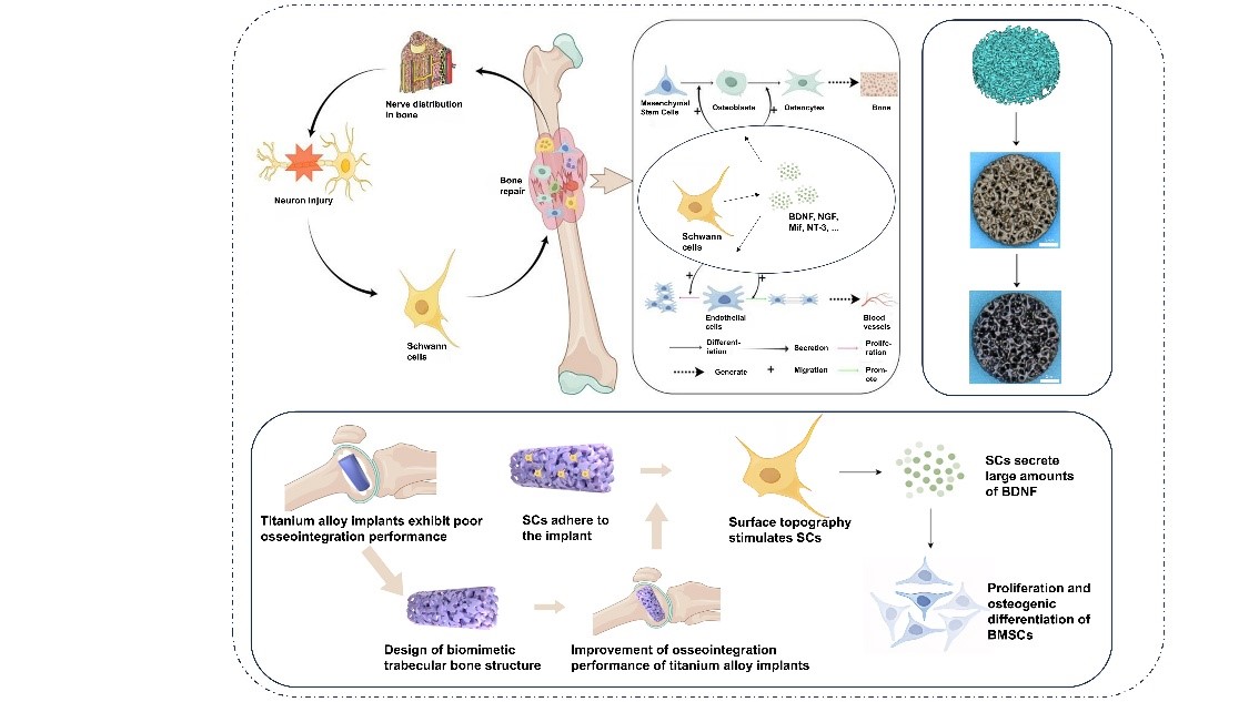
Using selective laser melting, a metal three-dimensional (3D) printing technique, we developed bionic trabecular titanium alloy scaffolds with a micro–nano composite porous structure to address the limitations of traditional titanium implants. By integrating bionic design principles with advanced metal 3D printing strategies, these scaffolds mimic the trabecular network of cancellous bone, reducing elastic modulus (to ~4 GPa) and mitigating stress shielding. The bioprinted scaffolds exhibited enhanced surface properties that promoted Schwann cell (SC) adhesion, elongation, and spindle-like morphology, forming cellular networks along the microporous architecture. In contrast, SCs on solid titanium scaffolds displayed a flattened morphology with limited functionality. Transcriptomic analysis revealed that the scaffold’s micro–nano structure regulated SC behavior via the focal adhesion kinase-mitogen-activated protein kinase mechanotransduction pathway, enhancing the secretion of pro-osteogenic (e.g., platelet-derived growth factor with two A subunits) and pro-angiogenic (e.g., vascular endothelial growth factor) factors. Trabecular-like scaffold-conditioned medium significantly accelerated bone marrow mesenchymal stem cell proliferation, osteogenic differentiation, and endothelial cell angiogenesis, achieving a 36% higher healing rate compared to controls. While in vivo validation remains essential, our in vitro model isolates SC-driven mechanisms, avoiding systemic confounders. This study highlights the potential of 3D bioprinted scaffolds for personalized bone defect repair, offering a biomechanically and biologically optimized solution to enhance osseointegration.
- Clynes MA, Harvey NC, Curtis EM, Fuggle NR, Dennison EM, Cooper C. The epidemiology of osteoporosis. Br Med Bull. 2020;133(1):105-117. doi: 10.1093/bmb/ldaa005
- Beird HC, Bielack SS, Flanagan AM, et al. Osteosarcoma. Nat Rev Dis Primers. 2022;8(1):77. doi: 10.1038/s41572-022-00409-y
- Shi Y, Liu J, Du M, et al. Customized barrier membrane (titanium alloy, poly ether-ether ketone and unsintered hydroxyapatite/poly-l-lactide) for guided bone regeneration. Front Bioeng Biotechnol. 2022;10:916967. doi: 10.3389/fbioe.2022.916967
- Rout PK, Roy S, Ganguly S, Rathore DK. A review on properties of magnesium-based alloys for biomedical applications. Biomed Phys Eng Express. 2022;8(4):042002. doi: 10.1088/2057-1976/ac6d81
- Jiang P, Zhang Y, Hu R, et al. Advanced surface engineering of titanium materials for biomedical applications: From static modification to dynamic responsive regulation. Bioact Mater. 2023;27:15-57. doi: 10.1016/j.bioactmat.2023.03.006
- Li J, Zheng Y, Yu Z, et al. Surface-modified titanium and titanium-based alloys for improved osteogenesis: a critical review. Heliyon. 2024;10(1):e23779. doi: 10.1016/j.heliyon.2023.e23779
- Liu B, Wang H, Zhang N, Zhang M, Cheng CK. Femoral stems with porous lattice structures: a review. Front Bioeng Biotechnol. 2021;9:772539. doi: 10.3389/fbioe.2021.772539
- Zhang B, Li J, He L, Huang H, Weng J. Bio-surface coated titanium scaffolds with cancellous bone-like biomimetic structure for enhanced bone tissue regeneration. Acta Biomater. 2020;114:431-448. doi: 10.1016/j.actbio.2020.07.024
- Ziaie B, Velay X, Saleem W. Advanced porous hip implants: a comprehensive review. Heliyon. 2024;10(18): e37818. doi: 10.1016/j.heliyon.2024.e37818
- Hou C, An J, Zhao D, et al. Surface modification techniques to produce micro/nano-scale topographies on Ti-based implant surfaces for improved osseointegration. Front Bioeng Biotechnol. 2022;10:835008. doi: 10.3389/fbioe.2022.835008
- Salhotra A, Shah HN, Levi B, Longaker MT. Mechanisms of bone development and repair. Nat Rev Mol Cell Biol. 2020;21(11):696-711. doi: 10.1038/s41580-020-00279-w
- Damiati LA, El Soury M. Bone-nerve crosstalk: a new state for neuralizing bone tissue engineering–a mini review. Front Med (Lausanne). 2024;11:1386683. doi: 10.3389/fmed.2024.1386683
- Yuan Q, Liao D, Yang X, et al. Effect of implant surface microtopography on proliferation, neurotrophin secretion, and gene expression of Schwann cells. J Biomed Mater Res A. 2010;93(1):381-8. doi: 10.1002/jbm.a.32548
- Wang YY, Gong P, Zhang J. Effects of different implant surface properties on the biological behavior of Schwann cells. Hua Xi Kou Qiang Yi Xue Za Zhi. 2021;39(3): 279-285. doi: 10.7518/hxkq.2021.03.006
- Jing Z, Zhang T, Xiu P, et al. Functionalization of 3D-printed titanium alloy orthopedic implants: a literature review. Biomed Mater. 2020;15(5):052003. doi: 10.1088/1748-605X/ab9078
- Fan L, Chen S, Yang M, Liu Y, Liu J. Metallic materials for bone repair. Adv Healthc Mater. 2024;13(3):e2302132. doi: 10.1002/adhm.202302132
- Gao X, Zhao Y, Wang M, Liu Z, Liu C. Parametric design of hip implant with gradient porous structure. Front Bioeng Biotechnol. 2022;10:850184. doi: 10.3389/fbioe.2022.850184
- Arias-González F, Rodríguez-Contreras A, Punset M, et al. Laser-Deposited beta type Ti-42Nb alloy with anisotropic mechanical properties for pioneering biomedical implants with a very low elastic modulus. Materials (Basel). 2022;15(20):7172. doi: 10.3390/ma15207172
- Yang S, Jiang W, Ma X, et al. Nanoscale morphologies on the surface of 3D-printed titanium implants for improved osseointegration: a systematic review of the literature. Int J Nanomedicine. 2023;18:4171-4191. doi: 10.2147/ijn.S409033
- Gu Y, Sun Y, Shujaat S, Braem A, Politis C, Jacobs R. 3D-printed porous Ti6Al4V scaffolds for long bone repair in animal models: a systematic review. J Orthop Surg Res. 2022;17(1):68. doi: 10.1186/s13018-022-02960-6
- Shao H, Zhang Q, Sun M, et al. Effects of hydroxyapatite-coated porous titanium scaffolds functionalized by exosomes on the regeneration and repair of irregular bone. Front Bioeng Biotechnol. 2023;11:1283811. doi: 10.3389/fbioe.2023.1283811
- Niinomi M, Liu Y, Nakai M, Liu H, Li H. Biomedical titanium alloys with Young’s moduli close to that of cortical bone. Regen Biomater. 2016;3(3):173-85. doi: 10.1093/rb/rbw016
- Dai Y, Lu T, Li L, et al. Electrospun composite PLLA-PPSB nanofiber nerve conduits for peripheral nerve defects repair and regeneration. Adv Healthc Mater. 2024; 13(10):e2303539. doi: 10.1002/adhm.202303539
- Li X, He N, Li X, et al. Graphdiyne-loaded polycaprolactone nanofiber scaffold for peripheral nerve regeneration. J Colloid Interface Sci. 2023;646:399-412. doi: 10.1016/j.jcis.2023.05.054
- da Silva VA, Bobotis BC, Correia FF, et al. The impact of biomaterial surface properties on engineering neural tissue for spinal cord regeneration. Int J Mol Sci. 2023; 24(17):13642. doi: 10.3390/ijms241713642
- Wang Z, Zhao Y, Bai H, et al. Bioactive prosthesis interface compositing variable-stiffness hydrogels regulates stem cells fates to facilitate osseointegration through mechanotransduction. Int J Biol Macromol. 2024; 259(Pt 2):129073. doi: 10.1016/j.ijbiomac.2023.129073
- Park EJ, Truong VL, Jeong WS, Min WK. Brain-derived neurotrophic factor (BDNF) enhances osteogenesis and may improve bone microarchitecture in an ovariectomized rat model. Cells. 2024;13(6):518. doi: 10.3390/cells13060518
- Fitzpatrick V, Martín-Moldes Z, Deck A, et al. Functionalized 3D-printed silk-hydroxyapatite scaffolds for enhanced bone regeneration with innervation and vascularization. Biomaterials. 2021;276:120995. doi: 10.1016/j.biomaterials.2021.120995
- Wu Z, Pu P, Su Z, Zhang X, Nie L, Chang Y. Schwann cell-derived exosomes promote bone regeneration and repair by enhancing the biological activity of porous Ti6Al4V scaffolds. Biochem Biophys Res Commun. 2020;531(4):559-565. doi: 10.1016/j.bbrc.2020.07.094
- Wei X, Zhou W, Tang Z, et al. Magnesium surface-activated 3D printed porous PEEK scaffolds for in vivo osseointegration by promoting angiogenesis and osteogenesis. Bioact Mater. 2023;20:16-28. doi: 10.1016/j.bioactmat.2022.05.011
- Choudhary R, Bulygina I, Lvov V, et al. Mechanical, structural, and biological characteristics of polylactide/ wollastonite 3D printed scaffolds. Polymers (Basel). 2022;14(19):3932. doi: 10.3390/polym14193932
- Ait Said H, Mabroum H, Lahcini M, et al. Manufacturing methods, properties, and potential applications in bone tissue regeneration of hydroxyapatite-chitosan biocomposites: a review. Int J Biol Macromol. 2023; 243:125150. doi: 10.1016/j.ijbiomac.2023.125150
- Zhang Z, Lv Y, Harati J, et al. Submicron-grooved films modulate the directional alignment and biological function of schwann cells. J Funct Biomater. 2023;14(5):238. doi: 10.3390/jfb14050238
- Kong D, Wang Q, Huang J, et al. Design and manufacturing of biomimetic scaffolds for bone repair inspired by bone trabeculae. Comput Biol Med. 2023;165:107369. doi: 10.1016/j.compbiomed.2023.107369