

Osteogenesis induced by magnetic responsive composite scaffolds under a static magnetic field

Currently, traditional osteogenesis methods face significant challenges in terms of therapeutic efficiency and biocompatibility, particularly in the context of bone repair where higher precision and efficacy are required. In this study, we fabricated a novel composite scaffold composed of polycaprolactone (PCL), polydopamine (PDA), and iron oxide nanoparticles (IONPs) and investigated its osteogenic potential. The incorporation of IONPs imparts magnetic responsiveness to the scaffold, thereby enabling the application of an external magnetic field to stimulate osteogenesis. Characterization of the scaffold confirmed its structural integrity, porosity, and biocompatibility, whereas the inclusion of PDA improved its hydrophilicity and cell adhesion properties. In vitro studies demonstrated that an external magnetic field significantly enhanced cell proliferation, osteogenic differentiation, and mineral deposition of osteoprogenitor cells cultured on the scaffolds. Furthermore, in vivo evaluation revealed that when the scaffold was exposed to magnetic stimulation, bone regeneration was accelerated, and integration of the defect site was improved. The magnetic-field-mediated approach proposed in this study effectively enhanced the osteogenic rate by augmenting the magnetic responsiveness of IONPs and combining the biocompatibility and cell-adhesion-promoting functions of PCL/PDA. This method offers a more controllable and biologically responsive alternative strategy for bone tissue regeneration with considerable potential for clinical applications.
1. Introduction
Large bone defects caused by bone disease, tumor resection, or prosthesis repair cannot be adequately resolved using physiological regeneration processes. Surgical methods for treating bone defects include autografts, allografts, and synthetic material grafting.1 However, these methods are insufficient for a complete repair of bone defects, necessitating the urgent development of novel biomaterial scaffolds for tissue engineering and regenerative medicine applications. Bone tissue-engineered scaffolds are temporary implants that promote tissue ingrowth and new bone regeneration by optimizing their geometry, mechanical properties, and biological performance to enhance healing. Several biomaterials have been developed for bone tissue engineering, including biometals such as lithium (Li),2 magnesium (Mg),3 and zinc (Zn).4 Moreover, scaffolds containing various bioactive substances are important tools for the regeneration of bone defects.5,6
In recent years, polycaprolactone (PCL), a biodegradable polymer, has gained attention as a promising material for bone tissue engineering due to its excellent biocompatibility, minimal inflammatory response, and ability to provide space for the formation of new bone tissue.7 Furthermore, during bone repair, scaffold materials can provide adhesion sites for cell attachment.8,9 Given these advantages, PCL was selected as the primary material for scaffold construction in this study. However, PCL also has certain limitations, such as a lack of active ligands on its surface, which leads to ineffective cell adhesion and subsequent cellular events. Its lack of bioactivity impedes cell proliferation and differentiation.10 Additionally, largepore PCL scaffolds produced by 3D printing devices may lead to the rapid depletion of nutrients, thus hindering the accumulation of extracellular matrix (ECM).11 Therefore, enhancing the surface modification of PCL scaffolds to improve cell interactions is a key focus of our current research. Recent efforts have focused on incorporating bioactive components, with polydopamine (PDA)-based modification emerging as a straightforward and versatile method for surface modification. This approach, derived from biomimetic techniques, is simple to prepare and has gradually become a research hotspot in the field of tissue engineering.12,13 PDA-based surface modification strategies offer excellent physical and chemical properties, thereby promoting cell growth and differentiation.14,15
Magnetic nanoparticles (MNPs), such as iron oxide nanoparticles (IONPs), have gained attention in bone tissue engineering because of their ability to respond to external magnetic fields, which can regulate cellular behavior under magnetic stimulation.16,17 However, concerns have been raised regarding the cytotoxic effects of IONPs, as many studies have not thoroughly screened the ion concentrations within composite scaffolds.
This study explored the synergistic effects of magnetic fields and nanoparticles on bone repair using PCL/PDA/ IONPs (PPI) composite materials under static magnetic field (SMF) stimulation. Research indicates that a static magnetic field, as an external physical stimulus, can promote bone-implant integration, increase bone density and calcium content, and accelerate fracture healing by altering the cellular microenvironment.18 Under static magnetic field exposure, MNPs significantly enhance the osteogenic potential of bone marrow mesenchymal stem cells (BMSCs) while inhibiting osteoclast activity.19 Numerous in vitro cell culture studies have also demonstrated significant changes in cellular behavior when subjected to external magnetic field stimulation.20,21 For instance, the simple application of static magnetic fields using magnets has been shown to accelerate osteogenic differentiation of rat osteoblasts and human mesenchymal stem cells in vitro. In addition, static magnetic fields can regulate intracellular ion channels and signaling pathways, thereby promoting the expression of osteogenesis-related genes. For example, under the influence of SMF, MNP-composite scaffolds significantly enhance the calcification ability of BMSCs.22,23
Several studies have shown that biological materials such as hydroxyapatite, bioactive glass, and iron sulfide (FeS) can be used to promote bone defect repair. Hydroxyapatite, a natural bone-matrix mineral, is widely used for bone repair. However, hydroxyapatite materials exhibit poor mechanical properties and degrade slowly in vivo, potentially leading to long-term burden.24 Bioactive glass also faces similar issues in osteogenesis.25 FeS materials, as iron-based compounds, have been investigated for bone repair in recent years, with studies showing that they can promote the proliferation and differentiation of osteoblasts under magnetic field stimulation. However, the application of FeS materials is hindered by the cytotoxicity of their degradation products, and their long-term effects in clinical applications remain uncertain.26 Therefore, although these materials have potential in the field of bone regeneration and osteogenesis, their limitations in practical applications must still be addressed.
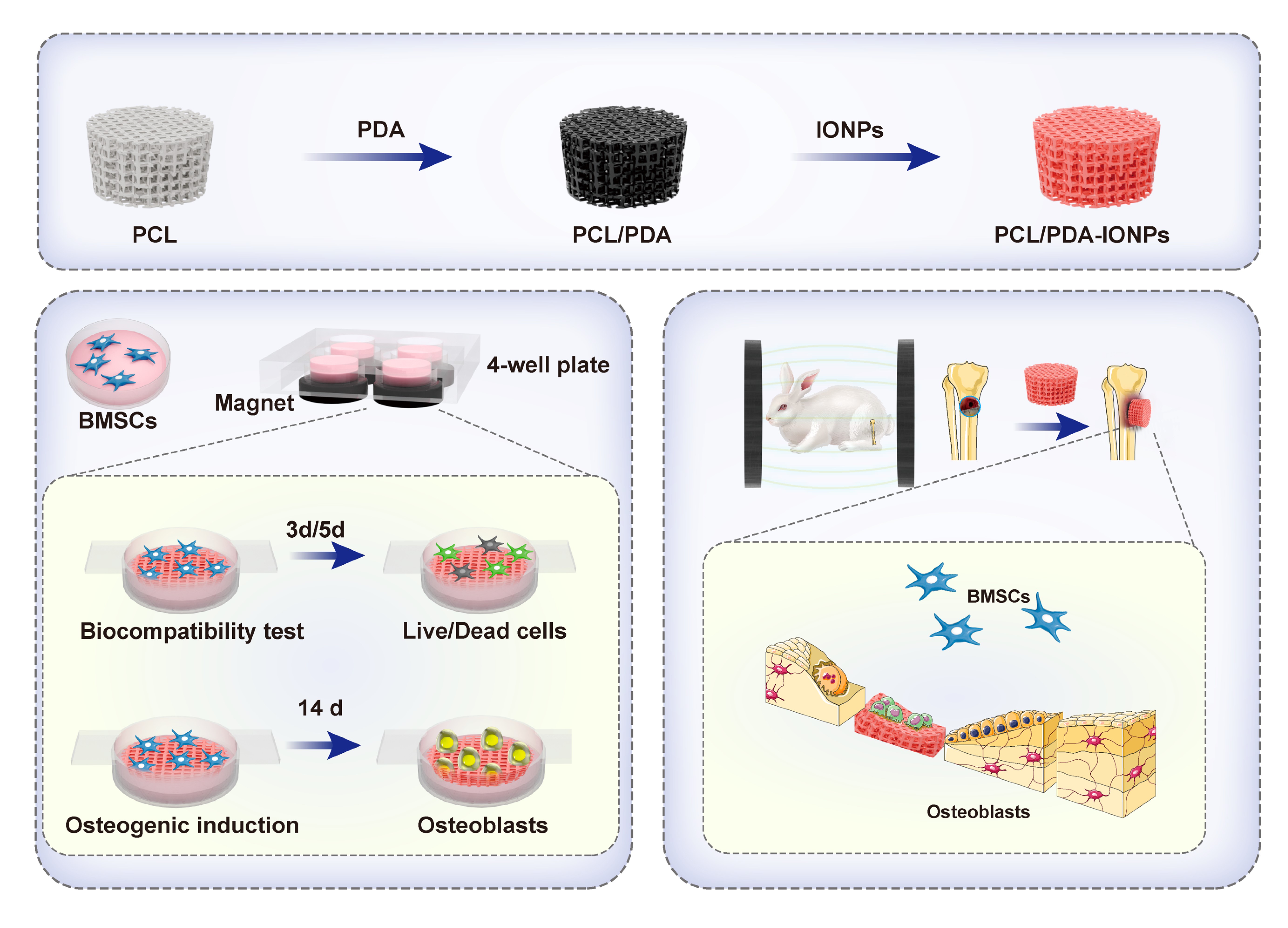
Although PPI composite materials have shown some progress in promoting osteogenesis, optimizing their performance to enhance bioactivity and improve longterm biocompatibility remains a key focus of current research. In this study, PCL scaffolds were printed using 3D printing technology, followed by surface modification with PDA to form PCL/PDA scaffolds. IONPs were successfully coated onto the scaffold surface, resulting in the development of PPI composite scaffolds. Subsequently, in vitro experiments were conducted using rabbit BMSCs (rBMSCs) to determine the optimal concentration of IONPs for PPI composite scaffolds. The osteogenic properties of PPI composite scaffolds with and without SMF application were also assessed. Finally, the in vivo osteogenic potential of the PPI composite scaffolds was evaluated using regular ultrasound examinations, micro-computed tomography (CT) imaging, and histopathological analysis (Figure 1). This study presents a new therapeutic method for the regeneration and healing of bone defects. Through innovative design and optimization of the structure, composition, and functional properties of biomaterials, this method significantly enhances bone conductivity, bone inductivity, and bioactivity, thereby effectively promoting bone tissue regeneration and functional reconstruction. It holds great promise as a potential treatment for bone defects and has significant clinical application prospects.

Figure 1. The experimental flow chart of this study. Abbreviations: BMSCs, bone marrow mesenchymal stem cells; IONPs, iron oxide nanoparticles; PCL, polycaprolactone; PDA, polydopamine.
2. Materials and methods
2.1. Preparation of scaffolds
2.1.1. Preparation of PCL scaffolds
A porous PCL scaffold model was designed using computeraided design (CAD) with a radius of 2.5 mm, height of 1 mm, and pore angles of 0°, 90°, and 180°. PCL (molecular weight = 65,000; Sigma, USA) was printed using the melt extrusion method, in which PCL pellets were placed in the printer’s extrusion chamber and melted at a controlled temperature of 110 ± 5 °C. The material was then extruded layer-by-layer along the z-axis to form a cylindrical model with a needle movement speed of 4 mm/s.
2.1.2. Preparation of PCL/PDA scaffolds
A total of 200 μL of 1.5 M Tris-HCl (ST789, Beyotime, China) was combined with 29.8 mL of water to achieve a final concentration of 10 mol/L solution. Subsequently, 600 mg of dopamine (DOPA) (62-31-7, Sigma, USA) was added to this solution, and the scaffold was immersed in the mixture, which was stirred in the dark for 24 h. After incubation, the scaffold was removed, washed thrice with phosphate-buffered saline (PBS) (KGL2207-500, KeyGEN, China), and immersed in 75% ethanol for 2 h. The scaffolds were then rinsed with sterilized deionized water and subjected to ultraviolet (UV) irradiation in a culture incubator for 2 h. Finally, the scaffold was washed three times with PBS, cultured in a low-glucose complete medium for 12 h, and sterilized to prepare the PCL/PDA scaffold.
2.1.3. Synthesis of IONPs
A glucan-based modification method was used to prepare a carboxymethylated sorbitol glucan (PSC) coating on IONPs. The classic chemical coprecipitation method was improved by utilizing an alternating current magnetic field (ACMF)-induced internal heating model to synthesize IONPs. Briefly, 200 mg of PSC (Sigma, USA) was dissolved in 10 mL of deionized water in a round-bottomed plastic tube and fixed in an ACMF-inducing device. Then, 6.0 mg FeCl3 (451649; Sigma, USA) and 30 mg FeCl2 (939935; Sigma, USA) were added. Subsequently, 1 g of 28% (w/v) ammonium hydroxide (Sigma, USA) was added, followed by vigorous mechanical stirring. The mixture was immediately subjected to medium-frequency radiofrequency heating to generate heat via the ACMF. The colloidal mixture was heated to 80°C by adjusting the magnetic field strength and maintained at that temperature for 1 h. Finally, the mixture was cooled to room temperature and purified by six cycles of ultrafiltration using a 100 kDa membrane (Sigma, USA), yielding a solution concentration of 23 mg/mL.
2.1.4. Preparation and storage of PPI composite scaffolds
After preparation, the PCL/PDA scaffolds were immersed in a nano-solution with concentrations of 57.5, 115, 230, 460, 920, and 1840 μg/mL, which had been filtered through a sterile filter. The scaffolds were sealed with a sealing film and placed on a shaker for 3 days. The scaffolds were removed and washed three times with deionized water to obtain the PPI scaffolds. The prepared PPI scaffolds were stored in sterile, light-protected containers at room temperature. Table S1, Supporting Information summarizes the nomenclature for the different scaffold compositions, IONP concentrations, and the application specifics of SMF.
2.2. Characterization of composite scaffolds
2.2.1. Scanning electron microscope (SEM)
The PCL, PCL/PDA, and PPI scaffolds were gold-coated by sputtering for 45 s at a current of 10 mA. Surface morphology and elemental composition were examined using SEM (Hitachi Regulus 8100, CanScan, Japan), and energy-dispersive spectroscopy (EDS) mapping was performed. Morphological images were captured at an accelerating voltage of 3 kV and EDS mapping was conducted at an accelerating voltage of 20 kV using an SE2 secondary detector.
2.2.2. Static water contact angle
At room temperature, a droplet of water was placed on the surface of each scaffold (n = 3) and images were captured using a contact angle goniometer (JY-82, Chengde Dingsheng Testing Equipment, China). The contact angle was measured as the angle formed between the liquid– solid and liquid–air interfaces. Each scaffold was measured three times, and the average value was recorded as the final result.
2.2.3. Compression testing
The compressive-strain properties of the PCL, PCL/PDA, and PPI scaffolds were tested using a universal testing machine (Chengde Dingsheng Testing Machine Testing Equipment Co., Ltd., China). The loading speed was set to 1.0 mm/min.
2.2.4. Iron release experiment
The in vitro iron release behavior of the PPI scaffolds (n = 3) was assessed using inductively coupled plasma mass spectrometry (ICP-MS; Agilent 7700X, Agilent Technologies, USA). The samples were rinsed three times with PBS and subsequently placed into centrifuge tubes containing 10 mL of Hanks’ solution (Shanghai Renjie Biotech Company, China), followed by incubation at 37 °C. At designated time points (days 1, 3, 5, 7, 14, 21, and 28), the supernatants were collected and 10 mL of fresh PBS was immediately added to each centrifuge tube. The supernatants were stored at 4 °C, and the cumulative iron release at each specific time point was calculated.
2.2.5. Vibrating sample magnetometer (VSM)
The PPI scaffolds were placed in a machine (LakeShore 7404, USA) to measure their hysteresis. The pole diameter was 5 cm, and the magnetic moment measurement range was from 5 × 10⁻⁷ emu to 10³ emu.
2.3. In vitro experiments
2.3.1. Establishing the SMF in vitro
The magnetic field setup and intensity were adopted from a previous study.27 Specifically, the setup consisted of a neodymium iron boron (Nd2Fe14B) magnet disk (1 mm thick, 15 mm diameter; Shenzhen Magician Technology, China) and a 4-well plastic culture plate to create the SMF exposure system. A magnet was placed beneath each well to expose the culture to the north pole of the magnetic field. The required magnetic field intensity was determined by adjusting the distance between the magnet and the culture plate. A Gauss meter (TM-701, Kanetec, Japan) was used to measure the SMF intensity on each plate. All the wells of the culture plates were simultaneously exposed to the same magnetic field. During exposure, adjacent culture plates were separated by more than 50 mm to eliminate interference from neighboring magnetic fields. In the SMF-exposure group, the cells were continuously exposed to SMF throughout the day in either basal or osteogenic medium. The magnetic flux density was monitored with a Gauss meter, and the average flux was maintained at 50 mT. In the non-exposed control group, a non-disk Nd2Fe14B magnet was placed beneath the culture plate. The control and experimental plates were placed in the same incubator, ensuring that the flux density in the control wells did not exceed 0.05 mT, which corresponds to the natural Earth magnetic field level.
2.3.2. Cell seeding
The PCL and PCL/PDA scaffolds were immersed in 70% ethanol for 2 h and then rinsed with sterile double-distilled water. The scaffolds were dried under UV light in a laminar flow hood for subsequent use. PPI composite scaffolds were prepared under sterile conditions and stored in a laminar flow hood. The scaffolds were placed in low-adhesion 24-well plates, and third-passage osteoblasts and rBMSCs were evenly seeded onto their surfaces in each group (n = 5). After incubation for 4 h, a culture medium was added to enhance cell adhesion to the scaffold surface. The seeding density is provided in detail in the subsequent steps.
2.3.3. Scaffold adhesion test
Sterile PCL, PCL/PDA, and PPI scaffolds with varying concentrations were placed on a low-adhesion plate (n = 5). A 100 μL suspension of 1 × 10⁶ osteoblast cells was added to each well and incubated at 37 °C with 5% CO2 for 4 h. Subsequently, 200 μL of Dulbecco’s Modified Eagle Medium (DMEM) culture medium (KGM31600-500, KeyGEN) was added to each well, and the cells were incubated for an additional 24 h. Following the removal of the medium, the cells that adhered to the scaffold were digested using 0.25% trypsin (KGY0012, KeyGEN). The digestion process was halted by adding 100 μL of DMEM, and the resulting cell solution was collected and centrifuged at 1500 rpm. The pellet was then resuspended in DMEM for counting. The cell adhesion rate for each scaffold was calculated using the following formula: scaffold adhesion rate = (number of cells adhered to the scaffold/number of cells seeded on the scaffold) × 100%. This method was also applied to determine the cell adhesion rates for the remaining scaffolds.
2.3.4. Cell Counting Kit-8 (CCK-8) assay
rBMSCs (5 × 10⁴ cells/well) were seeded onto the surfaces of PCL, PCL/PDA, and PPI scaffolds at concentrations of 57.5, 115, 230, 460, 920, and 1840 μg/mL (n = 5). Following incubation in the culture chamber for 1, 3, 5, and 7 days, cell toxicity was assessed using the CCK-8 assay kit (KGA317, Jiangsu Kaiji Bio, China). The CCK-8 working solution was prepared by adding 10% CCK-8 (v/v) to low-glucose complete DMEM. After discarding the original medium from each well, 100 μL of fresh CCK-8 working solution was added, and the wells were incubated for 1.5 h in the culture chamber. Subsequently, 80 μL of the reaction solution from each well was transferred to a 96-well plate, and the optical density (OD) at 450 nm was measured using a microplate reader (Model 680, Bio-Rad, USA). The experimental data are presented as the mean ± standard deviation.
2.3.5. LIVE/DEAD staining
rBMSCs, at a density of 5 × 10⁴ cells per well, were seeded onto the surfaces of PCL, PCL/PDA, and PPI scaffold groups containing 230, 460, and 920 μg/mL concentrations (n = 5). Following a 3-day culture period in an incubator, LIVE/DEAD staining (Life Technologies, USA) was conducted on the scaffold/cell composites in accordance with the manufacturer’s instructions. Images were captured using a laser confocal microscope (FV3000, Olympus Corporation, Japan), where live cells appeared green and dead cells appeared red. Cell growth on the scaffolds was then evaluated.
2.3.6. Alkaline phosphatase (ALP) staining and detection
rBMSCs were seeded at a density of 5 × 10⁴ cells per well onto the surfaces of PCL, PCL/PDA, 460/PPI, and 460/ PPI+SMF group scaffolds and cultured in an incubator for 7 days (n = 5). ALP staining was conducted using an ALP kit (KGA353, KeyGEN BioTECH, China), and the results were observed under a microscope. In addition, cells were seeded according to the method outlined in the CCK-8 protocol, achieving a cell density of 1 × 10⁵ cells/mL (n = 5). After 7 days of culture, the detection solution was added in accordance with the instructions provided with the ALP Kit (P0321S, Beyotime), and absorbance was measured at 405 nm.
2.3.7. Alizarin Red staining
Cells were seeded according to the protocol outlined in the CCK-8 method at a density of 1 × 10⁵ cells/mL and cultured for a duration of 7 days (n = 5). The cells were subsequently washed three times with PBS, fixed with paraformaldehyde (KGC3121-500, KeyGEN) for 30 min, and washed again with PBS. Then, Alizarin Red staining solution (C0148S, KeyGEN BioTECH) was added and incubated for 15 min. After three additional washes with PBS, cells were examined for calcium deposition using an inverted microscope.
2.3.8. Prussian blue staining
Following the sterilization of each scaffold group, the scaffolds were soaked in low-sugar DMEM complete medium at a ratio of 0.1 g/mL, in accordance with ISO 10993-12 standards. They were then incubated for 3 days before collection for subsequent use. rBMSCs were seeded at a density of 5 × 10⁴ cells per well. Once the cells adhered, extracts from the different groups were added to the corresponding well plates for cell culture (n = 5). After culturing in an incubator for 7 days, a Prussian blue kit (G1420, Beijing Solebao) was used for detection. Under the microscope, cell nuclei stained red, while IONPs were stained blue.
2.3.9. Immunofluorescence staining
Cells were seeded according to the method outlined in the CCK-8 protocol at a density of 1 × 10⁵/mL and cultured for 7 days. Subsequently, the cells were washed three times with PBS, fixed with paraformaldehyde for 30 min, and washed again three times with cold PBS (5 min each). The cells were then permeabilized with Triton X-100 (KGA1508-10, KeyGEN) in PBS at room temperature for 20 min. After permeabilization, the cells were washed thrice with PBS (5 min each) and incubated with a blocking solution (P0235, Beyotime) at room temperature for 30 min. The blocking solution was discarded, and the primary antibody of collagen 1 (COL-1; ab270993, Abcam, UK) and runt-related transcription factor 2 (RUNX2; ab92336, Abcam, UK) was added. The samples were incubated overnight at 4 °C. After incubation, the primary antibody was removed and the samples were washed three times with PBS-Tween (PBST) (5 min each). Next, the diluted secondary antibody (SA00012-1, Proteintech, USA) was added, and the cells were incubated at room temperature for 1 h in the dark. The secondary antibody was discarded and the samples were washed with PBST. DAPI was added to stain the cell nuclei and the samples were incubated in the dark for 5 min. Finally, the samples were washed three times with PBST (5 min each) and observed under a fluorescence microscope to capture images.
2.3.10. Quantitative real-time polymerase chain reaction (qRT-PCR)
After 7 and 14 days of culture, qRT-PCR (TransGen Biotech, USA) was conducted to assess the mRNA expression levels of osteogenesis-specific genes, including COL1, RUNX2, vascular endothelial growth factor (VEGF), and CD31 (n = 5). Total RNA was extracted using a TransZol Up Plus RNA Kit (TransGen Biotech) following the manufacturer’s instructions. RNA concentration was measured using a multifunctional microplate reader (Synergy 2, BioTek, USA) at a wavelength of 260 nm. qRT-PCR was performed using the TransScript® II Green One-Step qRT-PCR SuperMix (AQ311, TransGen Biotech). The target gene primer sequences used in this study are listed in Table 1.
Table 1. Primer sequences for quantitative real-time polymerase chain reaction (qRT-PCR)
| Genes | 5’-3’ | Sequence |
|---|---|---|
| GAPDH (Rabbit) | Forward | TCACCATCTTCCAGGAGCGA |
| Reverse | CACAATGCCGAAGTGGTCGT | |
| COL-1 (Rabbit) | Forward | CAGGACTTTGAGGAGGATGGT |
| Reverse | TTGGTAGCAGTAGGGGTATGG | |
| RUNX2 (Rabbit) | Forward | CCGAAATGCCTCTGCTGTTAT |
| Reverse | TCTGTCTGTGCCTTCTGGGTT | |
| VEGF (Rabbit) | Forward | TTCAACGTCACCATGCAGAT |
| Reverse | AAATGCTTTCTCCGCTCTGA | |
| CD31 (Rabbit) | Forward | TTCACACAGCAAGCTTCTGG |
| Reverse | AACAATGAGGTCTGGGAGGAC |
2.4. In vivo experiments
2.4.1. Surgical procedure
All animal experiments were conducted in accordance with the guidelines for animal experimentation in China and were approved by the Ethics Committee of the First Affiliated Hospital of Nanjing University (approval number: DWSY-22135264). Adult female New Zealand white rabbits (~2.5 kg) were randomly assigned to four groups with five animals per group. A bone defect with a diameter of 6 mm and a depth of 4.5 mm was created on the medial condyle of the tibia. Scaffolds (blank control, PCL, PCL/PDA, PPI, and PPI+SMF) were implanted, and the incision was closed. Postoperatively, penicillin (Henan Xinxiang Huaxing Pharmaceutical factory, China) was administered intramuscularly at a dose of 200,000 U/ day for 3 days. Twelve weeks postoperatively, all rabbits were euthanized and tibial samples were collected for further analysis.
2.4.2. In vivo SMF setup
The rabbits were housed in cages with permanent magnets fixed on opposite sides. These magnets provided SMF within the cage. Figure S1, Supporting Information illustrates the arrangement of the magnets. The magnetic field strength was measured using a Tesla meter (Lake Shore 7407, USA). As shown in Figure S2, Supporting Information, the magnetic field strength in the plane of the cage ranged from 0.05 to 0.2 mT, and the field strength on the sides of the cage ranged from 50 to 60 mT. The rabbits were exposed to the magnetic field throughout the day.
2.4.3. Conventional and contrast-enhanced ultrasound examination
Conventional and contrast-enhanced ultrasonography examinations were performed at 8 and 12 weeks, respectively. The Mindray MX 7 color Doppler ultrasound diagnostic device (Mindray, China) was utilized. The instrument was equipped with contrast-enhanced ultrasound quantitative analysis software and an L13-3Ns linear array high-frequency probe operating at a frequency of 6–9 MHz with a mechanical index of 0.14. The frequency of the conventional ultrasonography was fixed at 8.4 MHz. SonoVue (Bracco, Italy) was used as the contrast agent. Regarding anesthesia, the anesthetics Suwenxin (Dunhua City Shengda Animal Drugs, China) and Shutai50 (Virbac, French) were mixed in a 1:1 ratio and administered via intramuscular injection at a dosage of 0.1 mL/kg using a sterile syringe. After ensuring that the animal was calm enough to establish intravenous access, the appropriate position was adjusted for hair removal and the modeling area was fully exposed. A 24 G intravenous catheter (Qisheng company, China) was inserted along the ear vein to establish intravenous access. For quantitative analysis, the images were processed by a senior ultrasound physician with over 10 years of experience and by two graduate students. Images were analyzed using the TIC-analysis function of Mindray MX7 for quantitative assessments of different groups. The enhancement intensity (strength enhancement, ΔSI), defined as the difference between peak intensity (PI) and basic intensity (BI), as well as time to peak (TTP), were measured.
2.4.4. Micro-CT scanning
The animals were euthanized 12 weeks post-surgery, and tibial specimens were harvested. Micro-CT scanning (Bruker SkyScan1176, Billerica, MA, USA) was performed and data, including the relative bone volume fraction (BV/ TV), were analyzed using MIMICS software (Materialise, Belgium) to assess new bone formation.
2.4.5. Histopathological evaluation
After micro-CT scanning, specimens were fixed in 4% paraformaldehyde and decalcified with diluted nitric acid. The specimens were then embedded in paraffin, sectioned, and stained with hematoxylin-eosin (H&E: Beyotime, China); Masson’s trichrome (Beyotime, China) for analysis (n = 5).
2.5. Statistical analysis
All data were analyzed using a one-way analysis of variance (ANOVA) followed by the least significant difference method to determine significant differences. Statistical analysis was performed using GraphPad Prism (version 8.0; GraphPad Software, USA). Data are presented as mean ± standard deviation and statistical significance was set at p < 0.05.
3. Results
3.1. Scaffold preparation and characterization
In this study, the PCL particles were placed in the extrusion chamber of a 3D printer and melted to print scaffolds. After modification with PDA, the PCL/PDA scaffold was formed. The IONPs were loaded onto the surface of the PCL/PDA scaffold using an impregnation method to prepare a PPI composite scaffold (Figure 2A). First, the surfaces of the scaffolds were characterized. Figure 2B–M shows the macroscopic, microscopic, and surface morphologies of the PCL, PCL/PDA, and PPI scaffolds. Figure 2B–D shows the overall appearance; after the self-assembly of PDA, the surface color of the PCL scaffold darkened, but the scaffold’s diameter, height, and fiber diameter remained unchanged. SEM images (Figure 2E–M) showed that the surface of the PCL scaffold was smooth and crack-free, while the PCL/PDA scaffold displayed dispersed PDA particles. The surface of the PPI scaffold showed distinct differences from that of the PCL/PDA scaffold. Elemental mapping images (Figure 2N–P) revealed no significant differences in the distribution of carbon and oxygen between the PCL/ PDA and PPI scaffolds. However, iron accumulation was observed on the surface of the PPI scaffold, confirming the successful loading of IONPs.

Figure 2. Scaffold preparation and characterization: (A) scaffold preparation process; (B-M) scaffold general and scanning electron microscopy, (N-P) surface element content analysis; compared between lined groups, n=3. Scale bars: B–D: 2mm; E-G:500µm; H-J: 200µm; K-M: 50µm.
Hydrophilicity was evaluated using the water contact angle (Figure 3A). The water contact angle of the PCL scaffold was 59.9°, whereas the PCL/PDA and PPI scaffolds exhibited water contact angles of 0°, indicating that the unmodified PCL scaffold was hydrophobic and had poor water absorption. After PDA coating, the water contact angle remained low, and the water droplets spread rapidly, significantly increasing the hydrophilicity of the composite scaffold. To ensure the reliability of bone healing, scaffolds implanted during surgery must possess sufficient mechanical strength. Compression tests (Figure 3B) showed that the scaffolds coated with magnetic IONPs significantly improved the maximum bending strength and toughness. The concentration of IONPs used to prepare the PPI composite scaffold was 1840 μg/mL. The release curve (Figure 3C) shows that iron ions were released rapidly in the first 5 days, with approximately 73.64% of the ions released. After this period, the release rate decreased significantly, with a cumulative release rate of 86.09% after 28 days. Magnetic properties were measured using VSM (Figure 3D). The coercive force and remnant magnetization were almost zero, indicating that the magnetic stent has superparamagnetism and obvious magnetic response after magnetic coating. The saturation magnetization is about 1.908164 emu/g.

Figure 3. Scaffold characterization. (A) VSM; (B) hydrophilic testing; (C) iron release experiment results for PPI; (D) compression experiment results for PPI. ****p < 0.0001, compared between lined groups, n = 3. The error bars denote the standard deviation. Abbreviations: IONPs, iron oxide nanoparticles; PCL, polycaprolactone; PDA, polydopamine; PPI, PCL/PDA/IONPs composite material; VSM, vibrating sample magnetometer.
3.2. In vitro cell response
Figure 4A illustrates the in vitro cell compatibility testing procedure for the composite scaffolds. As shown in the scaffold adhesion experiment (Figure 4B), the PCL group exhibited poor cell adhesion, indicating a lack of biological activity. After PDA self-assembly, the adhesion of the scaffold improved to some extent. Following the loading of IONPs, the scaffold’s adhesion further increased, suggesting that the material not only enhances scaffold adhesion but may also promote cell proliferation to some extent. Previous studies have indicated that a pure IONPs solution of 100 μg/mL is appropriate for further osteogenic evaluation.28 Since IONPs were loaded onto the scaffold using an impregnation method in this study, the concentration screening was carried out accordingly. Based on these results, the selected concentrations were tested as described above. The CCK-8 assay demonstrated the proliferation of rBMSCs on the PCL-based scaffolds. As shown in Figure 4C and D, the OD values, which reflect cell numbers, gradually increased over time for both stem cells and osteoblasts, indicating that the scaffolds were nontoxic. Overall, the PPI scaffold showed a significant increase in cell numbers compared to the PCL and PCL/ PDA scaffolds, with the 460 μg/mL scaffold exhibiting the most pronounced effect in promoting cell proliferation. Figure 4E shows that after 7 and 14 days of seeding and culture, the PPI group scaffold exhibited more viable (green) mesenchymal stem cell adhesion than the PCL and PCL/PDA groups, with fewer dead cells (red). Based on these findings, we selected the 460 μg/mL PPI scaffold as the optimal concentration for further evaluation of osteogenic effects. Furthermore, an external SMF was applied, as shown in Figure 5A and B. After applying the external magnetic field, the cell numbers showed an increasing trend, and the 460/PPI+SMF group had significantly more cells than the other groups, regardless of whether osteoblasts or rBMSCs were cultured. LIVE/ DEAD staining (Figure 5C) revealed that the addition of an external magnetic field further promoted cell proliferation.

Figure 4. The cytocompatibility test of composite scaffold. (A) Flow chart of cytocompatibility testing; (B) Stent adhesion testing; CCK-8 assay results for rBMSCs (C) and osteoblasts (D); (E) LIVE/DEAD staining. *p < 0.05, **p < 0.01, ***p < 0.001, ****p < 0.0001, compared between lined groups, n = 5. The error bars denote the standard deviation. Scale bars (E): 200 μm. Abbreviations: BMSCs, bone marrow mesenchymal stem cells; IONPs, iron oxide nanoparticles; OD, optical density; PCL, polycaprolactone; PDA, polydopamine; PPI, PCL/PDA/IONPs composite material; rBMSCs, rat BMSCs.

Figure 5. The cytocompatibility of the composite scaffold after SMF application. CCK-8 assay results for rBMSCs (A) and osteoblasts (B); (C) LIVE/DEAD staining. *p < 0.05, **p < 0.01, ***p < 0.001, ****p < 0.0001, compared between lined groups, n = 5. The error bars denote the standard deviation. Scale bars: 200 μm. Abbreviations: CCK-8, cell counting kit-8; IONPs, iron oxide nanoparticles; OD, optical density; PCL, polycaprolactone; PDA, polydopamine; PPI, PCL/PDA/IONPs composite material; rBMSCs, rat bone marrow mesenchymal stem cells; SMF, static magnetic field.
To further validate the osteogenic differentiation results of the PCL-based composite scaffolds, a series of studies were conducted. Figure 6A shows the in vitro osteogenic testing procedure for the composite scaffolds. The scaffold extracts were used to culture cells, and Prussian blue staining was applied to the cells incubated with IONPs. This method is based on the reaction between Perl’s solution in the reagent kit and IONPs within the cells, resulting in a blue color observable under a microscope. Figure 6B shows that the IONPs were evenly distributed within the cells, and after the application of the SMF, the 460/PPI+SMF group exhibited a higher uptake rate. This further indicates that the composite scaffold at this concentration has no significant toxic effects on the cells. Therefore, to evaluate early osteogenesis, we performed both quantitative and qualitative ALP assays. Figure 6C shows the results of ALP staining. Owing to the black opacity of the scaffold, ALP deposition by cells in the scaffold pores can be assessed. After adding the SMF, the stained image became darker, indicating that the external magnetic field enhanced the osteogenic differentiation ability of stem cells. Figure 6D shows the quantitative expression of ALP at the unit level (μmol/h/mg protein), and the results reveal that the unmodified PCL scaffold exhibited lower ALP activity due to its lack of biological activity. Compared with the PCL/PDA and 460/PPI groups, the 460/PPI+SMF scaffold significantly upregulated the osteogenic differentiation ability of mesenchymal stem cells in the early stages.

Figure 6. The osteogenic differentiation ability of the scaffolds. (A) Flow charts for bone testing; (B) Prussian blue dyeing; (C) ALP staining; (D) quantitative detection of ALP; (E) Alizarin red staining; (F) Statistics on the number of alizarin red calcium nodules. *p < 0.05, **p < 0.01, ***p < 0.001, ****p < 0.0001, compared between lined groups, n = 5. The error bars denote the standard deviation. Scale bars (B): 25 μm; (C and E): 100 μm. Abbreviations: ALP, alkaline phosphatase; IONPs, iron oxide nanoparticles; OD, optical density; PCL, polycaprolactone; PDA, polydopamine; PPI, PCL/PDA/IONPs composite material; SMF, static magnetic field.
Matrix mineralization was used to assess the osteogenic potential of the composite scaffolds. Alizarin Red staining, indicated by dark red spots, was used to confirm calcium deposition in vitro. As shown in Figure 6E, only a few calcium nodules were visible in the PCL and PCL/PDA groups. However, after loading the IONPs, the number of calcium nodules significantly increased compared to that in the PCL and PCL/PDA groups. Furthermore, owing to the synergistic effect of the SMF, the 460/PPI+SMF scaffold group had more calcium nodules. Figure 6F summarizes the number of calcium nodules in different scaffold groups, revealing that the 460/PPI+SMF group had significantly more nodules than the other groups.
To further investigate the osteogenic differentiation of rBMSCs on PCL-based scaffolds, immunofluorescence and RT-qPCR analyses were performed to detect the expression of osteogenic-related genes, including COL1 and RUNX2, after 7 and 14 days of co-culture. As shown in Figure 7A, fewer cells were present in the PCL/ PDA and 460/PPI groups, whereas the 460/PPI+SMF scaffold showed higher osteogenic protein expression. Figure 7B and C indicates that the PPI+SMF scaffold exhibited significantly higher gene expression, further demonstrating that the addition of SMF stimulates bone mineralization. Figure S3, Supporting Information demonstrates that the 460/PPI composite scaffold played a significant role in promoting angiogenesis, and its ability to enhance blood vessel formation was further improved with the addition of an external magnetic field.

Figure 7. The detection of bone-associated genes and proteins for different composite scaffold groups. (A) Immunostaining of COL-1 and RUNX2; relative expression of COL1 (B) and RUNX2 (C) on days 7 and 14; *p < 0.05, **p < 0.01, ***p < 0.001, ****p < 0.0001, compared between lined groups, n = 5. The error bars denote the standard deviation. Scale bars (A): 50 μm. Abbreviations: COL-1, Collagen 1; IONPs, iron oxide nanoparticles; OD, optical density; PCL, polycaprolactone; PDA, polydopamine; PPI, PCL/PDA/IONPs composite material; RUNX2, runt-related transcription factor 2; SMF, static magnetic field.
3.3. In vivo bone regeneration
Figure 8A illustrates the procedure for the in vivo osteogenesis study of the composite scaffolds. Figure 8B shows the two-dimensional ultrasound results, which demonstrate the conditions of the defect sites in each group. At week 8, the blank group showed a clear defect with a low echogenicity. The PCL/PDA, 460/PPI, and 460/PPI+SMF scaffolds showed clear imaging with discontinuities in the bone cortex and a mesh-like high echogenic appearance with posterior echo attenuation. At week 12, the blank group exhibited a low echogenicity, and the 460/PPI+SMF group showed significantly smaller defect pores with irregular bone surface morphology and enhanced echogenic reflection with posterior echo attenuation. No obvious scaffold shadows were detected in any group; however, the defect sizes were still observable. The inner diameters of the bone defects were measured and analyzed (Figure 8D). At week 8, all groups showed some growth compared to the initial defect, with the 460/ PPI+SMF group showing a significant reduction in bone defects and effective repair. At week 12, the defects had further reduced, with the 460/PPI+SMF group showing a significantly smaller defect compared to other groups, and the difference was statistically significant. Thus, it can be concluded that the 460/PPI+SMF group exhibited a strong bone repair capability. Figure 8C shows contrastenhanced ultrasound imaging. At week 8, the blank group exhibited significant contrast-agent filling around the defect area with no enhancement in the center. The PCL/ PDA and 460/PPI groups exhibited low enhancement. The PPI+SMF group showed significantly greater enhancement along the scaffold shape after the contrast agent injection. After week 12, with the recovery of bone defects, bone formation was visible on the surface, and ultrasound imaging showed sound attenuation, which affected detection. We further performed a quantitative analysis of contrast-enhanced ultrasound imaging (Figure 8E and F). The ΔSI chart revealed that, both at week 8 and week 12, the 460/PPI+SMF group had significantly higher values than other groups. The TTP comparison also showed that the 460/PPI+SMF group exhibited statistically significant differences compared to the other groups.

Figure 8. Ultrasound examination at 8 and 12 weeks after implantation. (a) and (b) were used for conventional ultrasound to show the inner diameter of bone defect and quantitative analysis, and (c)–€ contrast-enhanced ultrasound was used for quantitative analysis of blood perfusion in each group. *p < 0.05, **p < 0.01, ***p < 0.001, ****p < 0.0001, compared between lined groups, n = 5. The error bars denote the standard deviation. Abbreviations: BMSCs, bone marrow mesenchymal stem cells; IONPs, iron oxide nanoparticles; PCL, polycaprolactone; PDA, polydopamine; PPI, PCL/PDA/IONPs composite material; SMF, static magnetic field; ΔSI, strength enhancement; TTP, time to peak.
At 12 weeks postoperatively, rabbit tibias were collected and in vivo osteogenesis was evaluated using micro-CT imaging, as shown in Figure 9A. Because PCL scaffolds do not appear on the micro-CT images, the grayish-white image in the defect area represents the newly formed mineralized bone tissue. At week 12, the control group exhibited no new bone formation in the defective area. The PCL/PDA and 460/PPI groups showed small amounts of bone deposition due to the scaffold support. In the 460/PPI+SMF group, clear bone growth was observed within the scaffold gaps, with thicker new bone tissue forming around the scaffold. The newly formed bone tissue integrated well with the surrounding tissue. The blank group, which lacked scaffold support, showed no bone tissue density in the cavity. The 460/PPI+SMF group exhibited significantly more calcified scaffold-like bone tissue compared to the other groups. The micro-CT images were then quantitatively analyzed, and as shown in Figure 9B, the BV/TV in the experimental groups was close to that of normal bone. Therefore, the micro-CT data clearly indicate that the 460/PPI+SMF scaffold showed the highest osteogenesis efficiency.

Figure 9. Evaluation of bone defect repair at 12 weeks after composite material implantation. (A) Microscopic CT images, yellow arrows indicate the location of the bone defect; (B) the relative bone volume fraction based on microscopic CT data; (C) H&E and Masson trichome staining images of the postoperative tissue section from the bone defect area. *p < 0.05, **p < 0.01, ***p < 0.001, ****p < 0.0001, compared between lined groups, n = 5. The error bars denote the standard deviation. Scale bars (A): 5 mm; (C): 100 μm. Abbreviations: CT, computed tomography; H&E, hematoxylin and eosin; IONPs, iron oxide nanoparticles; PCL, polycaprolactone; PDA, polydopamine; PPI, PCL/PDA/IONPs composite material; SMF, static magnetic field.
After micro-CT scanning, H&E and Masson’s trichome staining were performed to further evaluate the bone repair in each scaffold group. Figure 9C shows that, in the control group, fibrous tissue formed near the bone defect with minimal healing. Fibrous tissue is unfavorable for bone growth and healing. The PCL/PDA group showed new bone tissue formation along the scaffold surface, however, fibrous tissue was also formed, inhibiting bone growth. In the 460/PPI group, noticeable bone matrix and collagen fiber deposition were observed. In the 460/PPI+SMF group, a large number of osteoblasts and collagen deposits were visible, with the cells arranged densely and in an orderly pattern.
4. Discussion
In this study, we successfully fabricated a PPI composite scaffold by melting PCL particles to form the scaffold, followed by surface modification with PDA and loading with IONPs. Materials with water contact angles between 0° and 40° are considered ideal for bone-grafting structures.29 Surface characterization results showed that self-assembled PDA modification significantly improved the hydrophilicity of the PCL scaffold, reducing its contact angle from 59.9° to nearly 0°, thus greatly enhancing its biocompatibility. The significant change in the water contact angle aligns with existing studies, which suggest that hydrophilic modification promotes cell adhesion and spreading on scaffold surfaces, a crucial factor for bone tissue repair.30 Nanomaterials have attracted increasing attention because of their unique physicochemical properties, excellent biocompatibility, and ability to control stem cell differentiation. Surgical implants must possess adequate mechanical properties to ensure reliable bone healing.31 The compressive stress–strain process can be divided into following two stages: the elastic stage and the plastic densification stage. In the initial linear elastic or approximately nonlinear elastic stage, stress is proportional to strain, during which the scaffold fibers undergo elastic deformation under compressive force. As the compressive load increased further, the internal pore structure of the scaffold collapsed and the PCL-based scaffold began to densify, marking the transition to the plastic densification stage. The bending strength of the PPI scaffold was significantly higher than that of the PCL and PCL/PDA scaffolds, likely because of the uniform distribution of IONPs, which enhanced the structural strength of the scaffold.32 Our results demonstrate that PPI scaffolds not only possess good biocompatibility but also possess sufficient mechanical properties to support the repair of bone defects, especially in clinical applications involving large bone defects. Monitoring the iron release curve of the PPI scaffold revealed a rapid release rate during the first 5 days (approximately 73.64%), followed by a slower release in the later stages, with the cumulative release rate reaching 86.09% at 28 days. This result indicates that the PPI scaffold could provide a high concentration of iron ions in the early stages, which promoted cell proliferation and osteogenic differentiation. In the later stages, a slower release rate helps to prolong the biological activity of the scaffold, continuously supporting cell growth and repair. Similar studies have shown that the release behavior of MNPs in scaffolds is closely related to their biological effects on cells.33 Therefore, the potential advantages of the PPI scaffold in bone repair lie not only in its enhanced bone growth but also in its sustained release of iron ions, which supports the continuous tissue repair process. The hysteresis curve test results revealed that the PPI scaffold exhibited distinct superparamagnetism, with a saturation magnetization of approximately 1.908 emu/g. These findings indicate that the scaffold could rapidly respond to and generate magnetism under an external magnetic field, further confirming the effective loading of IONPs in the PPI scaffold. This property enabled the PPI scaffold to better utilize its magnetic responsiveness under SMF, thereby enhancing osteoblast differentiation. Superparamagnetic materials, under the influence of a magnetic field, promote cell aggregation and directional growth.34 In this study, the PPI scaffold under SMF exposure promoted the proliferation and differentiation of osteoblasts, which may be attributed to changes in the distribution of IONPs in the magnetic field. The guiding effect of the magnetic particles may influence cell behavior by altering the intracellular biophysical environment. Therefore, magnetic scaffolds can serve as effective materials for bone repair, thereby enhancing osteogenesis.
In the in vitro cellular experiments, the cell adhesion capability of scaffolds serves as a critical parameter to evaluate their biological activity.35 The results demonstrated that the PCL scaffold exhibited poor cell adhesion, whereas both PCL/PDA and PPI scaffolds showed significantly improved cell adhesion, with the PPI scaffold, particularly when loaded with IONPs, exhibiting superior adhesion and proliferation capabilities. This outcome is consistent with findings in the literature, where the incorporation of IONPs has been shown to enhance the surface reactivity of scaffolds and facilitate stronger interactions with the cell membrane, thus promoting cellular proliferation.36 The CCK-8 assay further confirmed that the PPI scaffold significantly enhanced cell proliferation compared to both PCL and PCL/PDA scaffolds at a concentration of 460 μg/mL. This indicates that at appropriate concentrations, IONPs did not induce cytotoxicity but instead contributed to enhanced cell proliferation. Furthermore, the LIVE/ DEAD staining results revealed that the PPI scaffold group exhibited markedly higher cell viability than the other groups, thereby supporting the excellent biocompatibility and proliferation-promoting properties of the PPI scaffold. Prussian blue staining was used to visualize the distribution of IONPs within the cells. The results indicated a uniform distribution of IONPs and no detrimental effects on cell growth, further demonstrating the favorable biocompatibility of the material. Studies have shown that PCL degrades relatively slowly and lacks the biological signals necessary to promote cell growth, proliferation, and differentiation.37 Therefore, the use of PCL alone is insufficient to effectively promote the differentiation of osteoprogenitor cells and the deposition of bone matrix. Research has indicated that the incorporation of PDA significantly enhances the hydrophilicity and cell adhesion properties of the material. However, relying solely on the functional properties of PDA may not be sufficient to substantially promote osteogenic differentiation and mineral deposition.2 For instance, although PDA has the potential to facilitate osteogenesis, its role in bone repair is typically gradual and relies on the synergistic effects of other environmental factors.38
The use of external magnetic fields, including electromagnetic fields (EMF) and SMF, is an innovative technique that effectively promotes bone defect healing.39 Compared to EMF, SMF is more convenient, economical, and safer.40 Notably, SMF does not induce current or charge movement, thus avoiding thermal or electrical hazards to patients. The characteristics of the SMF include constant strength and direction. Based on the magnetic field strength, SMF can be classified into low magnetic fields (HyMF, <5 μT, commonly found in outer space), weak SMF (5 μT–1 mT, such as the Earth’s geomagnetic field), moderate SMF (1 mT–1 T, generated by permanent magnets), and high SMF. Through ALP staining and Alizarin Red staining, the results demonstrated that the 460/PPI+SMF group exhibited significantly better osteogenic differentiation than the other groups. Particularly, in Alizarin Red staining, the 460/PPI+SMF group showed greater calcium nodule deposition, indicating that the scaffold, under the influence of the magnetic field, effectively promoted osteogenic differentiation. Previous studies have extensively investigated the osteogenic potential of composite scaffolds under various intensities of SMF, with moderate SMF showing significantly enhanced osteogenesis.41,42 Studies suggest that in conventional cell culture systems, moderate- to-weak magnetic fields (with intensities between 10 and 50 mT) accelerate osteoblast differentiation, which is mainly due to the increased phosphorylation levels of p38.21,43,44 In contrast, excessively high magnetic field strengths may inhibit cellular activity, whereas very low field strengths fail to effectively promote osteoblast activity. Therefore, an appropriate magnetic field strength promotes a uniform distribution of ions on the scaffold surface, creating a more favorable microenvironment for osteoblasts. The magnetic field enhances the directional differentiation of osteoblasts and the deposition of the bone matrix by modulating ion distribution, thus accelerating bone tissue formation. Therefore, in this study, a magnetic field strength below 50 mT was chosen, which not only promotes cell proliferation and differentiation, but also avoids the negative effects of overly strong magnetic fields. We found that the 460/PPI+SMF group demonstrated significantly superior osteogenic differentiation compared to the other groups. In particular, the 460/PPI+SMF group exhibited more calcium nodule deposition, indicating that the scaffold under the magnetic field effectively promoted osteogenic differentiation. ALP is an extracellular enzyme synthesized and secreted by osteoblasts and serves as a critical marker of early osteogenic differentiation. During the osteogenic process, ALP hydrolyzes phosphate esters, facilitating the release of calcium ions and hydroxyapatite, both of which are essential for bone mineralization.45 Alizarin Red staining is a technique that binds to calcium ions in the bone matrix, producing red precipitates in mineralized regions. This method allows the direct observation of mineralization in cells or tissues, allowing for the assessment of whether osteoblasts have successfully differentiated into mature osteoblasts and formed a calcified bone matrix. Generally, a higher degree of mineralization is indicated by darker staining, reflecting a more complete osteogenic process.46 The biological effects of magnetic fields are primarily mediated through the regulation of cell membrane potential and ion channels. Research has shown that exposure to magnetic fields can activate calcium ion channels, resulting in an increase in intracellular calcium ion concentration, which in turn activates osteogenic signaling pathways, such as the Wnt/β-catenin pathway.47
Studies indicate that MNPs can directly regulate osteoblast behavior. The synergistic effects of surface functionalization and a magnetic field significantly influence osteoblast adhesion, proliferation, and differentiation. MNPs functionalized with hydroxyapatite are commonly used to mimic the inorganic components of bone tissue. These materials demonstrate excellent biocompatibility in experimental settings and notably upregulate the expression of osteogenesis-related genes, such as ALP, COL1A1, and RUNX2.48 Therefore, we performed gene and protein analyses for osteogenesis. In our immunofluorescence and qRT-PCR analyses, we examined the expression of RUNX2 because RUNX2 is a key regulator of osteoblast differentiation. Numerous studies have identified RUNX2 as a signaling molecule in various cell types; it regulates cellular functions through the mitogen-activated protein kinase (MAPK) signaling pathway, which is essential for modulating osteogenic differentiation in rBMSCs.49 Furthermore, RUNX2 promotes the expression of osteogenic proteins, including COL-1 and osteopontin.50 Consequently, we assessed the expression of COL-1, as the protein serves as a mineralization scaffold for the ECM, facilitating bone matrix accumulation. Previous studies demonstrated that IONPs modulate gene expression in rBMSC through a diverse range of molecular mechanisms. Activation of the MAPK signaling pathway leads to the upregulation of osteogenic-related genes such as RUNX2,51 which is consistent with our findings. Additionally, magnetic fields enhance the osteogenic differentiation of BMSCs while concurrently inhibiting adipogenic differentiation.40
Previous studies have confirmed that IONPs can promote bone formation, but there are also concerns about the toxicity and side effects of IONPs on the human body.21,52 The IONP-loaded composite scaffolds screened by in vitro experiments showed an obvious promotion of proliferation on the first day of cell implantation. Combined with iron release experiments, it was found that even after 5 days, the ion release rate reached 73.64%, and the cells on the scaffolds showed a significant growth trend. The most important aspect was the distribution and possible excretion/degradation of the composite scaffold in vivo. Several studies have employed MNPs or their composites and conducted in vivo investigations. Following the injection of MNPs into rats, the body distribution and degradation levels of MNPs were assessed.53 The predominant accumulation of MNPs was observed in the liver and spleen, with minimal accumulation in the brain, heart, kidneys, and lungs. Additionally, liver function showed a decline after 3 weeks, suggesting that MNPs could serve as drug delivery carriers.53 In a different study, MNPs were predominantly found in the liver and spleen following their injection into rats.54 After 30 days, the MNP quantity within the body significantly diminished, and by 90 days, it had decreased to the ppm level. This observation suggests that the concentration of MNPs gradually deteriorated over time. The research was conducted in a systematic manner, with local implantation conditions differing from those observed in this investigation. In the present study, degradation of the PPI composite scaffolds occurred at a relatively slow rate, and the leaching rate of the MNPs was gradual. Moreover, despite the release of the MNPs, the quantity of implanted tissue remained adequate to avoid systemic toxicity.
The continuity of the surface bone can be observed using grayscale ultrasound. Quantitative ultrasound analysis allows for the assessment of the healing status based on the fracture gap and callus formation. In this study, highfrequency probes and contrast agents were employed for the quantitative observation and analysis of microvessels, enabling the assessment of microvascular structures and blood flow perfusion. According to the bone regeneration process, by week 8, osteoblasts proliferate in the bone endosteum and periosteum, and new blood vessels begin to reconstruct the newly formed bone tissue.55 Following the injection of the contrast agent, the experimental group exhibited a marked enhancement in the defect area. In the control group, the absence of scaffold support resulted in a lack of new blood vessels at the defect site, leading to low contrast enhancement. In contrast, in the PCL/PDA and 460/PPI groups, where scaffold support was present, new blood vessels and bone deposition were observed, although the contrast enhancement was less pronounced than in the 460/PPI+SMF group. After 12 weeks, the grayscale ultrasound revealed an increase in reflection from the defect surface, followed by subsequent echo attenuation. Contrast imaging continued to demonstrate echo attenuation, likely due to the gradual maturation of the primary bone callus, which resulted in new bone covering the defect surface, influencing the ultrasound examination. Quantitative analysis of the contrastenhanced images was performed, comparing ΔSI and TTP. ΔSI represents the enhancement intensity of the contrast agent in the defect area, indirectly reflecting the number of red blood cells entering the site. TTP denotes the time at which the contrast agent reaches peak enhancement, indicating the rate at which red blood cells enter the defect site. Our comparison revealed that the ΔSI value for the 460/PPI+SMF group was significantly higher than that of the other groups, suggesting a rich vascular supply in the defect area. Furthermore, the TTP value was lower in this group than in the other groups, indicating a higher number of blood vessels, more extensive vascular pathways, and a more mature functional state.
Micro-CT imaging revealed that the bone repair in the blank group, which did not receive porous scaffold implantation, was inadequate. This deficiency is primarily attributed to the absence of a scaffold to support bone deposition, which results in minimal new bone formation within the defect area. By contrast, new bone formation was observed at the defect site in both the PCL/PDA and 460/PPI groups; however, the structure of the newly formed bone appeared disorganized. Previous in vivo studies have demonstrated that porous scaffolds facilitate superior bone repair compared to groups lacking scaffold implantation,56 a finding that aligns with the results observed in our experiment. Moreover, it has been reported that the implantation of a scaffold in the defect area creates a hypoxic environment, which further promotes bone growth.57 Bone formation was quantified by calculating the BV/TV. The BV/TV and trabecular number of the 460/PPI+SMF composite scaffold group were significantly higher than those of the blank, PCL/PDA, and 460/PPI groups. This indicates that the scaffold-induced osteogenic repair in the 460/PPI+SMF group was more effective. In vivo experiments confirmed that the 460/PPI+SMF composite scaffold exhibited osteoinductive effects, which is consistent with the in vitro findings, where BMSCs were shown to promote osteogenic differentiation.
The histopathological staining results further validated the observations obtained from micro-CT imaging. In the blank group, bone defects showed minimal healing with only fibrous tissue formation in the defect area, which was insufficient to support effective bone repair. In contrast, the 460/PPI+SMF group demonstrated a significant increase in new bone tissue, with notably improved tissue integration, indicating that the application of SMF effectively promoted early osteogenic repair.58 The PCL/PDA and 460/PPI groups also showed some bone formation, but the results were far less pronounced compared to the 460/PPI+SMF group. Previous studies have found that magnetic fields can enhance osteoblast adhesion, directional differentiation, and mineralization,59 which is consistent with the findings of this study. Simultaneously, IONPs, as magnetic nanomaterials, exhibit superparamagnetic properties that allow them to align under the influence of an external magnetic field, thereby optimizing the scaffold structure and improving cell adhesion. Studies have shown that NMPs can enhance interactions between scaffolds and cells, thus supporting the osteogenic process more effectively.60 In this study, the bone matrix deposition in the 460/ PPI+SMF group was closely associated with the magnetic enhancement effect of IONPs. An external magnetic field may promote interactions between osteoblasts, facilitating synthesis and mineralization of the bone matrix. This was reflected by a higher degree of mineralization and a more mature bone tissue structure, supporting this hypothesis.
Although the application of SMF and PPI composite scaffolds shows promising potential for promoting osteogenesis, several limitations remain in the current research. First, the strength and mode of action of SMF have not yet been fully standardized. Although good results can be achieved under laboratory conditions, the stability and controllability of SMF in practical applications require further validation. Second, common animal models exhibit significant differences in bone metabolism compared with humans, which may limit the clinical applicability of the research findings. Addressing these issues will provide a solid foundation for future studies and advance the practical applications of bone-tissue regeneration technologies. In future studies, we recommend optimizing the parameters for SMF usage, including exploring the potential of dynamic or combined magnetic fields, to enhance the overall performance of the scaffold. Additionally, we propose the necessity of validating the scaffold’s performance in more complex bone defect models, while also investigating the degradation behavior and mechanical properties during long-term implantation.
5. Conclusion
In this study, we successfully developed a PPI composite scaffold and investigated its potential to promote osteogenesis in the presence of an external magnetic field. The combination of PCL, PDA, and IONPs provided a scaffold with unique physicochemical properties, including enhanced mechanical strength, bioactivity, and responsiveness to magnetic fields.
Our results showed that the PDA coating further improved osteoblast biocompatibility and adhesion. The incorporation of IONPs rendered the scaffold magnetically responsive, allowing localized stimulation of cells and tissues. The application of an external magnetic field significantly improved the osteogenic performance of the composite scaffolds. In vitro experiments showed that the expression of the osteogenic markers COL-1 and RUNX2 was upregulated under the combined action of the scaffold and magnetic field. Furthermore, an in vivo evaluation confirmed enhanced new bone formation and mineralization, highlighting the potential of this system for bone tissue engineering applications.
These findings suggest that the synergistic integration of biomaterials and external magnetic fields is a promising strategy for improving bone-regeneration therapy. Future work will focus on optimizing the scaffold design, exploring long-term effects, and investigating potential clinical applications to maximize the therapeutic benefits of this approach.
1.Park J, Lee SJ, Jung TG, et al. Surface modification of a threedimensional polycaprolactone scaffold by polydopamine, biomineralization, and BMP-2 immobilization for potential bone tissue applications. Colloids Surf B Biointerfaces. 2021;199:111528.
doi: 10.1016/j.colsurfb.2020.111528
2.Li J, Yao Q, Xu Y, et al. Lithium chloride-releasing 3D printed scaffold for enhanced cartilage regeneration. Med Sci Monit. 2019;25:4041-4050.
doi: 10.12659/MSM.916918
3.Dong Q, Zhang M, Zhou X, et al. 3D-printed Mgincorporated PCL-based scaffolds: a promising approach for bone healing. Mater Sci Eng C Mater Biol Appl. 2021;129:112372.
doi: 10.1016/j.msec.2021.112372
4.Elahi N, Rizwan M. Progress and prospects of magnetic iron oxide nanoparticles in biomedical applications: a review. Artif Organs. 2021;45(11):1272-1299.
doi: 10.1111/aor.14027
5.Alcala-Orozco CR, Mutreja I, Cui X, et al. Hybrid biofabrication of 3D osteoconductive constructs comprising Mg-based nanocomposites and cell-laden bioinks for bone repair. Bone. 2022;154:116198.
doi: 10.1016/j.bone.2021.116198
6.Li J, Li L, Zhou J, et al. 3D printed dual-functional biomaterial with self-assembly micro-nano surface and enriched nano argentum for antibacterial and bone regeneration. Appl Mater Today. 2019;17:206-215.
doi: 10.1016/j.apmt.2019.06.012
7.Kong N, Yang H, Tian R, et al. An injectable self-adaptive polymer as a drug carrier for the treatment of nontraumatic early stage osteonecrosis of the femoral head. Bone Res. 2022;10(1):28.
doi: 10.1038/s41413-022-00196-y
8.Cheng YL, Chen YW, Wang K, et al. Enhanced adhesion and differentiation of human mesenchymal stem cells inside apatite-mineralized/poly(dopamine)-coated poly(epsilon-caprolactone) scaffolds by stereolithography. J Mater Chem B. 2016;4(38): 6307-6315.
doi: 10.1039/c6tb01377e
9.Lutter G, Puehler T, Cyganek L, et al. Biodegradable poly-ε-caprolactone scaffolds with ECFCs and iMSCs for tissueengineered heart valves. Int J Mol Sci. 2022;23(1):527.
doi: 10.3390/ijms23010527
10.Abdelaziz D, Hefnawy A, Al-Wakeel E, et al. New biodegradable nanoparticles-in-nanofiber-based membranes for guided periodontal tissue and bone regeneration with enhanced antibacterial activity. J Adv Res. 2021;28:51-62.
doi: 10.1016/j.jare.2020.06.014
11.Dikici S, Aldemir Dikici B, MacNeil S, et al. Decellularized extracellular matrix decorated PCL PolyHIPE scaffolds for enhanced cellular activity, integration, and angiogenesis. Biomater Sci. 2021;9(21):7297-7310.
doi: 10.1039/d1bm01262b
12.Wei P, Xu Y, Zhang H, et al. Continued sustained insulinreleasing PLGA nanoparticles modified 3D-Printed PCL composite scaffolds for osteochondral repair. Chem Eng J. 2021;422:130051.
doi: 10.1016/j.cej.2021.130051
13.Kao CY, Lin TL, Lin YH, et al. Synergistic effect of static magnetic fields and 3D-printed iron-oxide-nanoparticle-containing calcium silicate/poly-ε-caprolactone scaffolds for bone tissue engineering. Cells. 2022;11(24):3967.
14.Li L, Li J, Guo J, et al. 3D molecularly functionalized cell-free biomimetic scaffolds for osteochondral regeneration. Adv Funct Mater. 2019;29(6):1807356.
15.Liu Y, Peng L, Li L, et al. 3D-bioprinted BMSC-laden biomimetic multiphasic scaffolds for efficient repair of osteochondral defects in an osteoarthritic rat model. Biomaterials. 2021;279:121216.
doi: 10.1016/j.biomaterials.2021.121216
16.Ge R, Li X, Lin M, et al. Fe3O4@polydopamine composite theranostic superparticles employing packed Fe3O4 nanoparticles as the core. ACS Appl Mater Interfaces. 2016;8(35):22942-22952.
17.Karahaliloğlu Z, Yalçın E, Demirbilek M, et al., 2017, Magnetic silk fibroin e-gel scaffolds for bone tissue engineering applications. J Bioact Compat Polym. 2017;32(6):596-614.
18.Fini M, Cadossi R, Canè V, et al. The effect of pulsed electromagnetic fields on the osteointegration of hydroxyapatite implants in cancellous bone: a morphologic and microstructural in vivo study. J Orthop Res. 2002;20(4):756-763.
doi: 10.1016/s0736-0266(01)00158-9
19.Wu XD, Kang L, Tian J, et al. Exosomes derived from magnetically actuated bone mesenchymal stem cells promote tendon-bone healing through the miR-21-5p/ SMAD7 pathway. Mater Today Bio. 2022;15:100319.
doi: 10.1016/j.mtbio.2022.100319
20.Sun J, Liu X, Huang J, et al. Magnetic assembly mediated enhancement of differentiation of mouse bone marrow cells cultured on magnetic colloidal assemblies. Sci Rep. 2014;4:5125.
doi: 10.1038/srep05125
21.Huang Z, He Y, Chang X, et al. A magnetic iron oxide/ polydopamine coating can improve osteogenesis of 3D-printed porous titanium scaffolds with a static magnetic field by upregulating the TGFβ-Smads pathway. Adv Healthc Mater. 2020;9(14):e2000318.
22.Yan Z, Sun T, Tan W, et al. Magnetic field boosts the transmembrane transport efficiency of magnesium ions from PLLA bone scaffold. Small. 2023;19(40): e2301426.
23.Wang Q, Chen B, Ma F, et al. Magnetic iron oxide nanoparticles accelerate osteogenic differentiation of mesenchymal stem cells via modulation of long noncoding RNA INZEB2. Nano Res. 2016;10(2):626-642.
doi: 10.1007/s12274-016-1322-4
24.Abar E, Vandghanooni S, Torab A, et al. A comprehensive review on nanocomposite biomaterials based on gelatin for bone tissue engineering. Int J Biol Macromol. 2024; 254(Pt 1):127556.
doi: 10.1016/j.ijbiomac.2023.127556
25.García-Lamas L, Lozano D, Jiménez-Díaz, V, et al. Enriched mesoporous bioactive glass scaffolds as bone substitutes in critical diaphyseal bone defects in rabbits. Acta Biomater. 2024;180:104-114.
doi: 10.1016/j.actbio.2024.04.005
26.Zheng L, Zhuang Z, Li Y, et al. Bone targeting antioxidative nano-iron oxide for treating postmenopausal osteoporosis. Bioact Mater. 2022;14:250-261.
doi: 10.1016/j.bioactmat.2021.11.012
27.Kim EC, Leesungbok R, Lee SW, et al. Effects of moderateintensity static magnetic fields on human bone marrow-derived mesenchymal stem cells. Bioelectromagnetics. 2015;36(4):267-276.
doi: 10.1002/bem.21903
28.Liao W, Lu J, Wang Q, et al. Osteogenesis of iron oxide nanoparticle-labeled human precartilaginous stem cells in penetrating network table hydrogel. Front Bioeng Biotechnol. 2022;10:872149.
doi: 10.3389/fbioe.2022.872149
29.Fu C, Jiang Y, Yang X, et al. Mussel-inspired gold nanoparticles and PLGA/L-Lysine-g-graphene oxide composite scaffolds for bone defect repair. Int J Nanomedicine. 2021;16:6693-6718.
doi: 10.2147/ijn.S328390
30.Ghorbani F, Kim M, Monavari M, et al. Mussel-inspired polydopamine-decorated alginate dialdehyde-gelatin 3D printed scaffolds for bone tissue engineering applications. Front Bioeng Biotechnol. 2022;10:940070.
doi: 10.3389/fbioe.2022.940070
31.Li Y, Wu R, Yu L, et al. Rational design of nonstoichiometric bioceramic scaffolds via digital light processing: tuning chemical composition and pore geometry evaluation. J Biol Eng. 2021;15(1):1.
doi: 10.1186/s13036-020-00252-3
32.Gharibshahian M, Salehi M, Beheshtizadeh N, et al. Recent advances on 3D-printed PCL-based composite scaffolds for bone tissue engineering. Front Bioeng Biotechnol. 2023;11:1168504.
doi: 10.3389/fbioe.2023.1168504
33.Zhang X, Yan Q, Liu X, et al. LncRNA00638 promotes the osteogenic differentiation of periodontal mesenchymal stem cells from periodontitis patients under static mechanical strain. Stem Cell Res Ther. 2023;14(1):177.
doi: 10.1186/s13287-023-03404-6
34.Bedford EE, Méthivier CM, Pradier C, et al. Nanostructured and spiky gold shell growth on magnetic particles for SERS applications. Nanomaterials (Basel). 2020;10(11):2136.
doi: 10.3390/nano10112136
35.Song X, Liu X, Ma Y, et al. Synthesis of Ce/Gd@HA/PLGA scaffolds contributing to bone repair and MRI enhancement. Front Bioeng Biotechnol. 2022;10:834226.
doi: 10.3389/fbioe.2022.834226
36.Li X, Wei Z, Lv H, et al. Iron oxide nanoparticles promote the migration of mesenchymal stem cells to injury sites. Int J Nanomedicine. 2019;14:573-589.
doi: 10.2147/IJN.S184920
37.Wang S, Gu R, Wang F, et al. 3D-printed PCL/Zn scaffolds for bone regeneration with a dose-dependent effect on osteogenesis and osteoclastogenesis. Mater Today Bio. 2022;13:100202.
doi: 10.1016/j.mtbio.2021.100202
38.Li D, Zheng S, Wei P, et al. Synchronized long-term delivery of growth hormone and insulin-like growth factor 1 through poly (lactic-co-glycolic acid) nanoparticles on polycaprolactone scaffolds for enhanced osteochondral regeneration. Int J Biol Macromol. 2024;282(Pt 4):136781.
doi: 10.1016/j.ijbiomac.2024.136781
39.Xu J, Liu K, Chen T, et al. Rotating magnetic field delays human umbilical vein endothelial cell aging and prolongs the lifespan of Caenorhabditis elegans. Aging (Albany NY). 2019;11(22):10385-10408.
40.Chen G, Zhuo Y, Tao B, et al. Moderate SMFs attenuate bone loss in mice by promoting directional osteogenic differentiation of BMSCs. Stem Cell Res Ther. 2020;11(1):487.
doi: 10.1186/s13287-020-02004-y
41.Marycz K, Alicka M, Kornicka-Garbowska K, et al. Promotion through external magnetic field of osteogenic differentiation potential in adipose-derived mesenchymal stem cells: design of polyurethane/poly(lactic) acid sponges doped with iron oxide nanoparticles. J Biomed Mater Res B Appl Biomater. 2020;108(4):1398-1411.
doi: 10.1002/jbm.b.34488
42.Meng J, Xiao B, Zhang Y, et al. Super-paramagnetic responsive nanofibrous scaffolds under static magnetic field enhance osteogenesis for bone repair in vivo. Sci Rep. 2013;3:2655.
doi: 10.1038/srep02655
43.Mouthuy PA, Somogyi Škoc M, Čipak Gašparović A, et al. Investigating the use of curcumin-loaded electrospun filaments for soft tissue repair applications. Int J Nanomedicine. 2017;12:3977-3991.
doi: 10.2147/ijn.S133326
44.Zhao YZ, Chen R, Xue PP, et al. Magnetic PLGA microspheres loaded with SPIONs promoted the reconstruction of bone defects through regulating the bone mesenchymal stem cells under an external magnetic field. Mater Sci Eng C Mater Biol Appl. 2021;122:111877.
doi: 10.1016/j.msec.2021.111877
45.Fujita T, Azuma Y, Fukuyama R, et al. Runx2 induces osteoblast and chondrocyte differentiation and enhances their migration by coupling with PI3K-Akt signaling. J Cell Biol. 2004;166(1):85-95.
46.Madel MB, Ibáñez L, Ciucci T, et al. Dissecting the phenotypic and functional heterogeneity of mouse inflammatory osteoclasts by the expression of Cx3cr1. Elife. 2020;9:e54493.
doi: 10.7554/eLife.54493
47.Ruolan W, Liangjiao C, Longquan S. The mTOR/ULK1 signaling pathway mediates the autophagy-promoting and osteogenic effects of dicalcium silicate nanoparticles. J Nanobiotechnology. 2020;18(1):119.
doi: 10.1186/s12951-020-00663-w
48.Kaliannagounder VK, Hossain MA, Kim JH, et al. Magnetic hydroxyapatite composite nanoparticles for augmented differentiation of MC3T3-E1 cells for bone tissue engineering. Mar Drugs. 2023;21(2):85.
doi: 10.3390/md21020085
49.Liu TM, Lee EH. Transcriptional regulatory cascades in Runx2-dependent bone development. Tissue Eng Part B Rev. 2013;19(3):254-263.
doi: 10.1089/ten.TEB.2012.0527
50.Li W, Huang C, Ma T, et al. Low-frequency electromagnetic fields combined with tissue engineering techniques accelerate intervertebral fusion. Stem Cell Res Ther. 2021;12(1):143
doi: 10.1186/s13287-021-02207-x
51.Wang Q, Chen B, Cao M, et al. Response of MAPK pathway to iron oxide nanoparticles in vitro treatment promotes osteogenic differentiation of hBMSCs. Biomaterials. 2016;86:11-20.
doi: 10.1016/j.biomaterials.2016.02.004
52.Gokduman K, Bestepe F, Li L, et al. Dose-, treatment- and time-dependent toxicity of superparamagnetic iron oxide nanoparticles on primary rat hepatocytes. Nanomedicine (Lond). 2018;13(11):1267-1284.
53.Jain TK, Reddy MK, Morales MA, et al. Biodistribution, clearance, and biocompatibility of iron oxide magnetic nanoparticles in rats. Mol Pharm. 2008;5(2): 316-327.
doi: 10.1021/mp7001285
54.Levy M, Luciani N, Alloyeau D, et al. Long term in vivo biotransformation of iron oxide nanoparticles. Biomaterials. 2011;32(16):3988-3999.
doi: 10.1016/j.biomaterials.2011.02.031
55.Marsell R, Einhorn TA. The biology of fracture healing. Injury. 2011;42(6):551-555.
doi: 10.1016/j.injury.2011.03.031
56.Yang J, Gao H, Zhang D, et al. Static compressive behavior and material failure mechanism of trabecular tantalum scaffolds fabricated by laser powder bed fusion-based additive manufacturing. Int J Bioprint. 2022;8(1):438.
57.Karageorgiou V, Kaplan D. Porosity of 3D biomaterial scaffolds and osteogenesis. Biomaterials. 2005;26(27): 5474-5491.
doi: 10.1016/j.biomaterials.2005.02.002
58.Jiang P, Zhang Y, Zhu C, et al. Fe(3)O(4)/BSA particles induce osteogenic differentiation of mesenchymal stem cells under static magnetic field. Acta Biomater. 2016; 46:141-150.
doi: 10.1016/j.actbio.2016.09.020
59.Chen H, Sun J, Wang Z, et al. Magnetic cell-scaffold interface constructed by superparamagnetic IONP enhanced osteogenesis of adipose-derived stem cells. ACS Appl Mater Interfaces. 2018;10(51):44279-44289
60.Yang J, Wu J, Guo Z, et al. Iron oxide nanoparticles combined with static magnetic fields in bone remodeling. Cells. 2022;11(20):3298.