

Physicochemical properties of gelatin/ hyaluronic acid-printed hydrogel incorporating thymoquinone: In vitro evaluation of biocompatibility for wound healing

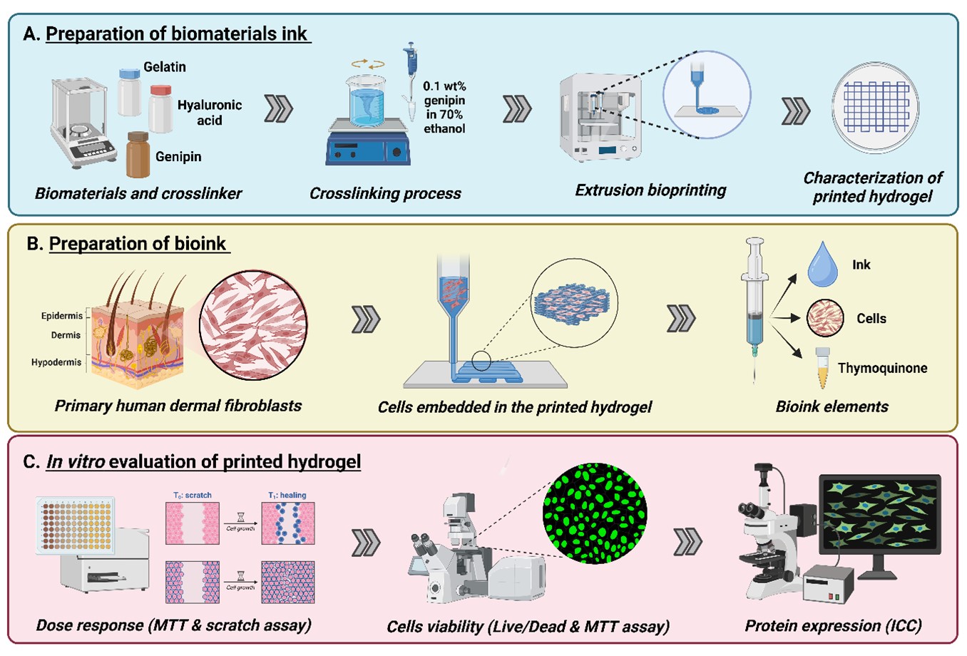
Wound healing is a multifaceted biological process that necessitates the development of advanced materials that can effectively support tissue regeneration and repair. The fabrication of bioengineered wound dressings has evolved significantly, with three-dimensional (3D) bioprinting emerging as a promising method to produce personalized, structurally stable hydrogels. In this study, we leveraged extrusion-based 3D bioprinting technology to develop gelatin-hyaluronic acid (GEL-HA) hydrogels, incorporating thymoquinone (TQ), a bioactive compound known for its regenerative properties. The use of 3D printing allowed for precise control over the scaffold’s architecture, optimizing its compressive strength and resilience while creating a bioactive, biocompatible platform for wound healing applications. This enables precise control over their architecture and mechanical properties to enhance wound healing where it offers promising potential as biocompatible scaffolds for wound healing applications due to their favorable physicochemical properties and ability to promote cell proliferation and migration. GEL-HA hydrogels were fabricated with varying HA concentrations (0.1–1.0 wt%), and the effects on the gelation process and physical characteristics were evaluated. Results showed that the ideal gelation temperature for the GEL-HA hydrogel was 22 °C, with the inclusion of HA reducing polymerization time. The printed hydrogels exhibited high water retention (>1000%) and satisfactory mechanical properties, with a degree of crosslinking of up to 40.21%. Furthermore, the hydrogels demonstrated a low biodegradation rate (less than 0.300 mg/h) and favorable water vapor transmission rate (WVTR) in a range of 2000– 3000 gm–2day–1, which are crucial for maintaining a moist environment for wound healing. The incorporation of TQ further enhanced the biocompatibility and cellular proliferation of human dermal fibroblasts (HDFs). Cell viability assays indicated that TQ promoted HDFs growth at concentrations of 0.005–0.1 μg/mL without toxicity. Moreover, the wound scratch assay demonstrated that TQ facilitated cell migration, with the optimum concentration of 0.1 μg/mL showing the most significant effect. The GEL-HA-TQ hydrogel also supported HDFs attachment and proliferation, as confirmed by live and dead cell staining, also with Ki67 level assessment, and collagen type-I immunocytochemistry. These findings suggest that GEL-HA hydrogels, combined with TQ, provide a promising and biocompatible platform for wound healing. It effectively promotes cell viability, migration, and extracellular matrix synthesis, which could be beneficial in regenerative medicine and tissue engineering applications.
- Almadani YH, Vorstenbosch J, Davison PG, Murphy AM. Wound healing: a comprehensive review. Semin Plast Surg. 2021;35(3):141-144. doi: 10.1055/s-0041-1731791
- Baron JM, Glatz M, Proksch E. Optimal support of wound healing: new insights. Dermatology. 2020;236(6):593-600. doi: 10.1159/000505291
- Kaur G, Narayanan G, Garg D, et al. Biomaterials-based regenerative strategies for skin tissue wound healing. ACS Applied Bio Mater. 2022;5(5):2069-2106. doi: 10.1021/acsabm.2c00035
- Ansari M, Darvishi A. A review of the current state of natural biomaterials in wound healing applications. Front Bioeng Biotechnol. 2024;12:1309541. doi: 10.3389/fbioe.2024.1309541
- Fadilah NIM, Isa ILM, Zaman WSWK, Tabata Y, Fauzi MB. The effect of nanoparticle-incorporated natural-based biomaterials towards cells on activated pathways: a systematic review. Polymers (Basel). 2022;14(3):476. doi: 10.3390/polym14030476
- Md Fadilah NI, Shahabudin NA, Mohd Razif RA, et al. Discovery of bioactive peptides as therapeutic agents for skin wound repair. J Tissue Eng. 2024;15: 20417314241280359. doi: 10.1177/20417314241280359
- Zarei F, Soleimaninejad M. Role of growth factors and biomaterials in wound healing. Artif Cells Nanomed Biotechnol. 2018;46(suppl 1):906-911. doi: 10.1080/21691401.2018.1439836
- Fadilah NIM, Fauzi MB, Maarof M. Effect of multiple-cycle collections of conditioned media from different cell sources towards fibroblasts in in vitro wound healing model. Pharmaceutics. 2024 ;16(6):767. doi: 10.3390/pharmaceutics16060767
- Md Fadilah NI, Mohd Abdul Kader Jailani MS, Badrul Hisham MAI, et al. Cell secretomes for wound healing and tissue regeneration: next generation acellular based tissue engineered products. J Tissue Eng. 2022;13:20417314221114273. doi: 10.1177/20417314221114273
- Zhong Y, Wei E-T, Wu L, et al. Novel biomaterials for wound healing and tissue regeneration. ACS Omega. 2024; 9(30):32268-32286. doi: 10.1021/acsomega.4c02775
- Fadilah NIM, Nizam NAAK, Fauzi MB. Antibacterial compounds-incorporated functional biomaterials for chronic wound healing application via 3D bioprinting: the mechanism of action. IJB. 2024;10(4):3372. doi: 10.36922/ijb.3372
- Fang H, Xu J, Ma H, et al. Functional materials of 3D bioprinting for wound dressings and skin tissue engineering applications: a review. Int J Bioprint. 2023; 9(5):757. doi: 10.18063/ijb.757
- Abuhamad AY, Masri S, Fadilah NIM, Alamassi MN, Maarof M, Fauzi MB. Application of 3D-printed bioinks in chronic wound healing: a scoping review. Polymers (Basel). 2024;16(17):2456. doi: 10.3390/polym16172456
- Tan SH, Ngo ZH, Sci DB, Leavesley D, Liang K. Recent advances in the design of three-dimensional and bioprinted scaffolds for full-thickness wound healing. Tissue Eng Part B Rev. 2022;28(1):160-181. doi: 10.1089/ten.TEB.2020.0339
- Khanna A. The new era of tissue engineering-3D printing of organs and tissue constructs. BME Horizon. 2024;2(1):112. doi: 10.37155/2972-449X-vol2(1)-112
- Masri S, Fadilah NIM, Hao LQ, et al. Multifunctionalised skin substitute of hybrid gelatin-polyvinyl alcohol bioinks for chronic wound: injectable vs. 3D bioprinting. Drug Deliv Transl Res. 2024;14(4):1005-1027. doi: 10.1007/s13346-023-01447-z
- Nizam AAK, Md Fadilah NI, Ahmad H, Maarof M, Fauzi MB. Injectable gelatin-palmitoyl-GDPH hydrogels as bioinks for future cutaneous regeneration: physicochemical characterization and cytotoxicity assessment. Polymers (Basel). 2024;17(1):41. doi: 10.3390/polym17010041
- Camci-Unal G, Cuttica D, Annabi N, Demarchi D, Khademhosseini A. Synthesis and characterization of hybrid hyaluronic acid-gelatin hydrogels. Biomacromolecules. 2013;14(4):1085-1092. doi: 10.1021/bm3019856
- Cao H, Wang J, Hao Z, Zhao D. Gelatin-based biomaterials and gelatin as an additive for chronic wound repair. Front Pharmacol. 2024;15:1398939. doi: 10.3389/fphar.2024.1398939
- Salih ARC, Farooqi HMU, Amin H, et al., Hyaluronic acid: comprehensive review of a multifunctional biopolymer. Future J Pharm Sci. 2024;10(1):63. doi: 10.1186/s43094-024-00636-y
- Lee SJ, Kim H-J, Choi EJ, et al. Immediately injectable modified gelatin and hyaluronic acid-based hydrogel encapsulating nano-hydroxyapatite and human adipose-derived MSCs for use as a bone filler in situ therapy. Carbohydr Polym Technol Appl. 2024;8:100625. doi: 10.1016/j.carpta.2024.100625
- Yang D, Chen H, Wei H, et al. Hydrogel wound dressings containing bioactive compounds originated from traditional Chinese herbs: a review. Smart Mater Med. 2024;5(1): 153-165. doi: 10.1016/j.smaim.2023.10.004
- López-Gutierrez J, Ramos-Payan R, Ayala-Hum A, et al. Biofunctionalization of hydrogel-based scaffolds for vascular tissue regeneration. Front Mater. 2023; 10. doi: 10.3389/fmats.2023.1168616
- Gu Q, Tomaskovic-Crook E, Wallace GG, Crook JM. Engineering human neural tissue by 3D bioprinting. Methods Mol Biol. 2018;1758:129-138. doi: 10.1007/978-1-4939-7741-3_10
- Abbas M, Gururani MA, Ali A, et al. Antimicrobial properties and therapeutic potential of bioactive compounds in Nigella sativa: a review. Molecules. 2024; 29(20):4914. doi: 10.3390/molecules29204914
- Sallehuddin N, Hao LQ, Wen APY, Fadilah NIM, Maarof M, Fauzi MB. Thymoquinone-incorporated collagee biomatrix: a promising approach for full-thickness wound healing. Pharmaceutics. 2024;16(11):1440. doi: 10.3390/pharmaceutics16111440
- Fan F, Saha S, Hanjaya-Putra D. Biomimetic hydrogels to promote wound healing. Front Bioeng Biotechnol. 2021;9:718377. doi: 10.3389/fbioe.2021.718377
- Masri S, Maarof M, Aziz IA, Idrus R, Fauzi MB. Performance of hybrid gelatin-PVA bioinks integrated with genipin through extrusion-based 3D bioprinting: An in vitro evaluation using human dermal fibroblasts. Int J Bioprint. 2023;9(3):677. doi: 10.18063/ijb.677
- Ng WC, Lokanathan Y, Fauzi MB, et al. In vitro evaluation of genipin-crosslinked gelatin hydrogels for vocal fold injection. Sci Rep. 2023;13(1):5128. doi: 10.1038/s41598-023-32080-y
- Phang SJ, Teh HX, Looi ML, et al. PVA/PVP nanofibres incorporated with Ecklonia cava phlorotannins exhibit excellent cytocompatibility and accelerate hyperglycaemic wound healing. Tissue Eng Regen Med. 2024; 21(2):243-260. doi: 10.1007/s13770-023-00590-5
- Amirrah IN, Zulkiflee I, Mohd Razip Wee MF, et al. Plasma-polymerised antibacterial coating of ovine tendon collagen type I (OTC) crosslinked with genipin (GNP) and dehydrothermal-crosslinked (DHT) as a cutaneous substitute for wound healing. Materials (Basel). 2023;16(7):2739. doi: 10.3390/ma16072739
- Salleh A, Mustafa N, Teow YH, et al. Dual-layered approach of ovine collagen-gelatin/cellulose hybrid biomatrix containing graphene oxide-silver nanoparticles for cutaneous wound healing: fabrication, physicochemical, cytotoxicity and antibacterial characterisation. Biomedicines. 2022;10(4):816. doi: 10.3390/biomedicines10040816
- Samadian H, Zamiri S, Ehterami A, et al., Electrospun cellulose acetate/gelatin nanofibrous wound dressing containing berberine for diabetic foot ulcer healing: in vitro and in vivo studies. Sci Rep. 2020;10(1):8312. doi: 10.1038/s41598-020-65268-7
- Maarof M, Chowdhury SR, Saim A, Bt Hj Idrus R, Lokanathan Y. Concentration dependent effect of human dermal fibroblast conditioned medium (DFCM) from three various origins on keratinocytes wound healing. Int J Mol Sci. 2020;21(8):2929. doi: 10.3390/ijms21082929
- Khaleghi M, Ahmadi E, Khodabandeh Shahraki M, Aliakbari F, Morshedi D. Temperature-dependent formulation of a hydrogel based on Hyaluronic acid-polydimethylsiloxane for biomedical applications. Heliyon. 2020;6(3):e03494. doi: 10.1016/j.heliyon.2020.e03494
- Wei P, Chen W, Song Q, et al. Superabsorbent hydrogels enhanced by quaternized tunicate cellulose nanocrystals with adjustable strength and swelling ratio. Cellulose. 2021;28(6):3723-3732. doi: 10.1007/s10570-021-03776-z
- Agubata CO, Mbah MA, Akpa PA, Ugwu G. Application of self-healing, swellable and biodegradable polymers for wound treatment. J Wound Care. 2021;30(Sup9a):IVi-IVx. doi: 10.12968/jowc.2021.30.Sup9a.IV
- Baseer A, Koenneke A, Zapp J, et al. Design and characterization of surface-crosslinked gelatin nanoparticles for the delivery of hydrophilic macromolecular drugs. Macromol Chem Phys. 2019;220(18):1900260. doi: 10.1002/macp.201900260
- Chang KC, Lin DJ, Wu YR, et al. Characterization of genipin-crosslinked gelatin/hyaluronic acid-based hydrogel membranes and loaded with hinokitiol: In vitro evaluation of antibacterial activity and biocompatibility. Mater Sci Eng C Mater Biol Appl. 2019;105:110074. doi: 10.1016/j.msec.2019.110074
- Zawani M, Maarof M, Tabata Y, Motta A, Fauzi MB. Quercetin-embedded gelastin injectable hydrogel as provisional biotemplate for future cutaneous application: optimization and in vitro evaluation. Gels. 2022;8(10):623. doi: 10.3390/gels8100623
- Liu L, Li X, Nagao M, Elias AL, Narain R, Chung HJ. A pH-indicating colorimetric tough hydrogel patch towards applications in a substrate for smart wound dressings. Polymers (Basel). 2017;9(11):558. doi: 10.3390/polym9110558
- Hazrati R, Davaran S, and Omidi Y. Bioactive functional scaffolds for stem cells delivery in wound healing and skin regeneration. React Funct Polym. 2022;174:105233. doi: 10.1016/j.reactfunctpolym.2022.105233
- Zhang M, Chu L, Chen J, et al. Asymmetric wettability fibrous membranes: Preparation and biologic applications. Compos Part B: Eng. 2023;269:111095. doi: 10.1016/j.compositesb.2023.111095
- Radakisnin R, Abdul Majid MS, Jamir MRM, Tahir MFM. Physical, thermal, and mechanical properties of highly porous polylactic acid/cellulose nanofibre scaffolds prepared by salt leaching technique. Nanotechnol Rev. 2021;10(1): 1469-1483. doi: 10.1515/ntrev-2021-0098
- Sallehuddin N, Md Fadilah NI, Hwei NM, et al. Characterization and cytocompatibility of collagen-gelatin-elastin (collagee) acellular skin substitute towards human dermal fibroblasts: in vitro assessment. Biomedicines. 2022;10(6):1327. doi: 10.3390/biomedicines10061327
- Gholamali I, Vu TT, Jo SH, Park SH, Lim KT. Exploring the progress of hyaluronic acid hydrogels: synthesis, characteristics, and wide-ranging applications. Materials (Basel). 2024;17(10):2439. doi: 10.3390/ma17102439
- Zhang S, Kang L, Hu S, et al. Carboxymethyl chitosan microspheres loaded hyaluronic acid/gelatin hydrogels for controlled drug delivery and the treatment of inflammatory bowel disease. Int J Biol Macromol. 2021;167:1598-1612. doi: 10.1016/j.ijbiomac.2020.11.117
- Song M, Furuya K. Fabrication and characterization of nanostructures on insulator substrates by electron-beam-induced deposition. Sci Technol Adv Mater. 2008;9(2):023002. doi: 10.1088/1468-6996/9/2/023002
- Miranda DG, Malmonge SM, Campos DM, Attik NG, Grosgogeat B, Gritsch K. A chitosan-hyaluronic acid hydrogel scaffold for periodontal tissue engineering. J Biomed Mater Res B Appl Biomater. 2016;104(8): 1691-1702. doi: 10.1002/jbm.b.33516
- Algahtani MS, Ahmad MZ, Shaikh IA, Abdel-Wahab BA, Nourein IH, Ahmad J. Thymoquinone loaded topical nanoemulgel for wound healing: formulation design and in-vivo evaluation. Molecules. 2021;26(13):3863. doi: 10.3390/molecules26133863.
- Trinh XT, Long NV, Van Anh LT, et al. A comprehensive review of natural compounds for wound healing: targeting bioactivity perspective. Int J Mol Sci. 2022;23(17): 9573. doi: 10.3390/ijms23179573
- Masri S, Fadilah NIM, Hao LQ, et al. Multifunctionalised skin substitute of hybrid gelatin-polyvinyl alcohol bioinks for chronic wound: injectable vs. 3D bioprinting. Drug Deliv Transl Res. 2024;14(4):1005-1027. doi: 10.1007/s13346-023-01447-z
- Thambirajoo M, Md Fadilah NI, Maarof M, et al. Functionalised sodium-carboxymethylcellulose-collagen bioactive bilayer as an acellular skin substitute for future use in diabetic wound management: the evaluation of physicochemical, cell viability, and antibacterial effects. Polymers (Basel). 2024;16(16):2252. doi: 10.3390/polym16162252
- Fadilah NIM, Phang SJ, Kamaruzaman N, et al. Antioxidant biomaterials in cutaneous wound healing and tissue regeneration: a critical review. Antioxidants (Basel). 2023;12(4):787. doi: 10.3390/antiox12040787
- Mohanto S, Narayana S, Merai KP, et al. Advancements in gelatin-based hydrogel systems for biomedical applications: a state-of-the-art review. Int J Biol Macromol. 2023; 253(Pt 5):127143. doi: 10.1016/j.ijbiomac.2023.127143
- Barrulas RV, Corvo MC. Rheology in product development: an insight into 3D printing of hydrogels and aerogels. Gels. 2023;9(12):986. doi: 10.3390/gels9120986
- Liu S, Li D, Wang Y, Zhou G, Ge K, Jiang L. Adhesive, antibacterial and double crosslinked carboxylated polyvinyl alcohol/chitosan hydrogel to enhance dynamic skin wound healing. Int J Biol Macromol. 2023; 228:744-753. doi: 10.1016/j.ijbiomac.2022.12.169
- Nguyen HM, Le TTN, Nguyen AT, et al. Biomedical materials for wound dressing: recent advances and applications. RSC Adv. 2023;13(8):5509-5528. doi: 10.1039/D2RA07673J
- Griveau L, Lafont M, le Goff H, et al. Design and characterization of an in vivo injectable hydrogel with effervescently generated porosity for regenerative medicine applications. Acta Biomater. 2022;140:324-337. doi: 10.1016/j.actbio.2021.11.036
- Balion Z, Sipailaite E, Stasyte G, et al. Investigation of cancer cell migration and proliferation on synthetic extracellular matrix peptide hydrogels. Front Bioeng Biotechnol. 2020;8:773. doi: 10.3389/fbioe.2020.00773
- Zhang Y, Xu Y, Gao J. The engineering and application of extracellular matrix hydrogels: a review. Biomaterials Sci. 2023; 11(11):3784-3799. doi: 10.1039/D3BM00183K