

Laser-induced forward transfer in picosecond regime for cell bioprinting

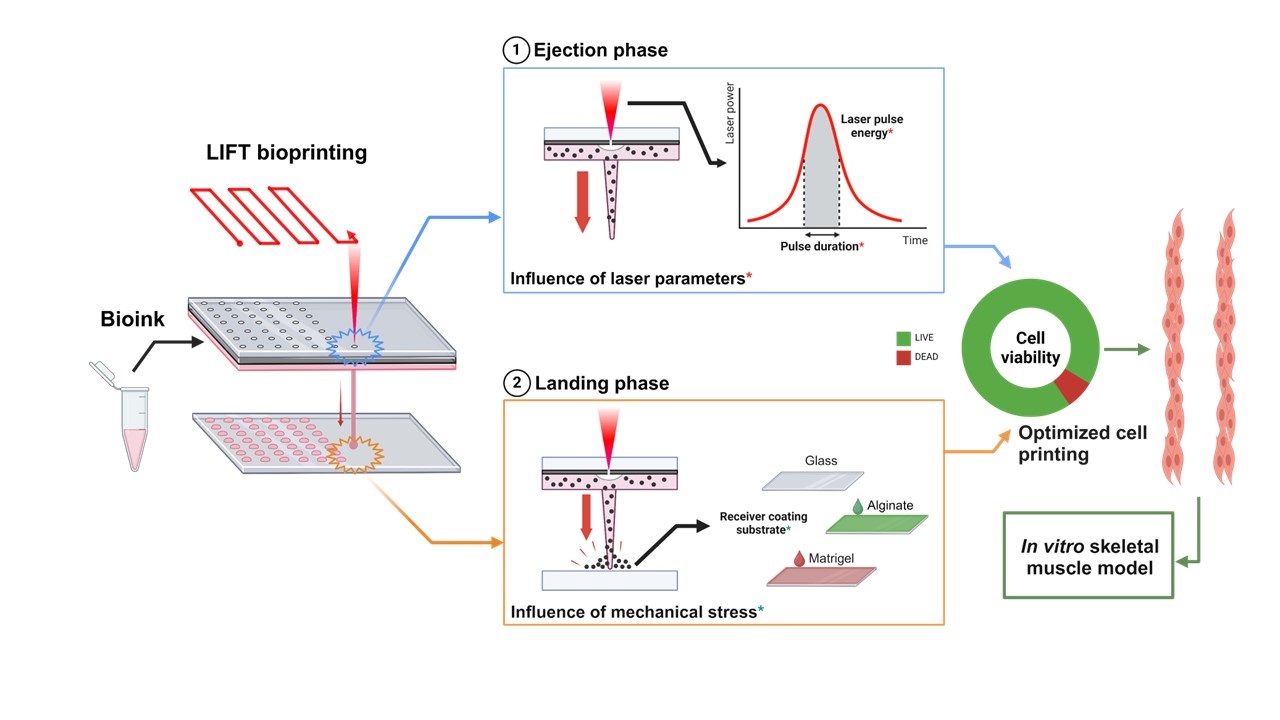
Based on interdisciplinary approaches, bioprinting methods aim to create and design highly organized 2D and 3D cultures. In this context, it has been more than a decade since laser-induced forward transfer (LIFT) was studied on a lab scale for its ability to transfer biomaterials, specifically bioink loaded with living cells, onto a substrate. Extreme physical and mechanical phenomena contribute to the jetting dynamic of the targeted bioink, raising a spontaneous biological question: does this process negatively affect the survival rate of transferred cells? This study demonstrates that laser pulse durations in the range of picoseconds to nanoseconds do not directly affect cell viability, indicating that LIFT is a valuable bioprinting method for transferring living cells. Moreover, we highlight the necessity of using hydrogel coatings on the surface of the receiver substrate to guarantee optimal post-printing viability of the cells. We demonstrate that the nature of the hydrogel also contributes to the resolution of the printed pattern. Among the tested materials, Matrigel demonstrated all the qualities required to ensure successful printing and should therefore be considered for future work. Overall, the results show the suitability of our LIFT setup for printing living cells in the picosecond regime with a high survival rate, paving the way for a wide range of biological applications.
- Tarassoli SP, Jessop ZM, Al-Sabah A, et al. Skin tissue engineering using 3D bioprinting: an evolving research field. J Plast Reconstr Aesthet Surg. 2018;71(5):615-623. doi: 10.1016/j.bjps.2017.12.006
- Guillemot F, Souquet A, Catros S, et al. High-throughput laser printing of cells and biomaterials for tissue engineering. Acta Biomater. 2010;6(7):2494-2500. doi: 10.1016/j.actbio.2009.09.029
- Zhu W, Ma X, Gou M, Mei D, Zhang K, Chen S. 3D printing of functional biomaterials for tissue engineering. Curr Opin Biotechnol. 2016;40:103-112. doi: 10.1016/j.copbio.2016.03.014
- Kačarević Ž, Rider P, Alkildani S, et al. An introduction to 3D bioprinting: possibilities, challenges and future aspects. Materials. 2018;11(11):2199. doi: 10.3390/ma11112199
- Li J, Chen M, Fan X, Zhou H. Recent advances in bioprinting techniques: approaches, applications and future prospects. J Transl Med. 2016;14(1):271. doi: 10.1186/s12967-016-1028-0
- Vinson BT, Sklare SC, Chrisey DB. Laser-based cell printing techniques for additive biomanufacturing. Curr Opin Biomed Eng. 2017;2:14-21. doi: 10.1016/j.cobme.2017.05.005
- Raees S, Ullah F, Javed F, et al. Classification, processing, and applications of bioink and 3D bioprinting: a detailed review. Int J Biol Macromol. 2023;232:123476. doi: 10.1016/j.ijbiomac.2023.123476
- Dey M, Ozbolat IT. 3D bioprinting of cells, tissues and organs. Sci Rep. 2020;10(1):14023. doi: 10.1038/s41598-020-70086-y
- Bohandy J, Kim BF, Adrian FJ. Metal deposition from a supported metal film using an excimer laser. J Appl Phys. 1986;60(4):1538-1539. doi: 10.1063/1.337287
- Bohandy J, Kim BF, Adrian FJ, Jette AN. Metal deposition at 532 nm using a laser transfer technique. J Appl Phys. 1988;63(4):1158-1162. doi: 10.1063/1.340023
- Rapp L, Cibert C, Nénon S, et al. Improvement in semiconductor laser printing using a sacrificial protecting layer for organic thin-film transistors fabrication. Appl Surf Sci. 2011;257(12):5245-5249. doi: 10.1016/j.apsusc.2010.10.147
- Rapp L, Constantinescu C, Delaporte P, Alloncle AP. Laser-induced forward transfer of polythiophene-based derivatives for fully polymeric thin film transistors. Org Electron. 2014;15(8):1868-1875. doi: 10.1016/j.orgel.2014.04.029
- Rapp L, Cibert C, Alloncle AP, et al. LIFT of organic materials for application in plastic micro-electronics. In: 3eme Russian-French Laser Physics Worshop for Young Scientist; 2008. Accessed December 3, 2024. https://hal.science/hal-00446484
- Colina M, Serra P, Fernández-Pradas JM, Sevilla L, Morenza JL. DNA deposition through laser induced forward transfer. Biosens Bioelectron. 2005;20(8):1638-1642. doi: 10.1016/j.bios.2004.08.047
- Serra P, Colina M, Fernández-Pradas JM, Sevilla L, Morenza JL. Preparation of functional DNA microarrays through laser-induced forward transfer. Appl Phys Lett. 2004;85(9):1639-1641. doi: 10.1063/1.1787614
- Fernández-Pradas JM, Colina M, Serra P, Domínguez J, Morenza JL. Laser-induced forward transfer of biomolecules. Thin Solid Films. 2004;453-454:27-30. doi: 10.1016/j.tsf.2003.11.154
- Barron JA, Spargo BJ, Ringeisen BR. Biological laser printing of three dimensional cellular structures. Appl Phys A. 2004;79(4-6):1027-1030. doi: 10.1007/s00339-004-2620-3
- Ringeisen BR, Kim H, Barron JA, et al. Laser printing of pluripotent embryonal carcinoma cells. Tissue Eng. 2004;10(3-4):483-491. doi: 10.1089/107632704323061843
- Guillotin B, Guillemot F. Cell patterning technologies for organotypic tissue fabrication. Trends Biotechnol. 2011;29(4):183-190. doi: 10.1016/j.tibtech.2010.12.008
- Koch L, Deiwick A, Chichkov B. Capillary-like formations of endothelial cells in defined patterns generated by laser bioprinting. Micromachines (Basel). 2021;12(12):1538. doi: 10.3390/mi12121538
- Koch L, Deiwick A, Schlie S, et al. Skin tissue generation by laser cell printing. Biotechnol Bioeng. 2012;109(7):1855-1863. doi: 10.1002/bit.24455
- Bosmans C, Rodriguez NG, Karperien M, et al. Towards single-cell bioprinting: micropatterning tools for organ-on-chip development. Trends Biotechnol. 2024;42(6):739-759. doi: 10.1016/j.tibtech.2023.11.014
- Hakobyan D, Médina C, Dusserre N, et al. Laser-assisted 3D bioprinting of exocrine pancreas spheroid models for cancer initiation study. Biofabrication. 2020;12(3):035001. doi: 10.1088/1758-5090/ab7cb8
- Sorkio A, Koch L, Koivusalo L, et al. Human stem cell based corneal tissue mimicking structures using laser-assisted 3D bioprinting and functional bioinks. Biomaterials. 2018;171:57-71. doi: 10.1016/j.biomaterials.2018.04.034
- Chang J, Sun X. Laser-induced forward transfer based laser bioprinting in biomedical applications. Front Bioeng Biotechnol. 2023;11:1255782. doi: 10.3389/fbioe.2023.1255782
- Curley JL, Sklare SC, Bowser DA, Saksena J, Moore MJ, Chrisey DB. Isolated node engineering of neuronal systems using laser direct write. Biofabrication. 2016;8(1):015013. doi: 10.1088/1758-5090/8/1/015013
- Colina M, Duocastella M, Fernández-Pradas JM, Serra P, Morenza JL. Laser-induced forward transfer of liquids: study of the droplet ejection process. J Appl Phys. 2006;99(8):084909. doi: 10.1063/1.2191569
- Fernández-Pradas JM, Florian C, Caballero-Lucas F, Sopeña P, Morenza JL, Serra P. Laser-induced forward transfer: propelling liquids with light. Appl Surf Sci. 2017;418:559-564. doi: 10.1016/j.apsusc.2016.10.197
- Gruene M, Unger C, Koch L, Deiwick A, Chichkov B. Dispensing pico to nanolitre of a natural hydrogel by laser-assisted bioprinting. BioMed Eng OnLine. 2011;10(1):19. doi: 10.1186/1475-925X-10-19
- Barron JA, Krizman DB, Ringeisen BR. Laser printing of single cells: statistical analysis, cell viability, and stress. Ann Biomed Eng. 2005;33(2):121-130. doi: 10.1007/s10439-005-8971-x
- Mézel C, Souquet A, Hallo L, Guillemot F. Bioprinting by laser-induced forward transfer for tissue engineering applications: jet formation modeling. Biofabrication. 2010;2(1):014103. doi: 10.1088/1758-5082/2/1/014103
- Koch L, Brandt O, Deiwick A, Chichkov B. Laser-assisted bioprinting at different wavelengths and pulse durations with a metal dynamic release layer: A parametric study. Int J Bioprint. 2017;3(1):001. doi: 10.18063/IJB.2017.01.001
- Vogel A, Busch S, Parlitz U. Shock wave emission and cavitation bubble generation by picosecond and nanosecond optical breakdown in water. J Acoust Soc Amer. 1996;100(1):148-165. doi: 10.1121/1.415878
- Duocastella M, Fernández-Pradas JM, Morenza JL, Serra P. Time-resolved imaging of the laser forward transfer of liquids. J Appl Phys. 2009;106(8):084907. doi: 10.1063/1.3248304
- Deng Y, Renaud P, Guo Z, Huang Z, Chen Y. Single cell isolation process with laser induced forward transfer. J Biol Eng. 2017;11(1):2. doi: 10.1186/s13036-016-0045-0
- Koch L, Kuhn S, Sorg H, et al. Laser printing of skin cells and human stem cells. Tissue Eng Part C Methods. 2010;16(5):847-854. doi: 10.1089/ten.tec.2009.0397
- Hopp B, Smausz T, Kresz N, et al. Survival and proliferative ability of various living cell types after laser-induced forward transfer. Tissue Eng. 2005;11(11-12):1817-1823. doi: 10.1089/ten.2005.11.1817
- Schiele NR, Chrisey DB, Corr DT. Gelatin-based laser direct-write technique for the precise spatial patterning of cells. Tissue Eng Part C Methods. 2011;17(3):289-298. doi: 10.1089/ten.tec.2010.0442
- Guillotin B, Souquet A, Catros S, et al. Laser assisted bioprinting of engineered tissue with high cell density and microscale organization. Biomaterials. 2010;31(28):7250-7256. doi: 10.1016/j.biomaterials.2010.05.055
- Karakaidos P, Kryou C, Simigdala N, Klinakis A, Zergioti I. Laser bioprinting of cells using UV and visible wavelengths: a comparative DNA damage study. Bioengineering. 2022;9(8):378. doi: 10.3390/bioengineering9080378
- Kryou C, Theodorakos I, Karakaidos P, Klinakis A, Hatziapostolou A, Zergioti I. Parametric study of Jet/Droplet formation process during LIFT printing of living Cell-Laden bioink. Micromachines. 2021;12(11):1408. doi: 10.3390/mi12111408
- Murru C, Duvert L, Magdinier F, et al. Assessment of laser-synthesized Si nanoparticle effects on myoblast motility, proliferation and differentiation: towards potential tissue engineering applications. Nanoscale Adv. 2024;6(8):2104-2112. doi: 10.1039/D3NA01020A
- Hanawa T. Biocompatibility of titanium from the viewpoint of its surface. Sci Technol Adv Mater. 2022;23(1):457-472. doi: 10.1080/14686996.2022.2106156
- Schindelin J, Arganda-Carreras I, Frise E, et al. Fiji: an open-source platform for biological-image analysis. Nat Methods. 2012;9(7):676-682. doi: 10.1038/nmeth.2019
- Zhang Z, Xu C, Xiong R, Chrisey DB, Huang Y. Effects of living cells on the bioink printability during laser printing. Biomicrofluidics. 2017;11(3):034120. doi: 10.1063/1.4985652
- Xiong R, Zhang Z, Chai W, Chrisey DB, Huang Y. Study of gelatin as an effective energy absorbing layer for laser bioprinting. Biofabrication. 2017;9(2):024103. doi: 10.1088/1758-5090/aa74f2
- Catros S, Guillotin B, Bačáková M, Fricain JC, Guillemot F. Effect of laser energy, substrate film thickness and bioink viscosity on viability of endothelial cells printed by laser-assisted bioprinting. Appl Surf Sci. 2011;257(12):5142-5147. doi: 10.1016/j.apsusc.2010.11.049
- Riester D. High speed photography of laser induced forward transfer (LIFT) of single and double-layered transfer layers for single cell transfer. JLMN. 2016;11(2):199-203. doi: 10.2961/jlmn.2016.02.0010
- Antoshin AA, Churbanov SN, Minaev NV, et al. LIFT-bioprinting, is it worth it? Bioprinting. 2019;15:e00052. doi: 10.1016/j.bprint.2019.e00052
- Liu X, Zu E, Chang X, et al. Bi-phasic effect of gelatin in myogenesis and skeletal muscle regeneration. Dis Model Mech. 2021;14(12):dmm049290. doi: 10.1242/dmm.049290
- Minaev NV, Yusupov VI, Zhigarkov VS, et al. Laser Printing of Gel Microdrops with Living Cells and Microorganisms. KnE Energy. 2018;3(3):23-31. doi: 10.18502/ken.v3i3.2010
- Ali M, Pages E, Ducom A, Fontaine A, Guillemot F. Controlling laser-induced jet formation for bioprinting mesenchymal stem cells with high viability and high resolution. Biofabrication. 2014;6(4):045001. doi: 10.1088/1758-5082/6/4/045001