

Extracellular matrix-inspired 4D SMART biomaterials for bioprinting in tissue engineering

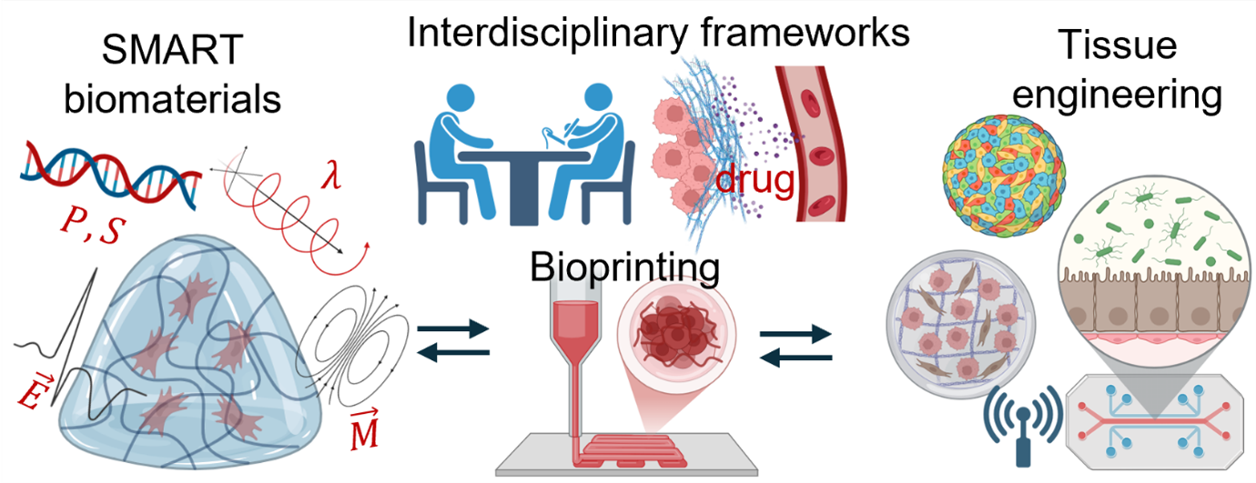
Tissue development and regeneration arise from a dynamic interplay among cells, the extracellular matrix (ECM), and surrounding biophysical and biochemical cues. These interactions form the basis for stimuli-responsive materials for advanced regenerative technologies (SMART) that drive innovation in four-dimensional (4D) bioprinting for tissue engineering. This review discusses the biophysical foundations of SMART materials, emphasizing native ECM components, their interactions, and organ-specific properties that inform biomimetic material design. We highlight recent advances in 4D SMART systems, including ionic self-healing, pH-, thermal-, hydration-, and magneto-responsive materials, and their roles in mimicking developmental and regenerative processes. This is followed by a comparative overview of these stimuli-responsive material classes, benchmarked against one another, native ECM performance, and clinical translation requirements, revealing persistent gaps in long-term stability, multi-stimuli integration, and regulatory feasibility. Together, these insights provide an interdisciplinary framework for designing adaptive, responsive biomaterials that guide tissue morphogenesis and advance the future of regenerative medicine.
- Crossley RM, Johnson S, Tsingos E, et al. Modeling the extracellular matrix in cell migration and morphogenesis: a guide for the curious biologist. Front Cell Dev Biol. 2024;12:1354132. doi: 10.3389/fcell.2024.1354132
- Martin R, Joung D. The promise and challenges of bioprinting in tissue engineering. Micromachines. 2024;15(12):1529. doi: 10.3390/mi15121529
- Lu P, Ruan D, Huang M, et al. Harnessing the potential of hydrogels for advanced therapeutic applications: current achievements and future directions. Signal Transduct Target Ther. 2024;9(1):166. doi: 10.1038/s41392-024-01852-x
- Dethe MR, Prabakaran A, Ahmed H, Agrawal M, Roy U, Alexander A. PCL-PEG copolymer based injectable thermosensitive hydrogels. J Control Release. 2022;343:217-236. doi: 10.1016/j.jconrel.2022.01.035
- Doberenz F, Zeng K, Willems C, Zhang K, Groth T. Thermoresponsive polymers and their biomedical application in tissue engineering – a review. J Mater Chem B. 2020;8(4):607-628. doi: 10.1039/C9TB02052G
- Pei Y, Chen J, Yang L, et al. The effect of pH on the LCST of poly(N-isopropylacrylamide) and poly(N-isopropylacrylamide-co-acrylic acid). J Biomat Sci Polymer Edn. 2004;15(5):585-594. doi: 10.1163/156856204323046852
- Correa S, Grosskopf AK, Lopez Hernandez H, et al. Translational applications of hydrogels. Chem Rev. 2021;121(18):11385-11457. doi: 10.1021/acs.chemrev.0c01177
- Taylor S, Mueller E, Jones LR, Makela AV, Ashammakhi N. Translational aspects of 3D and 4D printing and bioprinting. Adv Healthc Mater. 2024;13(27):2400463. doi: 10.1002/adhm.202400463
- Cao H, Duan L, Zhang Y, Cao J, Zhang K. Current hydrogel advances in physicochemical and biological response-driven biomedical application diversity. Signal Transduct Target Ther. 2021;6(1):426. doi: 10.1038/s41392-021-00830-x
- Di X, Gao X, Peng L, et al. Cellular mechanotransduction in health and diseases: from molecular mechanism to therapeutic targets. Signal Transduct Target Ther. 2023;8(1):282. doi: 10.1038/s41392-023-01501-9
- Ambattu LA, Yeo LY. Sonomechanobiology: vibrational stimulation of cells and its therapeutic implications. Biophys Rev. 2023;4(2):021301. doi: 10.1063/5.0127122
- Arif ZU, Khalid MY, Ahmed W, Arshad H. A review on four-dimensional (4D) bioprinting in pursuit of advanced tissue engineering applications. Bioprinting. 2022;27:e00203. doi: 10.1016/j.bprint.2022.e00203
- Zhou W, Chen Y, Roh T, et al. Multifunctional bioreactor system for human intestine tissues. ACS Biomater Sci Eng. 2018;4(1):231-239. doi: 10.1021/acsbiomaterials.7b00794
- Williamson A, Khoshmanesh K, Pirogova E, et al. Bioreactors: a regenerative approach to skeletal muscle engineering for repair and replacement. Adv Nanobiomed Res. 2024;4(10):2400030. doi: 10.1002/anbr.202400030
- Yarali E, Mirzaali MJ, Ghalayaniesfahani A, Accardo A, Diaz‐Payno PJ, Zadpoor AA. 4D printing for biomedical applications. Adv Mater. 2024;36(31):2402301. doi: 10.1002/adma.202402301
- Wang J, Wang Y, Wang R, et al. A review on 3D printing processes in pharmaceutical engineering and tissue engineering: applications, trends and challenges. Adv Mater Technol. 2025;10(2):2400620. doi: 10.1002/admt.202400620
- Joung D, Lavoie NS, Guo S, Park SH, Parr AM, McAlpine MC. 3D printed neural regeneration devices. Adv Funct Mater. 2020;30(1):1906237. doi: 10.1002/adfm.201906237
- Li YC, Zhang YS, Akpek A, Shin SR, Khademhosseini A. 4D bioprinting: the next-generation technology for biofabrication enabled by stimuli-responsive materials. Biofabrication. 2016;9(1):012001. doi: 10.1088/1758-5090/9/1/012001
- Michael PL, Lam YT, Mitchell TC, et al. Harnessing physiological shear stress in a perfusion bioreactor for enhanced endothelialization of small‐diameter vascular grafts. Adv Nanobiomed Res. 2025;5:2500025. doi: 10.1002/anbr.202500025
- Mierke CT. Viscoelasticity, like forces, plays a role in mechanotransduction. Front Cell Dev Biol. 2022;10:789841. doi: 10.3389/fcell.2022.789841
- Asadi Tokmedash M, Kim C, Chavda AP, Li A, Robins J, Min J. Engineering multifunctional surface topography to regulate multiple biological responses. Biomaterials. 2025;319:123136. doi: 10.1016/j.biomaterials.2025.123136
- Özkale B, Sakar MS, Mooney DJ. Active biomaterials for mechanobiology. Biomaterials. 2021;267:120497. doi: 10.1016/j.biomaterials.2020.120497
- Kayser LV, Lipomi DJ. Stretchable conductive polymers and composites based on PEDOT and PEDOT:PSS. Adv Mater. 2019;31(10):1806133. doi: 10.1002/adma.201806133
- Bryan AE, Krutko M, Rebholz S, et al. Development of a bioactive, piezoelectric PVDF-TrFE scaffold with evaluation of tissue reaction for potential in nerve repair. Biomater Sci. 2025;13(20):5769-5785. doi: 10.1039/D5BM01054C
- Mariano A, Lubrano C, Bruno U, Ausilio C, Dinger NB, Santoro F. Advances in cell-conductive polymer biointerfaces and role of the plasma membrane. Chem Rev. 2022;122(4):4552-4580. doi: 10.1021/acs.chemrev.1c00363
- Tonti OR, Larson H, Lipp SN, et al. Tissue-specific parameters for the design of ECM-mimetic biomaterials. Acta Biomater. 2021;132:83-102. doi: 10.1016/j.actbio.2021.04.017
- Valdoz JC, Johnson BC, Jacobs DJ, et al. The ECM: to scaffold, or not to scaffold, that is the question. Int J Mol Sci. 2021;22(23):12690. doi: 10.3390/ijms222312690
- Rahmati M, Silva EA, Reseland JE, A. Heyward C, Haugen HJ. Biological responses to physicochemical properties of biomaterial surface. Chem Soc Rev. 2020;49(15):5178-5224. doi: 10.1039/D0CS00103A
- Yamada KM, Doyle AD, Lu J. Cell–3D matrix interactions: recent advances and opportunities. Trends Cell Biol. 2022;32(10):883-895. doi: 10.1016/j.tcb.2022.03.002
- Hervé S, Miroshnikova YA. Biophysical determinants of nuclear shape and mechanics and their implications for genome integrity. Curr Opin Biomed Eng. 2024;30:100521. doi: 10.1016/j.cobme.2024.100521
- Miroshnikova YA, Wickström SA. Mechanical forces in nuclear organization. Cold Spring Harb Perspect Biol. 2022;14(1):a039685. doi: 10.1101/cshperspect.a039685
- Vermeulen S, Honig F, Vasilevich A, et al. Expanding biomaterial surface topographical design space through natural surface reproduction. Adv Mater. 2021;33(31):2102084. doi: 10.1002/adma.202102084
- Comelles J, Fernández-Majada V, Acevedo V, Rebollo- Calderon B, Martínez E. Soft topographical patterns trigger a stiffness-dependent cellular response to contact guidance. Mater Today Bio. 2023;19:100593. doi: 10.1016/j.mtbio.2023.100593
- Min K, Karuppannan SK, Tae G. The impact of matrix stiffness on hepatic cell function, liver fibrosis, and hepatocellular carcinoma-based on quantitative data. Biophys Rev. 2024;5(2):021306. doi: 10.1063/5.0197875
- Elosegui-Artola A, Gupta A, Najibi AJ, et al. Matrix viscoelasticity controls spatiotemporal tissue organization. Nat Mater. 2023;22(1):117-127. doi: 10.1038/s41563-022-01400-4
- Wu Y, Song Y, Soto J, et al. Viscoelastic extracellular matrix enhances epigenetic remodeling and cellular plasticity. Nat Commun. 2025;16(1):4054. doi: 10.1038/s41467-025-59190-7
- Wan S, Chen Y, Huang C, et al. Scalable ultrastrong MXene films with superior osteogenesis. Nature. 2024;634(8036):1103-1110. doi: 10.1038/s41586-024-08067-8
- Cheng Y, Wang Y, Wang Y, et al. Microenvironment-feedback regulated hydrogels as living wound healing materials. Nat Commun. 2025;16(1):6050. doi: 10.1038/s41467-025-60858-3
- Zhang C, Cai D, Liao P, et al. 4D printing of shape-memory polymeric scaffolds for adaptive biomedical implantation. Acta Biomater. 2021;122:101-110. doi: 10.1016/j.actbio.2020.12.042
- Benwood C, Chrenek J, Kirsch RL, et al. Natural biomaterials and their use as bioinks for printing tissues. Bioengineering. 2021;8(2):27. doi: 10.3390/bioengineering8020027
- Chen XB, Fazel Anvari-Yazdi A, Duan X, et al. Biomaterials/ bioinks and extrusion bioprinting. Bioact Mater. 2023;28:511-536. doi: 10.1016/j.bioactmat.2023.06.006
- Carvalho EM, Kumar S. Lose the stress: viscoelastic materials for cell engineering. Acta Biomater. 2023;163:146-157. doi: 10.1016/j.actbio.2022.03.058
- Fonseca AC, Melchels FP, Ferreira MJ, et al. Emulating human tissues and organs: a bioprinting perspective toward personalized medicine. Chem Rev. 2020;120(19):11093-11139. doi: 10.1021/acs.chemrev.0c00342
- Khalid MY, Otabil A, Mamoun OS, Askar K, Bodaghi M. Transformative 4D printed SMPs into soft electronics and adaptive structures: innovations and practical insights. Adv Mater Technol. 2025;10(19):e00309. doi: 10.1002/admt.202500309
- Mirasadi K, Yousefi MA, Jin L, et al. 4D printing of magnetically responsive shape memory polymers: toward sustainable solutions in soft robotics, wearables, and biomedical devices. Adv Sci. 2025:e13091. doi: 10.1002/advs.202513091
- Zhang W, Liu Y, Zhang H. Extracellular matrix: an important regulator of cell functions and skeletal muscle development. Cell Biosci. 2021;11(1):65. doi: 10.1186/s13578-021-00579-4
- Hynes RO. The extracellular matrix: not just pretty fibrils. Science. 2009;326(5957):1216-1219. doi: 10.1126/science.1176009
- Zhang S-y, Li X-y, Yang N, et al. Electrospun collagen nanofibers reduce inflammation, inhibit fibrosis, and promote wound healing on the ocular surface. ACS Appl Nano Mater. 2024;7(17):20267-20278. doi: 10.1021/acsanm.4c03180
- Orr SB, Chainani A, Hippensteel KJ, et al. Aligned multilayered electrospun scaffolds for rotator cuff tendon tissue engineering. Acta Biomater. 2015;24:117-126. doi: 10.1016/j.actbio.2015.06.010
- Kim HN, Jiao A, Hwang NS, et al. Nanotopography-guided tissue engineering and regenerative medicine. Adv Drug Deliv Rev. 2013;65(4):536-558. doi: 10.1016/j.addr.2012.07.014
- Putra VDL, Kilian KA, Knothe Tate ML. Biomechanical, biophysical and biochemical modulators of cytoskeletal remodelling and emergent stem cell lineage commitment. Commun Biol. 2023;6(1):75. doi: 10.1038/s42003-022-04320-w
- Pike DB, Cai S, Pomraning KR, et al. Heparin-regulated release of growth factors in vitro and angiogenic response in vivo to implanted hyaluronan hydrogels containing VEGF and bFGF. Biomaterials. 2006;27(30):5242-5251. doi: 10.1016/j.biomaterials.2006.05.018
- Zhao T, Huang Y, Zhu J, et al. Extracellular matrix signaling cues: biological functions, diseases, and therapeutic targets. MedComm. 2025;6(8):e70281. doi: 10.1002/mco2.70281
- Karamanos NK, Theocharis AD, Piperigkou Z, et al. A guide to the composition and functions of the extracellular matrix. FEBS J. 2021;288(24):6850-6912. doi: 10.1111/febs.15776
- Nicolas J, Magli S, Rabbachin L, Sampaolesi S, Nicotra F, Russo L. 3D extracellular matrix mimics: fundamental concepts and role of materials chemistry to influence stem cell fate. Biomacromolecules. 2020;21(6):1968-1994. doi: 10.1021/acs.biomac.0c00045
- Liu C, Pei M, Li Q, Zhang Y. Decellularized extracellular matrix mediates tissue construction and regeneration. Front Med. 2022;16(1):56-82. doi: 10.1007/s11684-021-0900-3
- Zhang X, Chen X, Hong H, Hu R, Liu J, Liu C. Decellularized extracellular matrix scaffolds: Recent trends and emerging strategies in tissue engineering. Bioact Mater. 2022;10:15-31. doi: 10.1016/j.bioactmat.2021.09.014
- Xie R, Yu X, Cao T, et al. Fibrous viscoelastic extracellular matrix assists precise neuronal connectivity. Adv Funct Mater. 2023;33(36):2301926. doi: 10.1002/adfm.202301926
- Karsdal MA, ed. Biochemistry of Collagens, Laminins and Elastin: Structure, Function and Biomarkers. 3rd ed. London. United Kingdom: Academic Press. An imprint of Elsevier; 2024.
- Wang Z, Liu H, Luo W, et al. Regeneration of skeletal system with genipin crosslinked biomaterials. J Tissue Eng. 2020;11:2041731420974861. doi: 10.1177/2041731420974861
- Despanie J, Dhandhukia JP, Hamm-Alvarez SF, MacKay JA. Elastin-like polypeptides: Therapeutic applications for an emerging class of nanomedicines. J Control Release 2016;240:93-108. doi: 10.1016/j.jconrel.2015.11.010
- Wang K, Meng X, Guo Z. Elastin structure, synthesis, regulatory mechanism and relationship with cardiovascular Diseases. Front Cell Dev Biol. 2021;9:596702. doi: 10.3389/fcell.2021.596702
- Dai X, Wu D, Xu K, Ming P, Cao S, Yu L. Viscoelastic mechanics: from pathology and cell fate to tissue regeneration biomaterial development. ACS Appl Mater Interfaces. 2025;17(6):8751-8770. doi: 10.1021/acsami.4c18174
- Tillman BW, Yazdani SK, Lee SJ, Geary RL, Atala A, Yoo JJ. The in vivo stability of electrospun polycaprolactone-collagen scaffolds in vascular reconstruction. Biomaterials. 2009;30(4):583-588. doi: 10.1016/j.biomaterials.2008.10.006
- Shao Y, Fu J. Integrated micro/nanoengineered functional biomaterials for cell mechanics and mechanobiology: a materials perspective. Adv Mater. 2014;26(10):1494-1533. doi: 10.1002/adma.201304431
- Cavalcanti‐Adam EA, Aydin D, Hirschfeld‐Warneken VC, Spatz JP. Cell adhesion and response to synthetic nanopatterned environments by steering receptor clustering and spatial location. HFSP J. 2008;2(5):276-285. doi: 10.2976/1.2976662
- Longstreth JH, Wang K. The role of fibronectin in mediating cell migration. Am J Physiol Cell Physiol. 2024;326(4):C1212-C1225. doi: 10.1152/ajpcell.00633.2023
- Parisi L, Toffoli A, Ghezzi B, Mozzoni B, Lumetti S, Macaluso GM. A glance on the role of fibronectin in controlling cell response at biomaterial interface. J Dent Sci Rev. 2020;56(1):50-55. doi: 10.1016/j.jdsr.2019.11.002
- Kupfer ME, Lin WH, Ravikumar V, et al. In situ expansion, differentiation, and electromechanical coupling of human cardiac muscle in a 3D bioprinted, chambered organoid. Circ Res. 2020;127(2):207-224. doi: 10.1161/CIRCRESAHA.119.316155
- Ahn S, Jain A, Kasuba KC, et al. Engineering fibronectin-templated multi-component fibrillar extracellular matrices to modulate tissue-specific cell response. Biomaterials. 2024;308:122560. doi: 10.1016/j.biomaterials.2024.122560
- Oliver‐Cervelló L, Martin‐Gómez H, Mas‐Moruno C. New trends in the development of multifunctional peptides to functionalize biomaterials. J Pept Sci. 2022;28(1):e3335. doi: 10.1002/psc.3335
- Hersel U, Dahmen C, Kessler H. RGD modified polymers: biomaterials for stimulated cell adhesion and beyond. Biomaterials. 2003;24(24):4385-4415. doi: 10.1016/S0142-9612(03)00343-0
- Xu X, Jha AK, Harrington DA, Farach-Carson MC, Jia X. Hyaluronic acid-based hydrogels: from a natural polysaccharide to complex networks. Soft Matter. 2012;8(12):3280. doi: 10.1039/c2sm06463d
- Bellis SL. Advantages of RGD peptides for directing cell association with biomaterials. Biomaterials. 2011;32(18):4205-4210. doi: 10.1016/j.biomaterials.2011.02.029
- Miner JH, Yurchenco PD. Laminin functions in tissue morphogenesis. Annu Rev Cell Dev Biol. 2004;20(1):255-284. doi: 10.1146/annurev.cellbio.20.010403.094555
- Mota C, Camarero-Espinosa S, Baker MB, Wieringa P, Moroni L. Bioprinting: from tissue and organ development to in vitro models. Chem Rev. 2020;120(19):10547-10607. doi: 10.1021/acs.chemrev.9b00789
- He L, Liao S, Quan D, et al. The influence of laminin-derived peptides conjugated to Lys-capped PLLA on neonatal mouse cerebellum C17.2 stem cells. Biomaterials. 2009;30(8):1578-1586. doi: 10.1016/j.biomaterials.2008.12.020
- Penton CM, Badarinarayana V, Prisco J, et al. Laminin 521 maintains differentiation potential of mouse and human satellite cell-derived myoblasts during long-term culture expansion. Skelet Muscle. 2016;6(1):44. doi: 10.1186/s13395-016-0116-4
- Aisenbrey EA, Murphy WL. Synthetic alternatives to matrigel. Nat Rev Mater. 2020;5(7):539-551. doi: 10.1038/s41578-020-0199-8
- Speer JE, Barcellona MN, Lu MY, et al. Development of a library of laminin-mimetic peptide hydrogels for control of nucleus pulposus cell behaviors. J Tissue Eng. 2021;12:20417314211021220. doi: 10.1177/20417314211021220
- Adams JC. Thrombospondins: conserved mediators and modulators of metazoan extracellular matrix. Int J Exp Pathol. 2024;105(5):136-169. doi: 10.1111/iep.12517
- Calabro NE, Kristofik NJ, Kyriakides TR. Thrombospondin-2 and extracellular matrix assembly. Biochim Biophys Acta. 2014;1840(8):2396-2402. doi: 10.1016/j.bbagen.2014.01.013
- Calabro NE, Barrett A, Chamorro-Jorganes A, et al. Thrombospondin-2 regulates extracellular matrix production, LOX levels, and cross-linking via downregulation of miR-29. Matrix Biol. 2019;82:71-85. doi: 10.1016/j.matbio.2019.03.002
- Bornstein P, Kyriakides TR, Yang Z, Armstrong LC, Birk DE. Thrombospondin 2 modulates collagen fibrillogenesis and angiogenesis. J Investig Dermatol Symp Proc. 2000;5(1):61-66. doi: 10.1046/j.1087-0024.2000.00005.x
- Xu ZY, Wang M, Shi JY, et al. Engineering a dynamic extracellular matrix using thrombospondin-1 to propel hepatocyte organoids reprogramming and improve mouse liver regeneration post-transplantation. Mater Today Bio. 2025;32:101700. doi: 10.1016/j.mtbio.2025.101700
- Kyriakides TR, MacLauchlan S. The role of thrombospondins in wound healing, ischemia, and the foreign body reaction. J Cell Commun Signal. 2009;3(3-4):215-225. doi: 10.1007/s12079-009-0077-z
- Chen D, Smith LR, Khandekar G, et al. Distinct effects of different matrix proteoglycans on collagen fibrillogenesis and cell-mediated collagen reorganization. Sci Rep. 2020;10(1):19065. doi: 10.1038/s41598-020-76107-0
- Iozzo RV, Schaefer L. Proteoglycan form and function: a comprehensive nomenclature of proteoglycans. Matrix Biol. 2015;42:11-55. doi: 10.1016/j.matbio.2015.02.003
- Schönherr E, Hausser HJ. Extracellular matrix and cytokines: a functional unit. J Immunol Res. 2000;7(2-4):89-101. doi: 10.1155/2000/31748
- Nguyen M, Panitch A. Proteoglycans and proteoglycan mimetics for tissue engineering. Am J Physiol Cell Physiol. 2022;322(4):C754-C761. doi: 10.1152/ajpcell.00442.2021
- Wei H, Cui J, Lin K, Xie J, Wang X. Recent advances in smart stimuli-responsive biomaterials for bone therapeutics and regeneration. Bone Res. 2022;10(1):17. doi: 10.1038/s41413-021-00180-y
- Perez S, Makshakova O, Angulo J, et al. Glycosaminoglycans: what remains to be deciphered? JACS Au. 2023;3(3):628-656. doi: 10.1021/jacsau.2c00569
- Wang Z, Wang Z, Lu WW, Zhen W, Yang D, Peng S. Novel biomaterial strategies for controlled growth factor delivery for biomedical applications. NPG Asia Mater. 2017;9(10):e435-e435. doi: 10.1038/am.2017.171
- Kempen DHR, Lu L, Heijink A, et al. Effect of local sequential VEGF and BMP-2 delivery on ectopic and orthotopic bone regeneration. Biomaterials. 2009;30(14):2816-2825. doi: 10.1016/j.biomaterials.2009.01.031
- Riley LA, Merryman WD. Cadherin-11 and cardiac fibrosis: a common target for a common pathology. Cell Signal. 2021;78:109876. doi: 10.1016/j.cellsig.2020.109876
- Koirala R, Priest AV, Yen CF, et al. Inside-out regulation of E-cadherin conformation and adhesion. Proc Natl Acad Sci U S A. 2021;118(30):e2104090118. doi: 10.1073/pnas.2104090118
- Hassan M, Mohanty AK, Wang T, Dhakal HN, Misra M. Current status and future outlook of 4D printing of polymers and composites—a prospective. Compos Part C Open Access. 2025;17:100602. doi: 10.1016/j.jcomc.2025.100602
- Daamen W, Veerkamp J, Vanhest J, Vankuppevelt T. Elastin as a biomaterial for tissue engineering. Biomaterials. 2007;28(30):4378-4398. doi: 10.1016/j.biomaterials.2007.06.025
- Zhu D, Wang H, Trinh P, Heilshorn SC, Yang F. Elastin-like protein-hyaluronic acid (ELP-HA) hydrogels with decoupled mechanical and biochemical cues for cartilage regeneration. Biomaterials. 2017;127:132-140. doi: 10.1016/j.biomaterials.2017.02.010
- Wang H, Zhu D, Paul A, et al. Covalently adaptable elastin‐like protein–hyaluronic acid (ELP–HA) hybrid hydrogels with secondary thermoresponsive crosslinking for injectable stem cell delivery. Adv Funct Mater. 2017;27(28):1605609. doi: 10.1002/adfm.201605609
- Sugawara-Narutaki A, Nakamura J, Ohtsuki C. Elastin-like hydrogels as tissue regeneration scaffolds. In: Hydrogels for Tissue Engineering and Regenerative Medicine. Amsterdam, Netherlands: Elsevier; 2024:65-77. doi: 10.1016/B978-0-12-823948-3.00018-X
- Nguyen LT, Odeleye AO, Chui C, Baudequin T, Cui Z, Ye H. Development of thermo-responsive polycaprolactone macrocarriers conjugated with poly(N-isopropyl acrylamide) for cell culture. Sci Rep. 2019;9(1):3477. doi: 10.1038/s41598-019-40242-0
- Trujillo S, Gonzalez-Garcia C, Rico P, et al. Engineered 3D hydrogels with full-length fibronectin that sequester and present growth factors. Biomaterials. 2020;252:120104. doi: 10.1016/j.biomaterials.2020.120104
- Namgung S, Kim T, Baik KY, Lee M, Nam J, Hong S. Fibronectin–carbon‐nanotube hybrid nanostructures for controlled cell growth. Small. 2011;7(1):56-61. doi: 10.1002/smll.201001513
- Shiwarski DJ, Tashman JW, Tsamis A, et al. Fibronectin-based nanomechanical biosensors to map 3D surface strains in live cells and tissue. Nat Commun. 2020;11(1):5883. doi: 10.1038/s41467-020-19659-z
- Silva GA, Czeisler C, Niece KL, et al. Selective differentiation of neural progenitor cells by high-epitope density nanofibers. Science. 2004;303(5662):1352-1355. doi: 10.1126/science.1093783
- Ishihara J, Ishihara A, Fukunaga K, et al. Laminin heparin-binding peptides bind to several growth factors and enhance diabetic wound healing. Nat Commun. 2018;9(1):2163. doi: 10.1038/s41467-018-04525-w
- Hayes AJ, Farrugia BL, Biose IJ, Bix GJ, Melrose J. Perlecan, a multi-functional, cell-instructive, matrix-stabilizing proteoglycan with roles in tissue development has relevance to connective tissue repair and regeneration. Front Cell Dev Biol. 2022;10:856261. doi: 10.3389/fcell.2022.856261
- Jha AK, Yang W, Kirn-Safran CB, Farach-Carson MC, Jia X. Perlecan domain I-conjugated, hyaluronic acid-based hydrogel particles for enhanced chondrogenic differentiation via BMP-2 release. Biomaterials. 2009;30(36): 6964-6975. doi: 10.1016/j.biomaterials.2009.09.009
- Kiani C, Chen L, Wu YJ, Yee AJ, Yang BB. Structure and function of aggrecan. Cell Res. 2002;12(1):19-32. doi: 10.1038/sj.cr.7290106
- Deng Z, Fan T, Xiao C, et al. TGF-β signaling in health, disease and therapeutics. Signal Transduct Target Ther. 2024;9(1):61. doi: 10.1038/s41392-024-01764-w
- Rahman MS, Akhtar N, Jamil HM, Banik RS, Asaduzzaman SM. TGF-β/BMP signaling and other molecular events: regulation of osteoblastogenesis and bone formation. Bone Res. 2015;3(1):15005. doi: 10.1038/boneres.2015.5
- Li Y, Liu Y, Bai H, et al. Sustained release of VEGF to promote angiogenesis and osteointegration of three-dimensional printed biomimetic titanium alloy implants. Front Bioeng Biotechnol. 2021;9:757767. doi: 10.3389/fbioe.2021.757767
- Schutte RJ, Xie L, Klitzman B, Reichert WM. In vivo cytokine-associated responses to biomaterials. Biomaterials. 2009;30(2):160-168. doi: 10.1016/j.biomaterials.2008.09.026
- Tu Z, Zhong Y, Hu H, et al. Design of therapeutic biomaterials to control inflammation. Nat Rev Mater. 2022;7(7): 557-574. doi: 10.1038/s41578-022-00426-z
- Barthel SR, Gavino JD, Descheny L, Dimitroff CJ. Targeting selectins and selectin ligands in inflammation and cancer. Expert Opin Ther Targets. 2007;11(11):1473-1491. doi: 10.1517/14728222.11.11.1473
- Pang X, He X, Qiu Z, et al. Targeting integrin pathways: mechanisms and advances in therapy. Signal Transduct Target Ther. 2023;8(1):1. doi: 10.1038/s41392-022-01259-6
- Hu M, Ling Z, Ren X. Extracellular matrix dynamics: tracking in biological systems and their implications. J Biol Eng. 2022;16(1):13. doi: 10.1186/s13036-022-00292-x
- Lu P, Weaver VM, Werb Z. The extracellular matrix: a dynamic niche in cancer progression. J Cell Biol. 2012;196(4):395-406. doi: 10.1083/jcb.201102147
- Estrach S, Vivier CM, Féral CC. ECM and epithelial stem cells: the scaffold of destiny. Front Cell Dev Biol. 2024;12:1359585. doi: 10.3389/fcell.2024.1359585
- Kozyrina AN, Piskova T, Di Russo J. Mechanobiology of epithelia from the perspective of extracellular matrix heterogeneity. Front Bioeng Biotechnol. 2020;8:596599. doi: 10.3389/fbioe.2020.596599
- Pfisterer K, Shaw LE, Symmank D, Weninger W. The extracellular matrix in skin inflammation and infection. Front Cell Dev Biol. 2021;9:682414. doi: 10.3389/fcell.2021.682414
- Wagenseil JE, Mecham RP. Vascular extracellular matrix and arterial mechanics. Physiol Rev. 2009;89(3):957-989. doi: 10.1152/physrev.00041.2008
- Burgess JK, Weiss DJ, Westergren-Thorsson G, et al. Extracellular matrix as a driver of chronic lung diseases. Am J Respir Cell Mol Biol. 2024;70(4):239-246. doi: 10.1165/rcmb.2023-0176PS
- Del Monte-Nieto G, Fischer JW, Gorski DJ, Harvey RP, Kovacic JC. Basic biology of extracellular matrix in the cardiovascular system, part 1/4. J Am Coll Cardiol. 2020;75(17):2169-2188. doi: 10.1016/j.jacc.2020.03.024
- Luo Y, Li N, Chen H, et al. Spatial and temporal changes in extracellular elastin and laminin distribution during lung alveolar development. Sci Rep. 2018;8(1):8334. doi: 10.1038/s41598-018-26673-1
- Mura M, Binnie M, Han B, et al. Functions of type II pneumocyte-derived vascular endothelial growth factor in alveolar structure, acute inflammation, and vascular permeability. Am J Pathol. 2010;176(4):1725-1734. doi: 10.2353/ajpath.2010.090209
- Ahmed DW, Tan ML, Liu Y, et al. Local photocrosslinking of native tissue matrix regulates lung epithelial cell mechanosensing and function. Nat Mater. 2025;24:1812–1825. doi: 10.1038/s41563-025-02329-0
- Merrilees MJ, Ching PS, Beaumont B, Hinek A, Wight TN, Black PN. Changes in elastin, elastin binding protein and versican in alveoli in chronic obstructive pulmonary disease. Respir Res. 2008;9(1):41. doi: 10.1186/1465-9921-9-41
- Ye X, Gaucher JF, Vidal M, Broussy S. A structural overview of vascular endothelial growth factors pharmacological ligands: from macromolecules to designed peptidomimetics. Molecules. 2021;26(22):6759. doi: 10.3390/molecules26226759
- Csapo R, Gumpenberger M, Wessner B. Skeletal muscle extracellular matrix – what do we know about its composition, regulation, and physiological roles? A narrative review. Front Physiol. 2020;11:253. doi: 10.3389/fphys.2020.00253
- Takala TE, Virtanen P. Biochemical composition of muscle extracellular matrix: the effect of loading. Scand Med Sci Sports. 2000;10(6):321-325. doi: 10.1034/j.1600-0838.2000.010006321.x
- Gillies AR, Lieber RL. Structure and function of the skeletal muscle extracellular matrix. Muscle Nerve. 2011;44(3):318-331. doi: 10.1002/mus.22094
- Stern MM, Myers RL, Hammam N, et al. The influence of extracellular matrix derived from skeletal muscle tissue on the proliferation and differentiation of myogenic progenitor cells ex vivo. Biomaterials. 2009;30(12):2393-2399. doi: 10.1016/j.biomaterials.2008.12.069
- Wachsmuth L, Söder S, Fan Z, Finger F, Aigner T. Immunolocalization of matrix proteins in different human cartilage subtypes. Histol Histopathol. 2006;(21):477-485. doi: 10.14670/HH-21.477
- Han B, Li Q, Wang C, et al. Decorin regulates the aggrecan network integrity and biomechanical functions of cartilage extracellular matrix. ACS Nano. 2019;13(10):11320-11333. doi: 10.1021/acsnano.9b04477
- McKee TJ, Perlman G, Morris M, Komarova SV. Extracellular matrix composition of connective tissues: a systematic review and meta-analysis. Sci Rep. 2019;9(1):10542. doi: 10.1038/s41598-019-46896-0
- Vainieri ML, Lolli A, Kops N, et al. Evaluation of biomimetic hyaluronic-based hydrogels with enhanced endogenous cell recruitment and cartilage matrix formation. Acta Biomater. 2020;101:293-303. doi: 10.1016/j.actbio.2019.11.015
- Fontcuberta-Rigo M, Nakamura M, Puigbò P. Phylobone: a comprehensive database of bone extracellular matrix proteins in human and model organisms. Bone Res. 2023;11(1):44. doi: 10.1038/s41413-023-00281-w
- Qin L, Yang S, Zhao C, et al. Prospects and challenges for the application of tissue engineering technologies in the treatment of bone infections. Bone Res. 2024;12(1):28. doi: 10.1038/s41413-024-00332-w
- Hua R, Ni Q, Eliason TD, et al. Biglycan and chondroitin sulfate play pivotal roles in bone toughness via retaining bound water in bone mineral matrix. Matrix Biol. 2020;94:95-109. doi: 10.1016/j.matbio.2020.09.002
- Lewns FK, Tsigkou O, Cox LR, Wildman RD, Grover LM, Poologasundarampillai G. Hydrogels and bioprinting in bone tissue engineering: creating artificial stem‐cell niches for in vitro models. Adv Mater. 2023;35(52):2301670. doi: 10.1002/adma.202301670
- Silva AC, Pereira C, Fonseca ACRG, Pinto-do-Ó P, Nascimento DS. Bearing my heart: the role of extracellular matrix on cardiac development, homeostasis, and injury response. Front Cell Dev Biol. 2021;8:621644. doi: 10.3389/fcell.2020.621644
- Barallobre-Barreiro J, Loeys B, Mayr M, Rienks M, Verstraeten A, Kovacic JC. Extracellular matrix in vascular disease, part 2/4. J Am Coll Cardiol. 2020;75(17): 2189-2203. doi: 10.1016/j.jacc.2020.03.018
- El-Husseiny HM, Mady EA, El-Dakroury WA, Doghish AS, Tanaka R. Stimuli-responsive hydrogels: smart state of-the-art platforms for cardiac tissue engineering. Front Bioeng Biotechnol. 2023;11:1174075. doi: 10.3389/fbioe.2023.1174075
- Pohl U. Connexins: key players in the control of vascular plasticity and function. Physiol Rev. 2020;100(2): 525-572. doi: 10.1152/physrev.00010.2019
- Davis MJ, Earley S, Li YS, Chien S. Vascular mechanotransduction. Physiol Rev. 2023;103(2):1247-1421. doi: 10.1152/physrev.00053.2021
- Lee TT, García JR, Paez JI, et al. Light-triggered in vivo activation of adhesive peptides regulates cell adhesion, inflammation and vascularization of biomaterials. Nat Mater. 2015;14(3):352-360. doi: 10.1038/nmat4157
- Amelung CD, Gerecht S. Cell–material interactions in vascular tissue engineering. Acc Mater Res. 2025;6(5):577-588. doi: 10.1021/accountsmr.4c00390
- Grigoryan B, Paulsen SJ, Corbett DC, et al. Multivascular networks and functional intravascular topologies within biocompatible hydrogels. Science. 2019;364(6439):458-464. doi: 10.1126/science.aav9750
- Suttkus A, Morawski M, Arendt T. Protective properties of neural extracellular matrix. Mol Neurobiol. 2016; 53(1):73-82. doi: 10.1007/s12035-014-8990-4
- Chelyshev YA, Kabdesh IM, Mukhamedshina YO. Extracellular matrix in neural plasticity and regeneration. Cell Mol Neurobiol. 2022;42(3):647-664. doi: 10.1007/s10571-020-00986-0
- Wareham LK, Baratta RO, Del Buono BJ, Schlumpf E, Calkins DJ. Collagen in the central nervous system: contributions to neurodegeneration and promise as a therapeutic target. Mol Neurodegener. 2024;19(1):11. doi: 10.1186/s13024-024-00704-0
- Weickenmeier J, De Rooij R, Budday S, Steinmann P, Ovaert TC, Kuhl E. Brain stiffness increases with myelin content. Acta Biomater. 2016;42:265-272. doi: 10.1016/j.actbio.2016.07.040
- Moshayedi P, Ng G, Kwok JC, et al. The relationship between glial cell mechanosensitivity and foreign body reactions in the central nervous system. Biomaterials. 2014;35(13):3919-3925. doi: 10.1016/j.biomaterials.2014.01.038
- Dityatev A, Schachner M, Sonderegger P. The dual role of the extracellular matrix in synaptic plasticity and homeostasis. Nat Rev Neurosci. 2010;11(11):735-746. doi: 10.1038/nrn2898
- Lam D, Enright HA, Cadena J, et al. Tissue-specific extracellular matrix accelerates the formation of neural networks and communities in a neuron-glia co-culture on a multi-electrode array. Sci Rep. 2019;9(1):4159. doi: 10.1038/s41598-019-40128-1
- Rozario T, DeSimone DW. The extracellular matrix in development and morphogenesis: a dynamic view. Dev Biol. 2010;341(1):126-140. doi: 10.1016/j.ydbio.2009.10.026
- Muir VG, Burdick JA. Chemically modified biopolymers for the formation of biomedical hydrogels. Chem Rev. 2021;121(18):10908-10949. doi: 10.1021/acs.chemrev.0c00923
- Joyce K, Fabra GT, Bozkurt Y, Pandit A. Bioactive potential of natural biomaterials: identification, retention and assessment of biological properties. Signal Transduct Target Ther. 2021;6(1):122. doi: 10.1038/s41392-021-00512-8
- Liu B, Li H, Meng F, et al. 4D printed hydrogel scaffold with swelling-stiffening properties and programmable deformation for minimally invasive implantation. Nat Commun. 2024;15(1):1587. doi: 10.1038/s41467-024-45938-0
- Ni C, Chen D, Yin Y, et al. Shape memory polymer with programmable recovery onset. Nature. 2023;622(7984):748-753. doi: 10.1038/s41586-023-06520-8
- Wang XQ, Chan KH, Lu W, et al. Macromolecule conformational shaping for extreme mechanical programming of polymorphic hydrogel fibers. Nat Commun. 2022;13(1):3369. doi: 10.1038/s41467-022-31047-3
- Yao X, Chen H, Qin H, Wu QH, Cong HP, Yu SH. Solvent-adaptive hydrogels with lamellar confinement cellular structure for programmable multimodal locomotion. Nat Commun. 2024;15(1):9254. doi: 10.1038/s41467-024-53549-y
- Wang M, Zhang P, Shamsi M, et al. Tough and stretchable ionogels by in situ phase separation. Nat Mater. 2022;21(3):359-365. doi: 10.1038/s41563-022-01195-4
- Kim J, Choi H, Kim Y, Song S. Thermo‐responsive nanocomposite bioink with growth‐factor holding and its application to bone regeneration. Small. 2023;19(9): 2203464. doi: 10.1002/smll.202203464
- Parimita S, Kumar A, Krishnaswamy H, Ghosh P. 4D printing of pH-responsive bilayer with programmable shape-shifting behaviour. Eur Polym J. 2025;222:113581. doi: 10.1016/j.eurpolymj.2024.113581
- Liu J, Huang YS, Liu Y, et al. Reconfiguring hydrogel assemblies using a photocontrolled metallopolymer adhesive for multiple customized functions. Nat Chem. 2024;16(6):1024-1033. doi: 10.1038/s41557-024-01476-2
- Chiesa I, Esposito A, Vozzi G, Gottardi R, De Maria C. 4D bioprinted self‐folding scaffolds enhance cartilage formation in the engineering of trachea. Adv Mater Technol. 2025;10(6):2401210. doi: 10.1002/admt.202401210
- Zhou B, Zou Y, You H, Zhang B, Lu X. Preparation and evaluation of 4D‐printed poly(L‐lactic) acid/silk fibroin polymer blends with enhanced mechanical properties and water‐induced shape memory effects. Adv Eng Mater. 2025;27(7):2402496. doi: 10.1002/adem.202402496
- Lai J, Xiong T, Chen S, et al. Facile single‐nanocomposite 4D bioprinting of dynamic hydrogel constructs with thickness‐controlled gradient. Adv Sci. 2025;12:e09449. doi: 10.1002/advs.202509449
- Miao W, Zou W, Jin B, et al. On demand shape memory polymer via light regulated topological defects in a dynamic covalent network. Nat Commun. 2020;11(1):4257. doi: 10.1038/s41467-020-18116-1
- Zhang L, Huang X, Cole T, et al. 3D-printed liquid metal polymer composites as NIR-responsive 4D printing soft robot. Nat Commun. 2023;14(1):7815. doi: 10.1038/s41467-023-43667-4
- Matsuura K, Inaba H. Photoresponsive peptide materials: spatiotemporal control of self-assembly and biological functions. Biophys Rev. 2023;4(4):041303. doi: 10.1063/5.0179171
- Butenko S, Nagalla RR, Guerrero-Juarez CF, et al. Hydrogel crosslinking modulates macrophages, fibroblasts, and their communication, during wound healing. Nat Commun. 2024;15(1):6820. doi: 10.1038/s41467-024-50072-y
- McCracken JM, Rauzan BM, Kjellman JCE, Su H, Rogers SA, Nuzzo RG. Ionic hydrogels with biomimetic 4D‐printed mechanical gradients: models for soft‐bodied aquatic organisms. Adv Funct Mater. 2019;29(28):1806723. doi: 10.1002/adfm.201806723
- Díaz‐Payno PJ, Kalogeropoulou M, Muntz I, et al. Swelling‐dependent shape‐based transformation of a human mesenchymal stromal cells‐laden 4D bioprinted construct for cartilage tissue engineering. Adv Healthc Mater. 2023;12(2):2201891. doi: 10.1002/adhm.202201891
- Araújo-Custódio S, Gomez-Florit M, Tomás AR, et al. Injectable and magnetic responsive hydrogels with bioinspired ordered structures. ACS Biomater Sci Eng. 2019;5(3):1392-1404. doi: 10.1021/acsbiomaterials.8b01179
- Daul B, Martin R, Glass P, et al. 3D printed magnetic origami scaffolds for guided tissue assembly. Adv Mater Interfaces. 2025;12:2400903. doi: 10.1002/admi.202400903
- Zhang H, Hua S, He C, et al. Application of 4D‐printed magnetoresponsive fogs hydrogel scaffolds in auricular cartilage regeneration. Adv Healthc Mater. 2025;14(9):2404488. doi: 10.1002/adhm.202404488
- Rajan Unnithan A, Krishnamoorthi Kaliannagounder V, Rao Alluri N, Park CH, Veluswamy P, Ramachandra Kurup Sasikala A. Design and application of piezoelectric conductive smart scaffold for noninvasive neural tissue regeneration via custom‐made in vitro mechano‐stimulator. Adv Nanobiomed Res. 2025;5:2500058. doi: 10.1002/anbr.202500058
- Ebrahimi S, Khoomortezaei S, Fan J, et al. Conducting polymer coatings for bioelectronic arthroscopy probes. Adv Healthc Mater. 2025;27:e02269. doi: 10.1002/adhm.202502269
- Liu W, Zhao H, Zhang C, et al. In situ activation of flexible magnetoelectric membrane enhances bone defect repair. Nat Commun. 2023;14(1):4091. doi: 10.1038/s41467-023-39744-3
- Yuan Y, Raheja K, Milbrandt NB, et al. Thermoresponsive polymers with LCST transition: synthesis, characterization, and their impact on biomedical frontiers. RSC Appl Polym. 2023;1(2):158-189. doi: 10.1039/D3LP00114H
- Qiao SL, Mamuti M, An HW, Wang H. Thermoresponsive polymer assemblies: from molecular design to theranostics application. Prog Polym Sci. 2022;131:101578. doi: 10.1016/j.progpolymsci.2022.101578
- Frazar EM, Shah RA, Dziubla TD, Hilt JZ. Multifunctional temperature‐responsive polymers as advanced biomaterials and beyond. J Appl Polym Sci. 2020;137(25):48770. doi: 10.1002/app.48770
- Heskins M, Guillet JE. Solution properties of poly(N-isopropylacrylamide). J Macromol Sci Part A. 1968;2(8):1441-1455. doi: 10.1080/10601326808051910
- Tang Z, Okano T. Recent development of temperature-responsive surfaces and their application for cell sheet engineering. Regen Biomater. 2014;1(1):91-102. doi: 10.1093/rb/rbu011
- Li Z, Zhu Y, Matson JB. pH-responsive self-assembling peptide-based biomaterials: designs and applications. ACS Appl Bio Mater. 2022;5(10):4635-4651. doi: 10.1021/acsabm.2c00188
- Vegad U, Patel M, Khunt D, Zupančič O, Chauhan S, Paudel A. pH stimuli-responsive hydrogels from non-cellulosic biopolymers for drug delivery. Front Bioeng Biotechnol. 2023;11:1270364. doi: 10.3389/fbioe.2023.1270364
- Qu X, Wirsén A, Albertsson AC. Novel pH-sensitive chitosan hydrogels: swelling behavior and states of water. Polymer. 2000;41(12):4589-4598. doi: 10.1016/S0032-3861(99)00685-0
- Zhu L, Bratlie KM. pH sensitive methacrylated chitosan hydrogels with tunable physical and chemical properties. Biochem Eng J. 2018;132:38-46. doi: 10.1016/j.bej.2017.12.012
- Ollier RC, Webber MJ. Mechanoresponsive hydrogels emerging from dynamic and non‐covalent interactions. Adv Mater. 2025;37(40):2507397. doi: 10.1002/adma.202507397
- Yang EC, Divine R, Miranda MC, et al. Computational design of non-porous pH-responsive antibody nanoparticles. Nat Struct Mol Biol. 2024;31(9):1404-1412. doi: 10.1038/s41594-024-01288-5
- Mintis DG, Mavrantzas VG. Effect of pH and molecular length on the structure and dynamics of short poly(acrylic acid) in dilute solution: detailed molecular dynamics study. J Phys Chem B. 2019;123(19):4204-4219. doi: 10.1021/acs.jpcb.9b01696
- Farasati Far B, Omrani M, Naimi Jamal MR, Javanshir S. Multi-responsive chitosan-based hydrogels for controlled release of vincristine. Commun Chem. 2023;6(1):28. doi: 10.1038/s42004-023-00829-1
- Zoe LH, David SR, Rajabalaya R. Chitosan nanoparticle toxicity: a comprehensive literature review of in vivo and in vitro assessments for medical applications. Toxicol Rep. 2023;11:83-106. doi: 10.1016/j.toxrep.2023.06.012
- Shin Y, Kim D, Hu Y, et al. pH-responsive succinoglycan-carboxymethyl cellulose hydrogels with highly improved mechanical strength for controlled drug delivery systems. Polymers. 2021;13(18):3197. doi: 10.3390/polym13183197
- Héraly F, Zhang M, Åhl A, Cao W, Bergström L, Yuan J. Nanodancing with moisture: humidity‐sensitive bilayer actuator derived from cellulose nanofibrils and reduced graphene oxide. Adv Intell Syst. 2022;4(1):2100084. doi: 10.1002/aisy.202100084
- Guo F, Kim F, Han TH, Shenoy VB, Huang J, Hurt RH. Hydration-responsive folding and unfolding in graphene oxide liquid crystal phases. ACS Nano. 2011;5(10):8019-8025. doi: 10.1021/nn2025644
- Formisano N, Van Der Putten C, Grant R, et al. Mechanical properties of bioengineered corneal stroma. Adv Healthc Mater. 2021;10(20):2100972. doi: 10.1002/adhm.202100972
- Pérez-Madrigal MM, Shaw JE, Arno MC, Hoyland JA, Richardson SM, Dove AP. Robust alginate/hyaluronic acid thiol-yne click-hydrogel scaffolds with superior mechanical performance and stability for load-bearing soft tissue engineering. Biomater Sci. 2020;8(1):405-412. doi: 10.1039/C9BM01494B
- Jamilludin MA, Dinatha IKH, Supii AI, Partini J, Kusindarta DL, Yusuf Y. Functionalized cellulose nanofibrils in carbonate-substituted hydroxyapatite nanorod-based scaffold from long-spined sea urchin (Diadema setosum) shells reinforced with polyvinyl alcohol for alveolar bone tissue engineering. RSC Adv. 2023;13(46):32444-32456. doi: 10.1039/D3RA06165E
- Lawless BM, Sadeghi H, Temple DK, Dhaliwal H, Espino DM, Hukins DWL. Viscoelasticity of articular cartilage: analysing the effect of induced stress and the restraint of bone in a dynamic environment. J Mech Behav Biomed Mater. 2017;75:293-301. doi: 10.1016/j.jmbbm.2017.07.040
- Lee HP, Gaharwar AK. Light‐responsive inorganic biomaterials for biomedical applications. Adv Sci. 2020;7(17):2000863. doi: 10.1002/advs.202000863
- Chen G, Cao Y, Tang Y, et al. Advanced near‐infrared light for monitoring and modulating the spatiotemporal dynamics of cell functions in living systems. Adv Sci. 2020;7(8):1903783. doi: 10.1002/advs.201903783
- Lu Y, Chen C, Li H, et al. Visible light-responsive hydrogels for cellular dynamics and spatiotemporal viscoelastic regulation. Nat Commun. 2025;16(1):1365. doi: 10.1038/s41467-024-54880-0
- Guo P, Dong L, Xue B, Cao Y, Yang J. From light to life: molecular mechanisms and macroscopic transformations in photoresponsive hydrogels. Polym Sci Technol. 2025;1:812−831. doi: 10.1021/polymscitech.5c00102
- Overchuk M, Weersink RA, Wilson BC, Zheng G. Photodynamic and photothermal therapies: synergy opportunities for nanomedicine. ACS Nano. 2023;17(9):7979-8003. doi: 10.1021/acsnano.3c00891
- Jayakumar MKG, Idris NM, Zhang Y. Remote activation of biomolecules in deep tissues using near-infrared-to-UV upconversion nanotransducers. Proc Natl Acad Sci U S A. 2012;109(22):8483-8488. doi: 10.1073/pnas.1114551109
- Liu TM, Conde J, Lipiński T, Bednarkiewicz A, Huang CC. Revisiting the classification of nir-absorbing/emitting nanomaterials for in vivo bioapplications. NPG Asia Mater. 2016;8(8):e295-e295. doi: 10.1038/am.2016.106
- Lee CH, Wu SB, Hong CH, Yu HS, Wei YH. Molecular mechanisms of UV-induced apoptosis and its effects on skin residential cells: the implication in UV-Based Phototherapy. Int J Mol Sci. 2013;14(3):6414-6435. doi: 10.3390/ijms14036414
- Meng F, Meyer CM, Joung D, Vallera DA, McAlpine MC, Panoskaltsis‐Mortari A. 3D bioprinted in vitro metastatic models via reconstruction of tumor microenvironments. Adv Mater. 2019;31(10):1806899. doi: 10.1002/adma.201806899
- He D, Zhao A, Su H, et al. An injectable scaffold based on temperature‐responsive hydrogel and factor‐loaded nanoparticles for application in vascularization in tissue engineering. J Biomed Mater Res. 2019;107(10): 2123-2134. doi: 10.1002/jbm.a.36723
- Bonnet S. Ruthenium-based photoactivated chemotherapy. J Am Chem Soc. 2023;145(43):23397-23415. doi: 10.1021/jacs.3c01135
- Munteanu AC, Notaro A, Jakubaszek M, et al. Synthesis, characterization, cytotoxic activity, and metabolic studies of ruthenium(II) polypyridyl complexes containing flavonoid ligands. Inorg Chem. 2020;59(7):4424-4434. doi: 10.1021/acs.inorgchem.9b03562
- António JPM, Gandioso A, Nemati F, et al. Polymeric encapsulation of a ruthenium( ii ) polypyridyl complex: from synthesis to in vivo studies against high-grade epithelial ovarian cancer. Chem Sci. 2023;14(2):362-371. doi: 10.1039/D2SC05693C
- Bolanta SO, Malijauskaite S, McGourty K, O’Reilly EJ. Synthesis of poly(acrylic acid)-cysteine-based hydrogels with highly customizable mechanical properties for advanced cell culture applications. ACS Omega. 2022;7(11):9108-9117. doi: 10.1021/acsomega.1c03408
- Suhail M, Liu JY, Hung MC, Chiu IH, Minhas MU, Wu PC. Preparation, in vitro characterization, and cytotoxicity evaluation of polymeric pH-responsive hydrogels for controlled drug release. Pharmaceutics. 2022;14(9):1864. doi: 10.3390/pharmaceutics14091864
- Leigh BL, Cheng E, Xu L, Derk A, Hansen MR, Guymon CA. Antifouling photograftable zwitterionic coatings on PDMS substrates. Langmuir. 2019;35(5):1100-1110. doi: 10.1021/acs.langmuir.8b00838
- Zhao C, Zheng J. Synthesis and characterization of poly(N -hydroxyethylacrylamide) for long-term antifouling ability. Biomacromolecules. 2011;12(11):4071-4079. doi: 10.1021/bm2011455
- Fernandes LC, Correia DM, Costa CM, Lanceros‐Mendez S. Recent advances in ionic liquid‐based hybrid materials for electroactive soft actuator applications. Macromol Mater Eng. 2025;310(2):2400279. doi: 10.1002/mame.202400279
- Datta D, Colaco V, Bandi SP, et al. Stimuli-responsive self-healing ionic gels: a promising approach for dermal and tissue engineering applications. ACS Biomater Sci Eng. 2025;11(3):1338-1372. doi: 10.1021/acsbiomaterials.4c02264
- Plaas AHK, Moran MM, Sandy JD, Hascall VC. Aggrecan and hyaluronan: the infamous cartilage polyelectrolytes – then and now. In: Connizzo BK, Han L, Sah RL, eds. Electromechanobiology of Cartilage and Osteoarthritis. Vol. 1402. Advances in Experimental Medicine and Biology. Cham, Switzerland: Springer International Publishing; 2023:3-29. doi: 10.1007/978-3-031-25588-5_1
- Ricka J, Tanaka T. Swelling of ionic gels: quantitative performance of the Donnan theory. Macromolecules. 1984;17(12):2916-2921. doi: 10.1021/ma00142a081
- Wei W. Hofmeister effects shine in nanoscience. Adv Sci. 2023;10(22):2302057. doi: 10.1002/advs.202302057
- Huang S, Zhao Z, Feng C, Mayes E, Yang J. Nanocellulose reinforced P(AAm-co-AAc) hydrogels with improved mechanical properties and biocompatibility. Compos A Appl Sci Manuf. 2018;112:395-404. doi: 10.1016/j.compositesa.2018.06.028
- Echeverria C, Fernandes SN, Godinho MH, Borges JP, Soares PIP. Functional stimuli-responsive gels: hydrogels and microgels. Gels. 2018;4(2):54. doi: 10.3390/gels4020054
- Chen H, Wei P, Qi Y, Xie Y, Huang X. Water-induced cellulose nanofibers/poly(vinyl alcohol) hydrogels regulated by hydrogen bonding for in situ water shutoff. ACS Appl Mater Interfaces. 2023;15(33):39883-39895. doi: 10.1021/acsami.3c07989
- Xu W, Newton MAA, Chen Z, Xin B. Design, characterization, and performance evaluation of novel PVA/CS/CNF/MOP TN ionic conductive hydrogels for flexible sensors. J Polym Sci. 2024;62(12):2744-2761. doi: 10.1002/pol.20240043
- Zhang H, Fu C, Yong LC, Sun N, Liu FG. Flexible and transparent PVA/CNF hydrogel with ultrahigh dielectric constant. ACS Appl Polym Mater. 2024;6(10):5706-5713. doi: 10.1021/acsapm.4c00302
- Xu Z, Chen H, Yang HB, et al. Hierarchically aligned heterogeneous core-sheath hydrogels. Nat Commun. 2025;16(1):400. doi: 10.1038/s41467-024-55677-x
- Zhang A, Yue Z, Grove B, et al. Highly‐soft, scalable, personalizable skin‐interfaced systems via self‐healing gels. Adv Funct Mater. 2025;35(50):e07821. doi: 10.1002/adfm.202507821
- Mistral J, Ve Koon KT, Fernando Cotica L, et al. Chitosan-coated superparamagnetic Fe3 O4 nanoparticles for magnetic resonance imaging, magnetic hyperthermia, and drug delivery. ACS Appl Nano Mater. 2024;7(7):7097-7110. doi: 10.1021/acsanm.3c06118
- Mammoto A, Mammoto T, Ingber DE. Mechanosensitive mechanisms in transcriptional regulation. J Cell Sci. 2012:125 (13): 3061–3073. doi: 10.1242/jcs.093005
- Lucariello M, Valicenti ML, Giannoni S, et al. Mechanobiology in action: biomaterials, devices, and the cellular machinery of force sensing. Biomolecules. 2025;15(6):848. doi: 10.3390/biom15060848
- Estelrich J, Busquets MA, Del Carmen Morán M. Effect of PEGylation on ligand-targeted magnetoliposomes: a missed goal. ACS Omega. 2017;2(10):6544-6555. doi: 10.1021/acsomega.7b00778
- Masoumi Godgaz S, Asefnejad A, Bahrami SH. Fabrication of PEGylated SPIONs-loaded niosome for codelivery of paclitaxel and trastuzumab for breast cancer treatment: in vivo study. ACS Appl Bio Mater. 2024;7(5):2951-2965. doi: 10.1021/acsabm.4c00027
- Carter TJ, Agliardi G, Lin F, et al. Potential of magnetic hyperthermia to stimulate localized immune activation. Small. 2021;17(14):2005241. doi: 10.1002/smll.202005241
- Moonesi Rad R, Daul B, Glass P, et al. 3D printed magnet‐infused origami platform for 3D cell culture assessments. Adv Mater Technol. 2023;8(8):2202204. doi: 10.1002/admt.202202204
- Yang D, Hu Y, Liu S, et al. Synthesis and assembly strategy of electroactive biomaterials and systems for soft tissue engineering applications. Chem. 2025;11(9):102596. doi: 10.1016/j.chempr.2025.102596
- Ning C, Zhou Z, Tan G, Zhu Y, Mao C. Electroactive polymers for tissue regeneration: Developments and perspectives. Prog Polym Sci. 2018;81:144-162. doi: 10.1016/j.progpolymsci.2018.01.001
- Chen AY, Pegg E, Chen A, Jin Z, Gu GX. 4D printing of electroactive materials. Adv Intell Syst. 2021;3(12):2100019. doi: 10.1002/aisy.202100019
- Grodzinsky AJ. Electromechanical and physicochemical properties of connective tissue. Crit Rev Biomed Eng. 1983;9(2):133-199. doi: 10.1016/0021-9290(87)90282-x.
- Pillay V, Tsai T, Choonara YE, et al. A review of integrating electroactive polymers as responsive systems for specialized drug delivery applications. J Biomed Mater Res. 2014;102(6):2039-2054. doi: 10.1002/jbm.a.34869
- Acharya R, Dutta SD, Patil TV, Ganguly K, Randhawa A, Lim KT. A review on electroactive polymer–metal composites: development and applications for tissue regeneration. J Funct Biomater. 2023;14(10):523. doi: 10.3390/jfb14100523
- Rivnay J, Inal S, Collins BA, et al. Structural control of mixed ionic and electronic transport in conducting polymers. Nat Commun. 2016;7(1):11287. doi: 10.1038/ncomms11287
- Deng X, Zhuang Y, Cui J, et al. Open challenges and opportunities in piezoelectricity for tissue regeneration. Adv Sci. 2025;12(38):e10349. doi: 10.1002/advs.202510349
- Filippi M, Badolato A, Georgopoulou A, et al. Bioprinting of piezoresistive organohydrogel networks for advanced real-time mechanosensing in engineered tissue models. Trends Biotechnol. 2025;43(10):2509-2538. doi: 10.1016/j.tibtech.2025.05.026
- Faber L, Yau A, Chen Y. Translational biomaterials of four-dimensional bioprinting for tissue regeneration. Biofabrication. 2024;16(1):012001. doi: 10.1088/1758-5090/acfdd0
- Narayana S, Gowda BHJ, Hani U, Ahmed MG, Asiri ZA, Paul K. Smart poly(N-isopropylacrylamide)-based hydrogels: a tour d’horizon of biomedical applications. Gels. 2025;11(3):207. doi: 10.3390/gels11030207
- Cui Z, Lee BH, Pauken C, Vernon BL. Degradation, cytotoxicity, and biocompatibility of NIPAAm‐based thermosensitive, injectable, and bioresorbable polymer hydrogels. J Biomed Mater Res. 2011;98A(2):159-166. doi: 10.1002/jbm.a.33093
- Allyn MM, Luo RH, Hellwarth EB, Swindle-Reilly KE. Considerations for polymers used in ocular drug delivery. Front Med. 2022;8:787644. doi: 10.3389/fmed.2021.787644
- Chu S, Shi X, Tian Y, Gao F. pH-responsive polymer nanomaterials for tumor therapy. Front Oncol. 2022;12:855019. doi: 10.3389/fonc.2022.855019
- Gao W, Chan JM, Farokhzad OC. pH-responsive nanoparticles for drug delivery. Mol Pharm. 2010;7(6):1913-1920. doi: 10.1021/mp100253e
- Linsley CS, Wu BM. Recent advances in light-responsive on-demand drug-delivery systems. Ther Deliv. 2017;8(2):89-107. doi: 10.4155/tde-2016-0060
- Verbroekken RMC, Savchak OK, Alofs TFJ, Schenning APHJ, Gumuscu B. Light-responsive liquid crystal surface topographies for dynamic stimulation of cells. ACS Appl Mater Interfaces. 2025;17(19):27871-27881. doi: 10.1021/acsami.5c02526
- Lungu CN, Gurau G, Mehedinti MC. Pro-angiogenic bioactive molecules in vascular morphogenesis: integrating endothelial cell dynamics. Curr Issues Mol Biol. 2025;47(10):851. doi: 10.3390/cimb47100851
- Bril M, Saberi A, Jorba I, et al. Shape‐morphing photoresponsive hydrogels reveal dynamic topographical conditioning of fibroblasts. Adv Sci. 2023;10(31):2303136. doi: 10.1002/advs.202303136
- Li C, Iscen A, Palmer LC, Schatz GC, Stupp SI. Light-driven expansion of spiropyran hydrogels. J Am Chem Soc. 2020;142(18):8447-8453. doi: 10.1021/jacs.0c02201
- Yue K, Trujillo-de Santiago G, Alvarez MM, Tamayol A, Annabi N, Khademhosseini A. Synthesis, properties, and biomedical applications of gelatin methacryloyl (GelMA) hydrogels. Biomaterials. 2015;73:254-271. doi: 10.1016/j.biomaterials.2015.08.045
- Nowak BP, Niehues M, Ravoo BJ. Magneto-responsive hydrogels by self-assembly of low molecular weight peptides and crosslinking with iron oxide nanoparticles. Soft Matter. 2021;17(10):2857-2864. doi: 10.1039/D0SM02049D
- Hu K, Yu T, Tang S, et al. Dual anisotropicity comprising 3D printed structures and magnetic nanoparticle assemblies: towards the promotion of mesenchymal stem cell osteogenic differentiation. NPG Asia Mater. 2021;13(1):19. doi: 10.1038/s41427-021-00288-x
- Malektaj H, Drozdov AD, deClaville Christiansen J. Mechanical properties of alginate hydrogels cross-linked with multivalent cations. Polymers. 2023;15(14):3012. doi: 10.3390/polym15143012
- Salari P, Easson GWD, Broz KS, Kelly MP, Tang SY. Effects of sustained tensile distraction on vertebrae and intervertebral disc growth: an in vivo study using a mouse tail model. J Bone Joint Surg Am. 2025;107(10):1107-1115. doi: 10.2106/JBJS.24.00224
- Freeman FE, Kelly DJ. Tuning alginate bioink stiffness and composition for controlled growth factor delivery and to spatially direct MSC fate within bioprinted tissues. Sci Rep. 2017;7(1):17042. doi: 10.1038/s41598-017-17286-1
- Meyers K, Lee BP, Rajachar RM. Electroactive polymeric composites to mimic the electromechanical properties of myocardium in cardiac tissue repair. Gels. 2021;7(2):53. doi: 10.3390/gels7020053
- Roshanbinfar K, Schiffer M, Carls E, et al. Electrically conductive collagen‐PEDOT:PSS hydrogel prevents post‐infarct cardiac arrhythmia and supports hiPSC‐cardiomyocyte function. Adv Mater. 2024;36(28):2403642. doi: 10.1002/adma.202403642
- Jebran AF, Seidler T, Tiburcy M, et al. Engineered heart muscle allografts for heart repair in primates and humans. Nature. 2025;639(8054):503-511. doi: 10.1038/s41586-024-08463-0
- Zhu J. Bioactive modification of poly(ethylene glycol) hydrogels for tissue engineering. Biomaterials. 2010;31(17):4639-4656. doi: 10.1016/j.biomaterials.2010.02.044
- Patel DK, Jung E, Priya S, Won SY, Han SS. Recent advances in biopolymer-based hydrogels and their potential biomedical applications. Carbohydr Polym. 2024;323:121408. doi: 10.1016/j.carbpol.2023.121408
- Sexton ZA, Rütsche D, Herrmann JE, et al. Rapid model-guided design of organ-scale synthetic vasculature for biomanufacturing. Science. 2025;388(6752):1198-1204. doi: 10.1126/science.adj6152
- Chen Z, Wang Y, Chen H, et al. A magnetic multi-layer soft robot for on-demand targeted adhesion. Nat Commun. 2024;15(1):644. doi: 10.1038/s41467-024-44995-9
- Pang X, Liang S, Wang T, et al. Engineering thermo-pH dual responsive hydrogel for enhanced tumor accumulation, penetration, and chemo-protein combination therapy. Int J Nanomed. 2020;15:4739-4752. doi: 10.2147/IJN.S253990
- Kumar K, Nain A. Emerging 4D fabrication of tubular structures and clinical challenges: critical perspective. ACS Mater Au. 2025;5(6):886-895. doi: 10.1021/acsmaterialsau.5c00101
- Huang G, Zhao Y, Chen D, et al. Applications, advancements, and challenges of 3D bioprinting in organ transplantation. Biomater Sci. 2024;12(6):1425-1448. doi: 10.1039/D3BM01934A
- Osouli-Bostanabad K, Masalehdan T, Kapsa RM, et al. Traction of 3D and 4D printing in the healthcare industry: from drug delivery and analysis to regenerative medicine. ACS Biomater Sci Eng. 2022;8(7):2764-2797. doi: 10.1021/acsbiomaterials.2c00094
- Ramesh S, Harrysson OLA, Rao PK, et al. Extrusion bioprinting: recent progress, challenges, and future opportunities. Bioprinting. 2021;21:e00116. doi: 10.1016/j.bprint.2020.e00116
- Li MX, Wei QQ, Mo HL, et al. Challenges and advances in materials and fabrication technologies of small-diameter vascular grafts. Biomater Res. 2023;27(1):58. doi: 10.1186/s40824-023-00399-2
- Sun J, Gong Y, Xu M, Chen H, Shao H, Zhou R. Coaxial 3D bioprinting process research and performance tests on vascular scaffolds. Micromachines. 2024;15(4):463. doi: 10.3390/mi15040463
- Damiati LA, Alsudir SA, Mohammed RY, et al. 4D printing in skin tissue engineering: a revolutionary approach to enhance wound healing and combat infections. Bioprinting. 2025;45:e00386. doi: 10.1016/j.bprint.2025.e00386
- Bonatti AF, Batoni E, Fortunato GM, Vitale-Brovarone C, Vozzi G, De Maria C. Robust design methodologies to engineer multimaterial and multiscale bioprinters. Bioprinting. 2024;44:e00372. doi: 10.1016/j.bprint.2024.e00372
- Albrecht FB, Schmidt FF, Schmidt C, Börret R, Kluger PJ. Robot‐based 6D bioprinting for soft tissue biomedical applications. Eng Life Sci. 2024;24(7):e2300226. doi: 10.1002/elsc.202300226
- Pettersson ABV, Ballardini RM, Mimler M, et al. Core legal challenges for medical 3D printing in the EU. Healthcare. 2024;12(11):1114. doi: 10.3390/healthcare12111114
- Gilbert F, O’Connell CD, Mladenovska T, Dodds S. Print me an organ? Ethical and regulatory issues emerging from 3D bioprinting in medicine. Sci Eng Ethics. 2018;24(1):73-91. doi: 10.1007/s11948-017-9874-6
- Ricles LM, Coburn JC, Di Prima M, Oh SS. Regulating 3D-printed medical products. Sci Transl Med. 2018;10(461):eaan6521. doi: 10.1126/scitranslmed.aan6521
- U.S. Food and Drug Administration. 21 CFR Part 1271— Human cells, tissues, and cellular and tissue-based products. Code of Federal Regulations; 2024. https://www.ecfr.gov/current/title-21/chapter-I/ subchapter-L/part-1271
- U.S. Food and Drug Administration. Classification of products as drugs and devices and additional product classification issues: Guidance for industry and FDA staff (Footnote 12); 2017 https://www.fda.gov/regulatory-information/search-fda-guidance-documents/classification-products-drugs-and-devices-and-additional-product-classification-issues
- Vijayavenkataraman S, Lu WF, Fuh JYH. 3D bioprinting – an ethical, legal and social aspects (ELSA) framework. Bioprinting. 2016;1-2:11-21. doi: 10.1016/j.bprint.2016.08.001
- Melvin T. The European medical device regulation–what biomedical engineers need to know. IEEE J Transl Eng Health Med. 2022;10:1-5. doi: 10.1109/JTEHM.2022.3194415
- European Parliament & Council. Regulation (EC) No 1394/2007 on advanced therapy medicinal products, OJ L. 2007;324:121–137. https://eur-lex.europa.eu/eli/reg/2007/1394/oj/eng
- Dwenger A, Straßburger J, Schwerdtfeger W. Regulation (EC) No. 1394/2007 on advanced therapy medicinal products: incorporation into national law. Bundesgesundheitsblatt Gesundheitsforschung Gesundheitsschutz. 2010;53(1):14-19. doi: 10.1007/s00103-009-0985-3
- Kim M, Kim YJ, Kim YS, et al. One-year results of ear reconstruction with 3d printed implants. Yonsei Med J. 2024;65(8):456. doi: 10.3349/ymj.2023.0444
- World Health Organization. Considerations in developing a regulatory framework for human cells and tissues and for advanced therapy medicinal products (TRS 1048, Annex 3); 2023. https://www.who.int/publications/m/item/considerations-in-developing-a-regulatory-framework-for-human-cells-and-tissues-and-for-advanced-therapy-medicinal-products
- Human Tissue Act 2004, c. 30. (2004). UK Public General Acts. https://www.legislation.gov.uk/ukpga/2004/30/contents