

Advances in nanomaterial-enhanced bioinks for bone tissue bioprinting

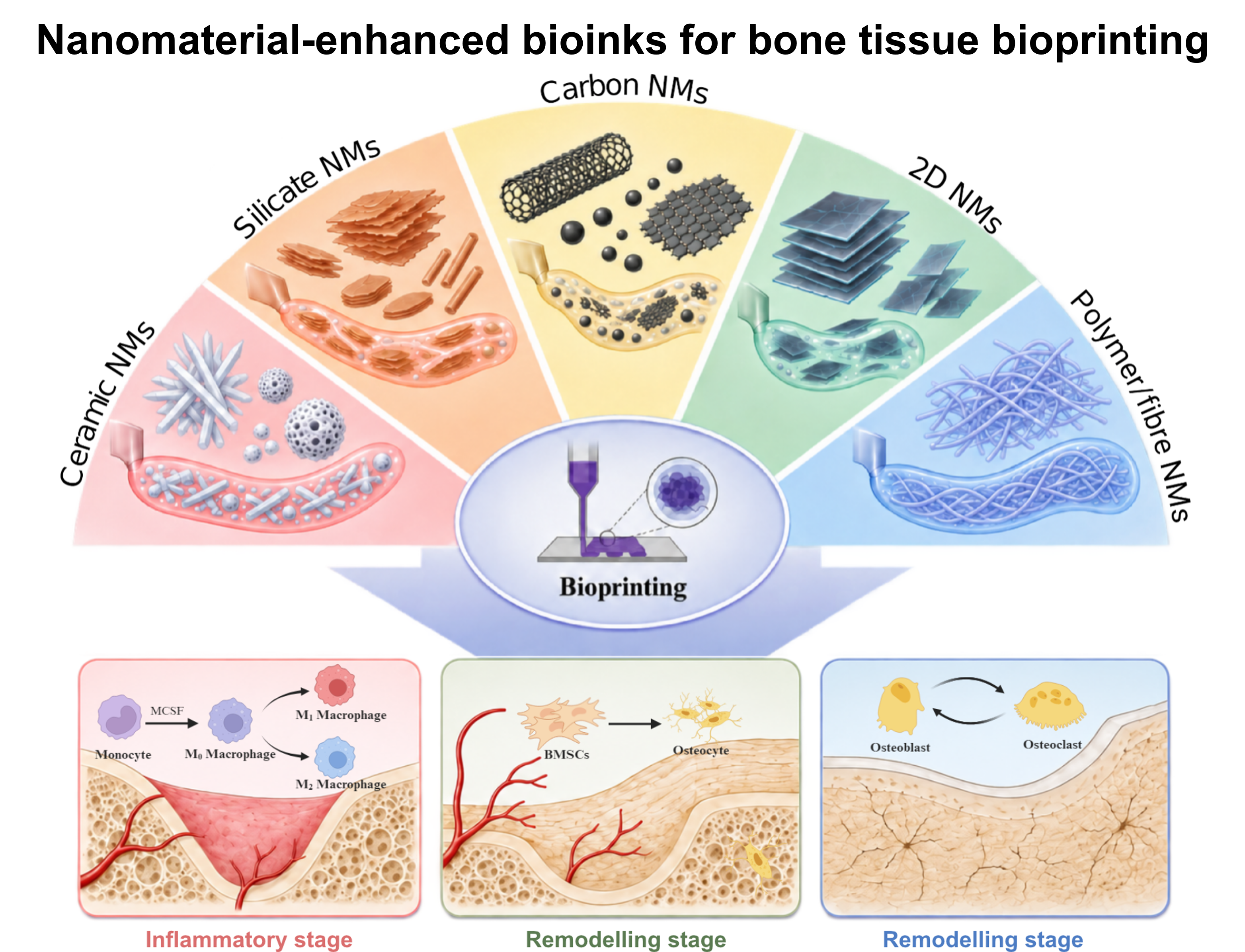
Three-dimensional bioprinting provides a promising strategy for constructing living bone substitutes with controlled architecture, cellular organisation, and regenerative function. However, conventional hydrogel bioinks often struggle to simultaneously achieve sufficient printability, structural stability, and biological activity for bone tissue formation. The incorporation of nanomaterials has therefore emerged as an effective approach to transform hydrogel bioinks into multifunctional composite systems capable of regulating both material properties and the cellular microenvironment. This review first outlines the dynamic process of bone repair, including inflammation, tissue formation, vascularisation, and remodelling, and discusses the requirements that these biological processes impose on bioink design. Major bioprinting technologies, including extrusion-based, inkjet, light-based, coaxial, and embedded bioprinting, are then summarised with particular attention to their compatibility with nanomaterial-containing bioinks. The review subsequently focuses on ceramic nanomaterials, silicates and nanoclays, carbon-based nanomaterials, two-dimensional nanomaterials, and polymeric or fibrous nanomaterials. Their roles in regulating rheological behaviour, print fidelity, mechanical stability, mineralisation, osteogenic differentiation, angiogenesis, and immunomodulation are critically discussed. Current evidence demonstrates that nanomaterial performance is strongly dependent on particle composition, concentration, size, morphology, surface chemistry, dispersion state, and interactions with the surrounding polymer network. Increasing nanomaterial content does not necessarily improve regenerative outcomes and may instead compromise extrusion, photocrosslinking, cellular activity, or long-term structural stability. Future development should therefore move beyond empirical nanoparticle incorporation towards coordinated optimisation of nanomaterial properties, bioink formulation, printing parameters, cellular composition, and degradation behaviour. Such an integrated design framework may enable the development of next-generation bioinks capable of more closely recapitulating the dynamic and multicellular microenvironment required for functional bone regeneration.
- de Carvalho ABG, Rahimnejad M, Oliveira R, et al. Personalized bioceramic grafts for craniomaxillofacial bone regeneration. Int J Oral Sci. 2024;16(1):62. doi: 10.1038/s41368-024-00327-7
- Lei C, Song J-h, Li S, et al. Advances in materials-based therapeutic strategies against osteoporosis. Biomaterials. 2023;296:122066. doi: 10.1016/j.biomaterials.2023.122066
- Migliorini F, La Padula G, Torsiello E, Spiezia F, Oliva F, Maffulli N. Strategies for large bone defect reconstruction after trauma, infections or tumour excision: a comprehensive review of the literature. Eur J Med Res. 2021;26(1):118. doi: 10.1186/s40001-021-00593-9
- Chen S, Wang M. Fabrication, Properties, and Applications of Scaffolds for Bone Tissue Regeneration. Adv Mater Technol. 2026;11(9):e01877. doi: 10.1002/admt.202501877
- Xie M, Shi Y, Zhang C, et al. In situ 3D bioprinting with bioconcrete bioink. Nat Commun. 2022;13(1):3597. doi: 10.1038/s41467-022-30997-y
- Qin Y, Jing Z, Zou D, et al. A metamaterial scaffold beyond modulus limits: enhanced osteogenesis and angiogenesis of critical bone defects. Nat Commun. 2025;16(1):2180. doi: 10.1038/s41467-025-57609-9
- Liu D, Liu J, Zhao P, et al. 3D Bioprinted Tissue-Engineered Bone with Enhanced Mechanical Strength and Bioactivities: Accelerating Bone Defect Repair through Sequential Immunomodulatory Properties. Adv Healthc Mater. 2024;13(30):e2401919. doi: 10.1002/adhm.202401919
- Wang J, Wu Y, Li G, et al. Engineering Large-Scale Self-Mineralizing Bone Organoids with Bone Matrix-Inspired Hydroxyapatite Hybrid Bioinks. Adv Mater. 2024;36(30):e2309875. doi: 10.1002/adma.202309875
- Zhu H, Monavari M, Zheng K, et al. 3D Bioprinting of Multifunctional Dynamic Nanocomposite Bioinks Incorporating Cu-Doped Mesoporous Bioactive Glass Nanoparticles for Bone Tissue Engineering. Small. 2022;18(12):e2104996. doi: 10.1002/smll.202104996
- Liu C, Dai T, Wu X, et al. 3D bioprinting of cell-laden nano-attapulgite/gelatin methacrylate composite hydrogel scaffolds for bone tissue repair. J Mater Sci Technol. 2023;135:111-125. doi: 10.1016/j.jmst.2022.07.011
- Kühl J, Krümpelmann SM, Hildebrandt L, et al. Nanomaterial-modified bioinks for DLP-based bioprinting of bone constructs: Impact on mechanical properties and mesenchymal stem cell function. Int J Bioprinting. 2024;10(6):4015. doi: 10.36922/ijb.4015
- Davern JW, Hipwood L, Bray LJ, Meinert C, Klein TJ. Addition of Laponite to gelatin methacryloyl bioinks improves the rheological properties and printability to create mechanically tailorable cell culture matrices. APL Bioeng. 2024;8(1):016101. doi: 10.1063/5.0166206
- Tavares MT, Gaspar VM, Monteiro MV, Farinha JPS, Baleizão C, Mano JF. GelMA/bioactive silica nanocomposite bioinks for stem cell osteogenic differentiation. Biofabrication. 2021;13(3):035012 doi: 10.1088/1758-5090/abdc86
- Chimene D, Miller L, Cross LM, Jaiswal MK, Singh I, Gaharwar AK. Nanoengineered Osteoinductive Bioink for 3D Bioprinting Bone Tissue. ACS Appl Mater Interfaces. 2020;12(14):15976-15988. doi: 10.1021/acsami.9b19037
- Liu B, Li J, Lei X, et al. 3D-bioprinted functional and biomimetic hydrogel scaffolds incorporated with nanosilicates to promote bone healing in rat calvarial defect model. Mater Sci Eng C Mater Biol Appl. 2020;112:110905. doi: 10.1016/j.msec.2020.110905
- Zhang J, Eyisoylu H, Qin XH, Rubert M, Muller R. 3D bioprinting of graphene oxide-incorporated cell-laden bone mimicking scaffolds for promoting scaffold fidelity, osteogenic differentiation and mineralization. Acta Biomater. 2021;121:637-652. doi: 10.1016/j.actbio.2020.12.026
- Lee SH, Kang MS, Jeon S, et al. 3D bioprinting of human mesenchymal stem cells-laden hydrogels incorporating MXene for spontaneous osteodifferentiation. Heliyon. 2023;9(3):e14490. doi: 10.1016/j.heliyon.2023.e14490
- Lafuente-Merchan M, Ruiz-Alonso S, Garcia-Villen F, et al. 3D Bioprinted Hydroxyapatite or Graphene Oxide Containing Nanocellulose-Based Scaffolds for Bone Regeneration. Macromol Biosci. 2022;22(11):e2200236. doi: 10.1002/mabi.202200236
- Montanari M, Korkeamaki JT, Campodoni E, et al. Effects of Magnesium-Doped Hydroxyapatite Nanoparticles on Bioink Formulation for Bone Tissue Engineering. ACS Appl Bio Mater. 2025;8(1):535-547. doi: 10.1021/acsabm.4c01418
- Chakraborty A, Roy A, Ravi SP, Paul A. Exploiting the role of nanoparticles for use in hydrogel-based bioprinting applications: concept, design, and recent advances. Biomater Sci. 2021;9(19):6337-6354. doi: 10.1039/d1bm00605c
- Cai Y, Chang SY, Gan SW, Ma S, Lu WF, Yen CC. Nanocomposite bioinks for 3D bioprinting. Acta Biomater. 2022;151:45-69. doi: 10.1016/j.actbio.2022.08.014
- Hull SM, Brunel LG, Heilshorn SC. 3D Bioprinting of Cell-Laden Hydrogels for Improved Biological Functionality. Adv Mater. 2022;34(2):e2103691. doi: 10.1002/adma.202103691
- Li N, Guo R, Zhang ZJ. Bioink Formulations for Bone Tissue Regeneration. Front Bioeng Biotechnol. 2021;9:630488. doi: 10.3389/fbioe.2021.630488
- Dang Y, Zhang Y, Luo G, et al. The decisive early phase of biomaterial-induced bone regeneration. Appl Mater Today. 2024;38:102236. doi: 10.1016/j.apmt.2024.102236
- Xu G, Chen J, Xiao Z, Xu J, Zhang L, Tang P. Bioaugmented osteosynthesis: precise monitoring and intervention of the bone healing microenvironment. Bone Res. 2025;13(1):95. doi: 10.1038/s41413-025-00466-5
- Molitoris KH, Huang M, Baht GS. Osteoimmunology of Fracture Healing. Curr Osteoporos Rep. 2024;22(3):330-339. doi: 10.1007/s11914-024-00869-z
- Newman H, Shih YV, Varghese S. Resolution of inflammation in bone regeneration: From understandings to therapeutic applications. Biomaterials. 2021;277:121114. doi: 10.1016/j.biomaterials.2021.121114
- Li C, Guo C, Fitzpatrick V, et al. Design of biodegradable, implantable devices towards clinical translation. Nat Rev Mater. 2019;5(1):61-81. doi: 10.1038/s41578-019-0150-z
- Burgan J, Rahmati M, Lee M, Saiz AM. Innate immune response to bone fracture healing. Bone. 2025;190:117327. doi: 10.1016/j.bone.2024.117327
- Yang L, Shi F, Cao F, et al. Neutrophils in Tissue Injury and Repair: Molecular Mechanisms and Therapeutic Targets. MedComm (2020). 2025;6(5):e70184. doi: 10.1002/mco2.70184
- Hachemi Y, Perrin S, Ethel M, et al. Multimodal analyses of immune cells during bone repair identify macrophages as a therapeutic target in musculoskeletal trauma. Bone Res. 2024;12(1):56. doi: 10.1038/s41413-024-00347-3
- Zou NY, Liu R, Huang M, et al. Age-related secretion of grancalcin by macrophages induces skeletal stem/progenitor cell senescence during fracture healing. Bone Res. 2024;12(1):6. doi: 10.1038/s41413-023-00309-1
- Yu X, Jiang S, Li D, Shen SGF, Wang X, Lin K. Osteoimmunomodulatory bioinks for 3D bioprinting achieve complete regeneration of critical-sized bone defects. Composites Part B: Engineering. 2024;273:111256. doi: 10.1016/j.compositesb.2024.111256
- Dutta SD, Ganguly K, Patil TV, Randhawa A, Lim KT. Unraveling the potential of 3D bioprinted immunomodulatory materials for regulating macrophage polarization: State-of-the-art in bone and associated tissue regeneration. Bioact Mater. 2023;28:284-310. doi: 10.1016/j.bioactmat.2023.05.014
- Xu Z, Qi X, Bao M, et al. Biomineralization inspired 3D printed bioactive glass nanocomposite scaffolds orchestrate diabetic bone regeneration by remodeling micromilieu. Bioact Mater. 2023;25:239-255. doi: 10.1016/j.bioactmat.2023.01.024
- Kuwahara ST, Liu S, Chareunsouk A, Serowoky M, Mariani FV. On the horizon: Hedgehog signaling to heal broken bones. Bone Res. 2022;10(1):13. doi: 10.1038/s41413-021-00184-8
- Jeffery EC, Mann TLA, Pool JA, Zhao Z, Morrison SJ. Bone marrow and periosteal skeletal stem/progenitor cells make distinct contributions to bone maintenance and repair. Cell Stem Cell. 2022;29(11):1547-1561 e6. doi: 10.1016/j.stem.2022.10.002
- Liu YL, Tang XT, Shu HS, Zou W, Zhou BO. Fibrous periosteum repairs bone fracture and maintains the healed bone throughout mouse adulthood. Dev Cell. 2024;59(9):1192-1209 e6. doi: 10.1016/j.devcel.2024.03.019
- Julien A, Kanagalingam A, Martinez-Sarra E, et al. Direct contribution of skeletal muscle mesenchymal progenitors to bone repair. Nat Commun. 2021;12(1):2860. doi: 10.1038/s41467-021-22842-5
- Sivaraj KK, Majev PG, Jeong HW, et al. Mesenchymal stromal cell-derived septoclasts resorb cartilage during developmental ossification and fracture healing. Nat Commun. 2022;13(1):571. doi: 10.1038/s41467-022-28142-w
- Bixel MG, Sivaraj KK, Timmen M, et al. Angiogenesis is uncoupled from osteogenesis during calvarial bone regeneration. Nat Commun. 2024;15(1):4575. doi: 10.1038/s41467-024-48579-5
- Zhai X, Ruan C, Ma Y, et al. 3D-Bioprinted Osteoblast-Laden Nanocomposite Hydrogel Constructs with Induced Microenvironments Promote Cell Viability, Differentiation, and Osteogenesis both In Vitro and In Vivo. Adv Sci (Weinh). 2018;5(3):1700550. doi: 10.1002/advs.201700550
- Cidonio G, Glinka M, Kim Y-H, et al. Nanoclay-based 3D printed scaffolds promote vascular ingrowth ex vivo and generate bone mineral tissue in vitro and in vivo. Biofabrication. 2020;12(3):035010. doi: 10.1088/1758-5090/ab8753
- Kim YH, Kanczler JM, Lanham S, et al. Biofabrication of nanocomposite-based scaffolds containing human bone extracellular matrix for the differentiation of skeletal stem and progenitor cells. Biodes Manuf. 2024;7(2):121-136. doi: 10.1007/s42242-023-00265-z
- Del Toro Runzer C, Balmayor ER, van Griensven M. Biologics for bone regeneration: advances in cell, protein, gene, and mRNA therapies. Bone Res. 2026;14(1):5. doi: 10.1038/s41413-025-00487-0
- Ma Q, Liang M, Wu Y, et al. Osteoclast-derived apoptotic bodies couple bone resorption and formation in bone remodeling. Bone Res. 2021;9(1):5. doi: 10.1038/s41413-020-00121-1
- Sims NA. Osteoclast-derived coupling factors: origins and state-of-play Louis V Avioli lecture, ASBMR 2023. J Bone Miner Res. 2024;39(10):1377-1385. doi: 10.1093/jbmr/zjae110
- Uenaka M, Yamashita E, Kikuta J, et al. Osteoblast-derived vesicles induce a switch from bone-formation to bone-resorption in vivo. Nat Commun. 2022;13(1):1066. doi: 10.1038/s41467-022-28673-2
- Fischetti T, Borciani G, Avnet S, et al. Incorporation/Enrichment of 3D Bioprinted Constructs by Biomimetic Nanoparticles: Tuning Printability and Cell Behavior in Bone Models. Nanomaterials (Basel). 2023;13(14):2040. doi: 10.3390/nano13142040
- Zhao Z, Chen X, Chen H, et al. Cerium-Containing Mesoporous Bioactive Glass Nanoparticles Reinforced 3D-Printed Bioceramic Scaffolds toward Enhanced Mechanical, Antioxidant, and Osteogenic Activities for Bone Regeneration. Adv Healthc Mater. 2025;14(9):e2404346. doi: 10.1002/adhm.202404346
- Zhang M, Zhai X, Ma T, et al. Sequential Therapy for Bone Regeneration by Cerium Oxide-Reinforced 3D-Printed Bioactive Glass Scaffolds. ACS Nano. 2023;17(5):4433-4444. doi: 10.1021/acsnano.2c09855
- Li X, Liu B, Pei B, et al. Inkjet Bioprinting of Biomaterials. Chem Rev. 2020;120(19):10793-10833. doi: 10.1021/acs.chemrev.0c00008
- Mota C, Camarero-Espinosa S, Baker MB, Wieringa P, Moroni L. Bioprinting: From Tissue and Organ Development to in Vitro Models. Chem Rev. 2020;120(19):10547-10607. doi: 10.1021/acs.chemrev.9b00789
- Leucht A, Volz AC, Rogal J, Borchers K, Kluger PJ. Advanced gelatin-based vascularization bioinks for extrusion-based bioprinting of vascularized bone equivalents. Sci Rep. 2020;10(1):5330. doi: 10.1038/s41598-020-62166-w
- Oh D, Shirzad M, Chang Kim M, Chung E-J, Nam SY. Rheology-informed hierarchical machine learning model for the prediction of printing resolution in extrusion-based bioprinting. Int J Bioprinting. 2023;9(6):1280. doi: 10.36922/ijb.1280
- Grijalva Garces D, Strauss S, Gretzinger S, et al. On the reproducibility of extrusion-based bioprinting: round robin study on standardization in the field. Biofabrication. 2023;16(1):015002. doi: 10.1088/1758-5090/acfe3b
- Zhang J, Wehrle E, Adamek P, et al. Optimization of mechanical stiffness and cell density of 3D bioprinted cell-laden scaffolds improves extracellular matrix mineralization and cellular organization for bone tissue engineering. Acta Biomater. 2020;114:307-322. doi: 10.1016/j.actbio.2020.07.016
- Zhang C, Elvitigala KCML, Mubarok W, Okano Y, Sakai S. Machine learning-based prediction and optimisation framework for as-extruded cell viability in extrusion-based 3D bioprinting. Virtual Phys Prototyp. 2024;19(1):e2400330. doi: 10.1080/17452759.2024.2400330
- Ma W, Lu H, Xiao Y, Wu C. Advancing organoid development with 3D bioprinting. Organoid Res. 2025;1(1):25040004. doi: 10.36922/or025040004
- Shen M, Wang L, Gao Y, et al. 3D bioprinting of in situ vascularized tissue engineered bone for repairing large segmental bone defects. Mater Today Bio. 2022;16:100382. doi: 10.1016/j.mtbio.2022.100382
- Sun X, Jiao X, Yang X, et al. 3D bioprinting of osteon-mimetic scaffolds with hierarchical microchannels for vascularized bone tissue regeneration. Biofabrication. 2022;14(3):035008. doi: 10.1088/1758-5090/ac6700
- Pitacco P, Sadowska JM, O'Brien FJ, Kelly DJ. 3D bioprinting of cartilaginous templates for large bone defect healing. Acta Biomater. 2023;156:61-74. doi: 10.1016/j.actbio.2022.07.037
- Wei Q, Peng Y, Chen W, et al. Three-dimensional bioprinting for musculoskeletal regeneration and disease modeling. Int J Bioprinting. 2024;10(1):1037. doi: 10.36922/ijb.1037
- Fan J, Ding Z, Cai Y, et al. Revolutionizing Bone Regeneration: Vascularized Bone Tissue Engineering with Advanced 3D Printing Technology. Aggregate. 2025;6(4):e731. doi: 10.1002/agt2.731
- Shyam Mohan T, Datta P, Nesaei S, Ozbolat V, Ozbolat IT. 3D Coaxial Bioprinting: Process Mechanisms, Bioinks and Applications. Prog Biomed Eng (Bristol). 2022;4(2):022003. doi: 10.1088/2516-1091/ac631c
- Shahabipour F, Tavafoghi M, Aninwene GE II, et al. Coaxial 3D bioprinting of tri-polymer scaffolds to improve the osteogenic and vasculogenic potential of cells in co-culture models. J Biomed Mater Res A. 2022;110(5):1077-1089. doi: 10.1002/jbm.a.37354
- Ghahri T, Salehi Z, Aghajanpour S, et al. Development of osteon-like scaffold-cell construct by quadruple coaxial extrusion-based 3D bioprinting of nanocomposite hydrogel. Biomater Adv. 2023;145:213254. doi: 10.1016/j.bioadv.2022.213254
- Zhang J, Suttapreyasri S, Leethanakul C, Samruajbenjakun B. Fabrication of vascularized tissue-engineered bone models using triaxial bioprinting. J Biomed Mater Res A. 2024;112(7):1093-1106. doi: 10.1002/jbm.a.37694
- Zhou K, Sun Y, Yang J, Mao H, Gu Z. Hydrogels for 3D embedded bioprinting: a focused review on bioinks and support baths. J Mater Chem B. 2022;10(12):1897-1907. doi: 10.1039/d1tb02554f
- Jalandhra GK, Molley TG, Hung TT, Roohani I, Kilian KA. In situ formation of osteochondral interfaces through "bone-ink" printing in tailored microgel suspensions. Acta Biomater. 2023;156:75-87. doi: 10.1016/j.actbio.2022.08.052
- Guo C, Wu J, Zeng Y, Li H. Construction of 3D bioprinting of HAP/collagen scaffold in gelation bath for bone tissue engineering. Regen Biomater. 2023;10:rbad067. doi: 10.1093/rb/rbad067
- Abaci A, Guvendiren M. 3D bioprinting of dense cellular structures within hydrogels with spatially controlled heterogeneity. Biofabrication. 2024;16(3):035027. doi: 10.1088/1758-5090/ad52f1
- Truong H, Abaci A, Gharacheh H, Guvendiren M. Embedded bioprinting of dense cellular constructs in bone allograft-enhanced hydrogel matrices for bone tissue engineering. Biomater Sci. 2025;13(12):3213-3222. doi: 10.1039/d4bm01616e
- Zhang H, Luo Y, Hu Z, et al. Cation-crosslinked κ-carrageenan sub-microgel medium for high-quality embedded bioprinting. Biofabrication. 2024;16(2):025009. doi: 10.1088/1758-5090/ad1cf3
- Fang Y, Guo Y, Wu B, et al. Expanding Embedded 3D Bioprinting Capability for Engineering Complex Organs with Freeform Vascular Networks. Adv Mater. 2023;35(22):e2205082. doi: 10.1002/adma.202205082
- Suntornnond R, Ng WL, Huang X, Yeow CHE, Yeong WY. Improving printability of hydrogel-based bio-inks for thermal inkjet bioprinting applications via saponification and heat treatment processes. J Mater Chem B. 2022;10(31):5989-6000. doi: 10.1039/d2tb00442a
- Zhu H, Li R, Li S, et al. Multi-physical field control piezoelectric inkjet bioprinting for 3D tissue-like structure manufacturing. Int J Bioprinting. 2024;10(3):2120. doi: 10.36922/ijb.2120
- Gao G, Schilling AF, Yonezawa T, Wang J, Dai G, Cui X. Bioactive nanoparticles stimulate bone tissue formation in bioprinted three-dimensional scaffold and human mesenchymal stem cells. Biotechnol J. 2014;9(10):1304-11. doi: 10.1002/biot.201400305
- Gao G, Schilling AF, Hubbell K, et al. Improved properties of bone and cartilage tissue from 3D inkjet-bioprinted human mesenchymal stem cells by simultaneous deposition and photocrosslinking in PEG-GelMA. Biotechnol Lett. 2015;37(11):2349-55. doi: 10.1007/s10529-015-1921-2
- Ablanedo Morales P, Rodriguez B, Furth ME, et al. Thermal inkjet bioprinting drastically alters cell phenotype. Biofabrication. 2023;15(3):031001. doi: 10.1088/1758-5090/acd3b3
- Keriquel V, Oliveira H, Remy M, et al. In situ printing of mesenchymal stromal cells, by laser-assisted bioprinting, for in vivo bone regeneration applications. Sci Rep. 2017;7(1):1778. doi: 10.1038/s41598-017-01914-x
- Ng WL, Bartolo P. 3D bioprinting of tissues and organs for systemic diseases and localized injuries. Mil Med Res. 2026;13(1):100006. doi: 10.1016/j.mmr.2026.100006
- Wang Z, Kumar H, Tian Z, et al. Visible Light Photoinitiation of Cell-Adhesive Gelatin Methacryloyl Hydrogels for Stereolithography 3D Bioprinting. ACS Appl Mater Interfaces. 2018;10(32):26859-26869. doi: 10.1021/acsami.8b06607
- Haghpanah Z, Mondal D, Momenbeitollahi N, et al. In vitro evaluation of bone cell response to novel 3D-printable nanocomposite biomaterials for bone reconstruction. J Biomed Mater Res A. 2024;112(10):1725-1739. doi: 10.1002/jbm.a.37719
- Tao J, Zhu S, Liao X, et al. DLP-based bioprinting of void-forming hydrogels for enhanced stem-cell-mediated bone regeneration. Mater Today Bio. 2022;17:100487. doi: 10.1016/j.mtbio.2022.100487
- Ren X, Wang J, Wu Y, et al. One-pot synthesis of hydroxyapatite hybrid bioinks for digital light processing 3D printing in bone regeneration. J Mater Sci Technol. 2024;188:84-97. doi: 10.1016/j.jmst.2024.01.001
- Hospodiuk M, Dey M, Sosnoski D, Ozbolat IT. The bioink: A comprehensive review on bioprintable materials. Biotechnol Adv. 2017;35(2):217-239. doi: 10.1016/j.biotechadv.2016.12.006
- Gupta T, Ghosh SB, Bandyopadhyay-Ghosh S, Sain M. Is it possible to 3D bioprint load-bearing bone implants? A critical review. Biofabrication. 2023;15(4):042003. doi: 10.1088/1758-5090/acf6e1
- Badhe RV, Chatterjee A, Bijukumar D, Mathew MT. Current advancements in bio-ink technology for cartilage and bone tissue engineering. Bone. 2023;171:116746. doi: 10.1016/j.bone.2023.116746
- Chiticaru EA, Ionita M. Commercially available bioinks and state-of-the-art lab-made formulations for bone tissue engineering: A comprehensive review. Mater Today Bio. 2024;29:101341. doi: 10.1016/j.mtbio.2024.101341
- Rasouli R, Sweeney C, Frampton JP. Heterogeneous and Composite Bioinks for 3D-Bioprinting of Complex Tissue. Biomed Mater Devices. 2025;3(1):108-126. doi: 10.1007/s44174-024-00171-7
- Hoppe A, Guldal NS, Boccaccini AR. A review of the biological response to ionic dissolution products from bioactive glasses and glass-ceramics. Biomaterials. 2011;32(11):2757-74. doi: 10.1016/j.biomaterials.2011.01.004
- Li Z, Xiang S, Lin Z, et al. Graphene oxide-functionalized nanocomposites promote osteogenesis of human mesenchymal stem cells via enhancement of BMP-SMAD1/5 signaling pathway. Biomaterials. 2021;277:121082. doi: 10.1016/j.biomaterials.2021.121082
- Niknam Z, Hosseinzadeh F, Shams F, et al. Recent advances and challenges in graphene-based nanocomposite scaffolds for tissue engineering application. J Biomed Mater Res A. 2022;110(10):1695-1721. doi: 10.1002/jbm.a.37417
- Li C, Sun F, Tian J, et al. Continuously released Zn(2+) in 3D-printed PLGA/beta-TCP/Zn scaffolds for bone defect repair by improving osteoinductive and anti-inflammatory properties. Bioact Mater. 2023;24:361-375. doi: 10.1016/j.bioactmat.2022.12.015
- Nitschke BM, Beltran FO, Hahn MS, Grunlan MA. Trends in bioactivity: inducing and detecting mineralization of regenerative polymeric scaffolds. J Mater Chem B. 2024;12(11):2720-2736. doi: 10.1039/d3tb02674d
- Wang Z, Sun Y, Li C. Advances in 3D printing technology for preparing bone tissue engineering scaffolds from biodegradable materials. Front Bioeng Biotechnol. 2024;12:1483547. doi: 10.3389/fbioe.2024.1483547
- Bharadwaj T, Chrungoo S, Verma D. Self-assembled chitosan/gelatin nanofibrous aggregates incorporated thermosensitive nanocomposite bioink for bone tissue engineering. Carbohydr Polym. 2024;324:121544. doi: 10.1016/j.carbpol.2023.121544
- Zhu Y, Yu X, Liu H, et al. Strategies of functionalized GelMA-based bioinks for bone regeneration: Recent advances and future perspectives. Bioact Mater. 2024;38:346-373. doi: 10.1016/j.bioactmat.2024.04.032
- Korkeamäki JT, Silen R, Syverud K, Mustafa K, Rashad A, Miettinen S. Optimizing gelatin methacryloyl-based bioinks with nanohydroxyapatite for 3D bioprinting in bone tissue engineering. Bioprinting. 2026;56:e00479. doi: 10.1016/j.bprint.2026.e00479
- Kang Y, Xu J, Meng L, et al. 3D bioprinting of dECM/Gel/QCS/nHAp hybrid scaffolds laden with mesenchymal stem cell-derived exosomes to improve angiogenesis and osteogenesis. Biofabrication. 2023;15(2):024103. doi: 10.1088/1758-5090/acb6b8
- Coşkun S, Akbulut SO, Sarikaya B, Cakmak S, Gumusderelioglu M. Formulation of chitosan and chitosan-nanoHAp bioinks and investigation of printability with optimized bioprinting parameters. Int J Biol Macromol. 2022;222(Pt A):1453-1464. doi: 10.1016/j.ijbiomac.2022.09.078
- Korkeamäki JT, Rashad A, Ojansivu M, et al. Systematic development and bioprinting of novel nanostructured multi-material bioinks for bone tissue engineering. Biofabrication. 2025;17(2):025005. doi: 10.1088/1758-5090/ada63b
- Allen NB, Abar B, Johnson L, Burbano J, Danilkowicz RM, Adams SB. 3D-bioprinted GelMA-gelatin-hydroxyapatite osteoblast-laden composite hydrogels for bone tissue engineering. Bioprinting. 2022;26:e00196. doi: 10.1016/j.bprint.2022.e00196
- Bhattacharyya A, Janarthanan G, Kim T, et al. Modulation of bioactive calcium phosphate micro/nanoparticle size and shape during in situ synthesis of photo-crosslinkable gelatin methacryloyl based nanocomposite hydrogels for 3D bioprinting and tissue engineering. Biomater Res. 2022;26(1):54. doi: 10.1186/s40824-022-00301-6
- Indurkar A, Heid S, Bauer J, et al. Amorphous calcium phosphate reinforced alginate-dialdehyde-gelatin (ADA-GEL) bioink for biofabrication of bone tissue scaffolds. Carbohydr Polym Technol Appl. 2025;9:100710. doi: 10.1016/j.carpta.2025.100710
- Wu YF, Wen YT, Salamanca E, et al. 3D-bioprinted alginate-based bioink scaffolds with beta-tricalcium phosphate for bone regeneration applications. J Dent Sci. 2024;19(2):1116-1125. doi: 10.1016/j.jds.2023.12.023
- Bhattacharyya A, Khatun MR, Narmatha S, Nagarajan R, Noh I. Modulation of 3D Bioprintability in Polysaccharide Bioink by Bioglass Nanoparticles and Multiple Metal Ions for Tissue Engineering. Tissue Eng Regen Med. 2024;21(2):261-275. doi: 10.1007/s13770-023-00605-1
- Sánchez-Rodríguez VH, Pérez-Cortez JE, Gallegos-Martínez S, et al. Light-based and cost-effective bioprinting of musculoskeletal GelMA constructs enriched with mesoporous bioactive glass nanoparticles. Int J Bioprinting. 2024;10(4):1830. doi: 10.36922/ijb.1830
- Kolan KCR, Saxena A, Bromet BA, et al. Bioprinting with adipose stem cells and hydrogel modified with bioactive glass. Int J Bioprinting. 2024;10(2):2057. doi: 10.36922/ijb.2057
- Kiaee G, Dimitrakakis N, Sharifzadeh S, et al. Laponite-Based Nanomaterials for Drug Delivery. Adv Healthc Mater. 2022;11(7):e2102054. doi: 10.1002/adhm.202102054
- Kim J, Choi HS, Kim YM, Song SC. Thermo-Responsive Nanocomposite Bioink with Growth-Factor Holding and its Application to Bone Regeneration. Small. 2023;19(9):e2203464. doi: 10.1002/smll.202203464
- Chandra DK, Reis RL, Kundu SC, Kumar A, Mahapatra C. Nanomaterials-Based Hybrid Bioink Platforms in Advancing 3D Bioprinting Technologies for Regenerative Medicine. ACS Biomater Sci Eng. 2024;10(7):4145-4174. doi: 10.1021/acsbiomaterials.4c00166
- Zhang H, Qin C, Zhang M, et al. Calcium silicate nanowires-containing multicellular bioinks for 3D bioprinting of neural-bone constructs. Nano Today. 2022;46:101584. doi: 10.1016/j.nantod.2022.101584
- Li W, Miao W, Liu Y, et al. Bioprinted Constructs that Mimic the Ossification Center Microenvironment for Targeted Innervation in Bone Regeneration. Adv Funct Mater. 2021;32(9):2109871. doi: 10.1002/adfm.202109871
- Yang J, Chen Z, Gao C, et al. A mechanical-assisted post-bioprinting strategy for challenging bone defects repair. Nat Commun. 2024;15(1):3565. doi: 10.1038/s41467-024-48023-8
- Liu C, Ni S, Wu X, et al. Strontium-Modified porous attapulgite composite hydrogel scaffold with advanced angiogenic and osteogenic potential for bone defect repair. Compos Part A-appl S. 2024;187:108492. doi: 10.1016/j.compositesa.2024.108492
- Liu C, Qin W, Wang Y, et al. 3D Printed Gelatin/Sodium Alginate Hydrogel Scaffolds Doped with Nano-Attapulgite for Bone Tissue Repair. Int J Nanomedicine. 2021;16:8417-8432. doi: 10.2147/IJN.S339500
- Zorron M, Cabrera AL, Sharma R, et al. Emerging 2D Nanomaterials-Integrated Hydrogels: Advancements in Designing Theragenerative Materials for Bone Regeneration and Disease Therapy. Adv Sci (Weinh). 2024;11(31):e2403204. doi: 10.1002/advs.202403204
- Das A, Roy M, Saha M. Recent advances in biomedical applications of carbon and graphene quantum dots: A review. Biotechnol Bioeng. 2024;121(5):1469-1485. doi: 10.1002/bit.28700
- Dutta SD, Ganguly K, Hexiu J, Randhawa A, Moniruzzaman M, Lim KT. A 3D Bioprinted Nanoengineered Hydrogel with Photoactivated Drug Delivery for Tumor Apoptosis and Simultaneous Bone Regeneration via Macrophage Immunomodulation. Macromol Biosci. 2023;23(9):e2300096. doi: 10.1002/mabi.202300096
- Jiang Y, Zhou D, Yang B. 3D bioprinted GelMA/GO composite induces osteoblastic differentiation. J Biomater Appl. 2022;37(3):527-537. doi: 10.1177/08853282221098235
- Bu W, Xu X, Wang Z, et al. Ascorbic Acid-PEI Carbon Dots with Osteogenic Effects as miR-2861 Carriers to Effectively Enhance Bone Regeneration. ACS Appl Mater Interfaces. 2020;12(45):50287-50302. doi: 10.1021/acsami.0c15425
- Geng B, Li P, Fang F, et al. Antibacterial and osteogenic carbon quantum dots for regeneration of bone defects infected with multidrug-resistant bacteria. Carbon. 2021;184:375-385. doi: 10.1016/j.carbon.2021.08.040
- Tang J, Hu J, Bai X, et al. Near-Infrared Carbon Dots With Antibacterial and Osteogenic Activities for Sonodynamic Therapy of Infected Bone Defects. Small. 2024;20(49):e2404900. doi: 10.1002/smll.202404900
- Wang Y, Li J, Li Y, Yang B. Biomimetic bioinks of nanofibrillar polymeric hydrogels for 3D bioprinting. Nano Today. 2021;39:101180. doi: 10.1016/j.nantod.2021.101180
- Nelson ML, Burgoyne H, Kouchi FR, et al. Graphene and Ti(3)C(2)T(x) MXene Nanomaterial-Infused Bioinks for Mechanical and Electrical Improvement of 3D Bioprinted Scaffolds. ACS Appl Bio Mater. 2026;9(2):1142-1155. doi: 10.1021/acsabm.5c02206
- Liu X, Miller AL II, Park S, et al. Two-Dimensional Black Phosphorus and Graphene Oxide Nanosheets Synergistically Enhance Cell Proliferation and Osteogenesis on 3D Printed Scaffolds. ACS Appl Mater Interfaces. 2019;11(26):23558-23572. doi: 10.1021/acsami.9b04121
- Nie R, Sun Y, Lv H, et al. 3D printing of MXene composite hydrogel scaffolds for photothermal antibacterial activity and bone regeneration in infected bone defect models. Nanoscale. 2022;14(22):8112-8129. doi: 10.1039/d2nr02176e
- Miao Y, Chen Y, Luo J, et al. Black phosphorus nanosheets-enabled DNA hydrogel integrating 3D-printed scaffold for promoting vascularized bone regeneration. Bioact Mater. 2023;21:97-109. doi: 10.1016/j.bioactmat.2022.08.005
- Xu Y, Xu C, He L, et al. Stratified-structural hydrogel incorporated with magnesium-ion-modified black phosphorus nanosheets for promoting neuro-vascularized bone regeneration. Bioact Mater. 2022;16:271-284. doi: 10.1016/j.bioactmat.2022.02.024
- Ojansivu M, Rashad A, Ahlinder A, et al. Wood-based nanocellulose and bioactive glass modified gelatin-alginate bioinks for 3D bioprinting of bone cells. Biofabrication. 2019;11(3):035010. doi: 10.1088/1758-5090/ab0692
- Bedell ML, Torres AL, Hogan KJ, et al. Human gelatin-based composite hydrogels for osteochondral tissue engineering and their adaptation into bioinks for extrusion, inkjet, and digital light processing bioprinting. Biofabrication. 2022;14(4):045012. doi: 10.1088/1758-5090/ac8768
- Li N, Cui J, Chi M, Thieringer FM, Sharma N. Building a better bone: The synergy of 2D nanomaterials and 3D printing for bone tissue engineering. Materials & Design. 2023;234:112362. doi: 10.1016/j.matdes.2023.112362
- Xu W, Molino BZ, Cheng F, et al. On Low-Concentration Inks Formulated by Nanocellulose Assisted with Gelatin Methacrylate (GelMA) for 3D Printing toward Wound Healing Application. ACS Appl Mater Interfaces. 2019;11(9):8838-8848. doi: 10.1021/acsami.8b21268
- Dar MA, Xie R, Liu J, et al. Current Paradigms and Future Challenges in Harnessing Nanocellulose for Advanced Applications in Tissue Engineering: A Critical State-of-the-Art Review for Biomedicine. Int J Mol Sci. 2025;26(4):1449. doi: 10.3390/ijms26041449
- Fadeel B, Farcal L, Hardy B, et al. Advanced tools for the safety assessment of nanomaterials. Nat Nanotechnol. 2018;13(7):537-543. doi: 10.1038/s41565-018-0185-0
- Liu JH, Yang ST, Wang H, Chang Y, Cao A, Liu Y. Effect of size and dose on the biodistribution of graphene oxide in mice. Nanomedicine (Lond). 2012;7(12):1801-12. doi: 10.2217/nnm.12.60
- Su Z, Chen W, Liang S, et al. Multifaceted Characterization for the Hepatic Clearance of Graphene Oxide and Size-Related Hepatic Toxicity. Molecules. 2024;29(6):1335. doi: 10.3390/molecules29061335
- Ou L, Song B, Liang H, et al. Toxicity of graphene-family nanoparticles: a general review of the origins and mechanisms. Part Fibre Toxicol. 2016;13(1):57. doi: 10.1186/s12989-016-0168-y
- Sui B, Liu X, Sun J. Biodistribution, inter-/intra-cellular localization and respiratory dysfunction induced by Ti(3)C(2) nanosheets: Involvement of surfactant protein down-regulation in alveolar epithelial cells. J Hazard Mater. 2021;402:123562. doi: 10.1016/j.jhazmat.2020.123562
- Li Y, Jiao H, Zhang H, et al. Biosafety consideration of nanocellulose in biomedical applications: A review. Int J Biol Macromol. 2024;265(Pt 1):130900. doi: 10.1016/j.ijbiomac.2024.130900
- Agarwal T, Onesto V, Banerjee D, et al. 3D bioprinting in tissue engineering: current state-of-the-art and challenges towards system standardization and clinical translation. Biofabrication. 2025;17(4):042003. doi: 10.1088/1758-5090/ade47a
- Perin F, Ouyang L, Lim KS, et al. Bioprinted Constructs in the Regulatory Landscape: Current State and Future Perspectives. Adv Mater. 2026;38(4):e04037. doi: 10.1002/adma.202504037
- Theus AS, Ning L, Jin L, Roeder RK, Zhang J, Serpooshan V. Nanomaterials for bioprinting: functionalization of tissue-specific bioinks. Essays Biochem. 2021;65(3):429-439. doi: 10.1042/EBC20200095
- Stanco D, Urban P, Tirendi S, Ciardelli G, Barrero J. 3D bioprinting for orthopaedic applications: Current advances, challenges and regulatory considerations. Bioprinting. 2020;20:None. doi: 10.1016/j.bprint.2020.e00103
- Meng X, Ziadlou R, Grad S, et al. Animal Models of Osteochondral Defect for Testing Biomaterials. Biochem Res Int. 2020;2020:9659412. doi: 10.1155/2020/9659412
- Li Y, Chen SK, Li L, Qin L, Wang XL, Lai YX. Bone defect animal models for testing efficacy of bone substitute biomaterials. J Orthop Translat. 2015;3(3):95-104. doi: 10.1016/j.jot.2015.05.002