

Three-dimensional bioprinting of biomimetic bilayer hydrogels with engineered mechanical and cellular heterogeneity for wound healing

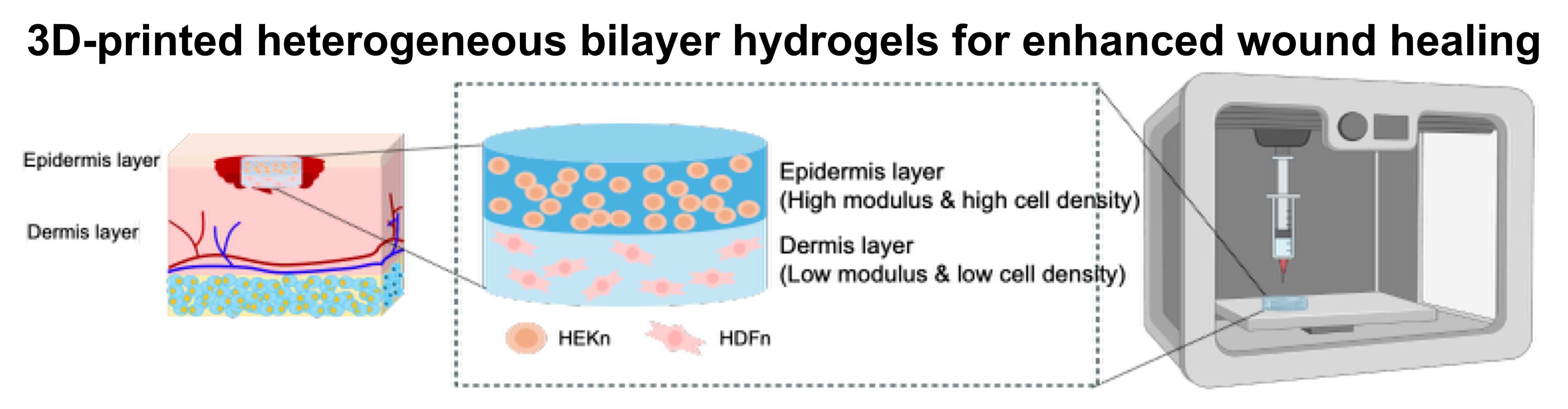
In view of the global burden of wound care on healthcare systems, there is an urgent need for advanced therapeutic solutions to promote wound healing. Although conventional bilayer dressings contain layers with predefined mechanical properties, their mechanical architecture is static and may be vulnerable to disruption during deformation. To address this limitation, we present a self-healing biomimetic bilayer hydrogel system that utilizes hyaluronic acid (HA) derivatives to emulate the mechanical and cellular heterogeneity of native skin tissue. The hydrogels were prepared by synthesizing carbodihydrazide-conjugated HA (HA-CDH) and crosslinking it with oxidized diol-modified HA in the presence of adipic acid dihydrazide. By adjusting the HA-CDH concentration, the bilayer hydrogel stiffness was optimized to effectively replicate the distinct mechanical properties of the epidermis and dermis. This bilayer structure with cell-specific stiffness and distinct cell types promoted cell-specific proliferation and upregulated key wound-healing markers, including genes encoding collagen type I and α-smooth muscle actin. Furthermore, the hydrogel exhibited excellent shear-thinning and dynamic self-healing properties, ensuring high shape fidelity during extrusion-based three-dimensional bioprinting of customized wound patches. In a full-thickness mouse wound model, these cell-laden bilayer patches promoted wound repair by significantly suppressing initial inflammatory responses, enhancing neovascularization, and facilitating balanced extracellular matrix remodeling without fibrotic thickening. The proposed self-healing bilayer hydrogel system stands as a promising, structurally dynamic solution for effective wound healing and has potential for further applications in tissue engineering.
- Chin SW, Azman AS, Tan JW. Incorporation of natural and synthetic polymers into honey hydrogel for wound healing: a review. Health Sci Rep. 2024;7(7):e2251. doi: 10.1002/hsr2.2251
- Sen CK. Human wound and its burden: updated 2022 compendium of estimates. Adv Wound Care (New Rochelle). 2023;12(12):657-670. doi: 10.1089/wound.2023.0150
- Davis M, Baird D, Hill D, Layher H, Akin R. Management of full-thickness skin grafts. Baylor Univ Med Cent Proc. 2021;34(6):683-686. doi: 10.1080/08998280.2021.1953867
- Zoio P, Ventura S, Leite M, Oliva A. Pigmented full-thickness human skin model based on a fibroblast-derived matrix for long-term studies. Tissue Eng Part C Methods. 2021;27(7):433-443. doi: 10.1089/ten.tec.2021.0069
- Chang DK, Louis MR, Gimenez A, Reece EM. The basics of integra dermal regeneration template and its expanding clinical applications. Semin Plast Surg. 2019;33(3):185-189. doi: 10.1055/s-0039-1693401
- Keirouz A, Chung M, Kwon J, Fortunato G, Radacsi N. 2D and 3D electrospinning technologies for the fabrication of nanofibrous scaffolds for skin tissue engineering: a review. Wiley Interdiscip Rev Nanomed Nanobiotechnol. 2020;12(4):e1626. doi: 10.1002/wnan.1626
- Ganier C, Rognoni E, Goss G, Lynch M, Watt FM. Fibroblast heterogeneity in healthy and wounded skin. Cold Spring Harb Perspect Biol. 2022;14(6):a041238. doi: 10.1101/cshperspect.a041238
- Kirsanova LV, Araviyskaya ER, Rybakova MG, Sokolovsky EV, Bogatenkov AI. Dermal fibroblasts in the focus of modern cosmetology: aging and response to cosmetic procedures: a review. Consilium Medicum. 2024;26(8):541-549. doi: 10.26442/20751753.2024.8.202913
- Bensa T, Tekkela S, Rognoni E. Skin fibroblast functional heterogeneity in health and disease. J Pathol. 2023;260(5):609-620. doi: 10.1002/path.6159
- Wei Q, Young J, Holle A, et al. Soft hydrogels for balancing cell proliferation and differentiation. ACS Biomater Sci Eng. 2020;6(8):4687-4701. doi: 10.1021/acsbiomaterials.0c00854
- Masri S, Zawani M, Zulkiflee I, et al. Cellular interaction of human skin cells towards natural bioink via 3D-bioprinting technologies for chronic wound: a comprehensive review. Int J Mol Sci. 2022;23(1):476. doi: 10.3390/ijms23010476
- Abbott A, Byrd J, Hielscher A. Abstract 3755: Generation of a sandwich based 3D hydrogel to support human mammary fibroblast viability and proliferation. Cancer Res. 2019;79(13_Supplement):3755. doi: 10.1158/1538-7445.AM2019-3755
- Ya C, Carrancá M, Sigaudo-Roussel D, Faure P, Fromy B, Debret R. Substrate softness promotes terminal differentiation of human keratinocytes without altering their ability to proliferate back into a rigid environment. Arch Dermatol Res. 2019;311(10):741-751. doi: 10.1007/s00403-019-01962-5
- Zhou X, Yu X, You T, et al. 3D printing-based hydrogel dressings for wound healing. Adv Sci (Weinh). 2024;11(47):e2404580. doi: 10.1002/advs.202404580
- Mai VT, Chatelin R, Courtial EJ, Boulocher C, Rieger R. Numerical modeling of hydrogel scaffold anisotropy during extrusion-based 3D printing for tissue engineering. Comput Methods Biomech Biomed Eng. 2026;29(7):1458-1477. doi: 10.1080/10255842.2024.2448557
- Sachdeo RA, Khanwelkar C, Shete A. 3D printing in wound healing: innovations, applications, and future directions. Cureus. 2024;16(12):e75331. doi: 10.7759/cureus.75331
- Kammona O, Tsanaktsidou E, Kiparissides C. Recent developments in 3D-(bio)printed hydrogels as wound dressings. Gels. 2024;10(2):147. doi: 10.3390/gels10020147
- Fu H, Zhang D, Zeng J, et al. Application of 3D-printed tissue-engineered skin substitute using innovative biomaterial loaded with human adipose-derived stem cells in wound healing. Int J Bioprint. 2023;9(2):674. doi: 10.18063/ijb.v9i2.674
- Huyan Y, Lian Q, Zhao T, Li D, He J. Pilot study of the biological properties and vascularization of 3D printed bilayer skin grafts. Int J Bioprint. 2020;6(1):246. doi: 10.18063/ijb.v6i1.246
- Wang L-M, Chang H, Zhang H, et al. Dynamic stiffness of polyelectrolyte multilayer films based on disulfide bonds for in situ control of cell adhesion. J Mater Chem B. 2015;3(38):7546-7553. doi: 10.1039/C5TB01151E
- Luo Z, Wang Y, Xu Y, Wang J, Yu Y. Modification and crosslinking strategies for hyaluronic acid-based hydrogel biomaterials. Smart Med. 2023;2(4):e20230029. doi: 10.1002/SMMD.20230029
- Chylińska N, Maciejczyk M. Hyaluronic acid and skin: its role in aging and wound-healing processes. Gels. 2025;11(4):281. doi: 10.3390/gels11040281
- Ahmadian E, Dizaj SM, Eftekhari A, et al. The potential applications of hyaluronic acid hydrogels in biomedicine. Drug Res. 2020;70(1):6-11. doi: 10.1055/a-0991-7585
- Yang H, Song L, Zou Y, et al. Role of hyaluronic acids and potential as regenerative biomaterials in wound healing. ACS Appl Bio Mater. 2020;4(1):311-324. doi: 10.1021/acsabm.0c01364
- Kim JK, Srinivasan P, Kim JH, et al. Structural and antioxidant properties of gamma irradiated hyaluronic acid. Food Chem. 2008;109(4):763-770. doi: 10.1016/j.foodchem.2008.01.038
- Seo M-D, Kang TJ, Lee CH, Lee A-Y, Noh M. HaCaT keratinocytes and primary epidermal keratinocytes have different transcriptional profiles of cornified envelope-associated genes to T helper cell cytokines. Biomol Ther (Seoul). 2012;20(2):171-176. doi: 10.4062/biomolther.2012.20.2.171
- Dussoyer M, Courtial EJ, Albouy M, Thépot A, Dos Santos M, Marquette CA. Mechanical properties of 3D bioprinted dermis: characterization and improvement. Int J Regen Med. 2019;2(1):2-5. doi: 10.31487/j.RGM.2019.01.001
- Vildanova RR, Sigaeva NN, Kukovinets OS, Kolesov SV. Preparation and rheological properties of hydrogels based on N-succinyl chitosan and hyaluronic acid dialdehyde. Polym Test. 2021;96:107120. doi: 10.1016/j.polymertesting.2021.107120
- Choh S-Y, Cross D, Wang C. Facile synthesis and characterization of disulfide-cross-linked hyaluronic acid hydrogels for protein delivery and cell encapsulation. Biomacromolecules. 2011;12(4):1126-1136. doi: 10.1021/bm101451k
- Goldberg MT, Han YP, Yan C, Shaw MC, Garner WL. TNF-α suppresses α-smooth muscle actin expression in human dermal fibroblasts: an implication for abnormal wound healing. J Invest Dermatol. 2007;127(11):2645-2655. doi: 10.1038/sj.jid.5700890
- Fan QG, Lewis DM, Tapley KN. Characterization of cellulose aldehyde using Fourier transform infrared spectroscopy. J Appl Polym Sci. 2001;82(5):1195-1202. doi: 10.1002/app.1953
- Kim HS, Kim C, Lee KY. Three-dimensional bioprinting of polysaccharide-based self-healing hydrogels with dual cross-linking. J Biomed Mater Res A. 2022;110(4):761-772. doi: 10.1002/jbm.a.37325
- Kim HS, Kim JS, Hwang J, Lee IY, Lee KY. Decoupling stiffness and toughness of self-healing hydrogels for complex tissue regeneration via 3D bioprinting. Chem Eng J. 2024;487:150551. doi: 10.1016/j.cej.2024.150551
- Mun CU, Kim HS, Kong M, Lee KY. Three-dimensional printing of hyaluronate-based self-healing ferrogel with enhanced stretchability. Colloids Surf B Biointerfaces. 2023;221:113004. doi: 10.1016/j.colsurfb.2022.113004
- Rana MM, De La Hoz Siegler H. Evolution of hybrid hydrogels: next-generation biomaterials for drug delivery and tissue engineering. Gels. 2024;10(4):216. doi: 10.3390/gels10040216
- Wu L, Zhou Y, Zhang Y, et al. Fast wound healing with a new functional hyaluronic acid dual network hydrogel. Gels. 2025;11(4):266. doi: 10.3390/gels11040266
- Zhai X, Hu H, Hu M, et al. A nano-composite hyaluronic acid-based hydrogel efficiently antibacterial and scavenges ROS for promoting infected diabetic wound healing. Carbohydr Polym. 2024;334:122064. doi: 10.1016/j.carbpol.2024.122064
- Chelu M, Calderon Moreno JM, Musuc AM, Popa M. Natural regenerative hydrogels for wound healing. Gels. 2024;10(9):547. doi: 10.3390/gels10090547
- Kong M, Kim HS, Hwang J, Park JS, Lee IY, Lee KY. Hyaluronic acid-based self-healing hydrogels with enhanced hydrolytic stability for 3D bioprinting in tissue engineering. Int J Bioprint. 2025;11(6):374-389. doi: 10.36922/IJB025370372
- Wu SD, Dai NT, Liao CY, Kang LY, Tseng YW, Hsu SH. Planar‐/Curvilinear‐Bioprinted tri‐cell‐laden hydrogel for healing irregular chronic wounds. Adv Healthc Mater. 2022;11(16):e2201021. doi: 10.1002/adhm.202201021
- Bártolo I, Reis RL, Marques AP, Cerqueira MT. Keratinocyte growth factor-based strategies for wound re-epithelialization. Tissue Eng Part B Rev. 2022;28(3):665-676. doi: 10.1089/ten.teb.2021.0030
- Pilcher BK, Dumin JA, Sudbeck BD, Krane SM, Welgus HG, Parks WC. The activity of collagenase-1 is required for keratinocyte migration on a type I collagen matrix. J Cell Biol. 1997;137(6):1445-1457. doi: 10.1083/jcb.137.6.1445
- Ramey‐Ward AN, Walthall HP, Smith S, Barrows TH. Human keratin matrices promote wound healing by modulating skin cell expression of cytokines and growth factors. Wound Repair Regen. 2024;32(3):257-267. doi: 10.1111/wrr.13137
- Singh D, Rai V, Agrawal DK. Regulation of collagen I and collagen III in tissue injury and regeneration. Cardiol Cardiovasc Med. 2023;7(1):5-16. doi: 10.26502/fccm.92920302
- Putra A, Alif I, Hamra N, et al. MSC-released TGF-β regulate α-SMA expression of myofibroblast during wound healing. J Stem Cells Regen Med. 2020;16(2):73-79. doi: 10.46582/jsrm.1602011
- Winstanley EW. Changes in epithelial thickness during the healing of excised full-thickness skin wounds. J Pathol. 1974;114(3):155-162. doi: 10.1002/path.1711140308
- Yang S-W, Geng Z-J, Ma K, Sun X-Y, Fu X-B. Comparison of the histological morphology between normal skin and scar tissue. J Huazhong Univ Sci Technolog Med Sci. 2016;36(2):265-269. doi: 10.1007/s11596-016-1578-7
- Shi Z, Yao C, Shui Y, Li S, Yan H. Research progress on the mechanism of angiogenesis in wound repair and regeneration. Front Physiol. 2023;14:1284981. doi: 10.3389/fphys.2023.1284981
- Van De Vlekkert D, Machado E, d'Azzo A. Analysis of generalized fibrosis in mouse tissue sections with Masson's trichrome staining. Bio-Protoc. 2020;10(10):e3629. doi: 10.21769/BioProtoc.3629
- Dadson K, Turdi S, Boo S, Hinz B, Sweeney G. Temporal and molecular analyses of cardiac extracellular matrix remodeling following pressure overload in adiponectin deficient mice. PLoS One. 2015;10(4):e0121049. doi: 10.1371/journal.pone.0121049
- Zhao P, Cai Z, Zhang X, et al. Hydrogen attenuates inflammation by inducing early M2 macrophage polarization in skin wound healing. Pharmaceuticals (Basel). 2023;16(6):885. doi: 10.3390/ph16060885
- Lin B, Dong K, Zhou S, Li X, Gao B. Hybrid biological macromolecules spider-silk fibroin optical patches for efficient wound healing. Int J Biol Macromol. 2024;280(4):135965. doi: 10.1016/j.ijbiomac.2024.135965
- Gounden V, Singh M. Hydrogels and wound healing: current and future prospects. Gels. 2024;10(1):43. doi: 10.3390/gels10010043