

Alginate-gelatin-silk fibroin-containing patches improve cardiac function in an in vivo myocardial infarction murine model

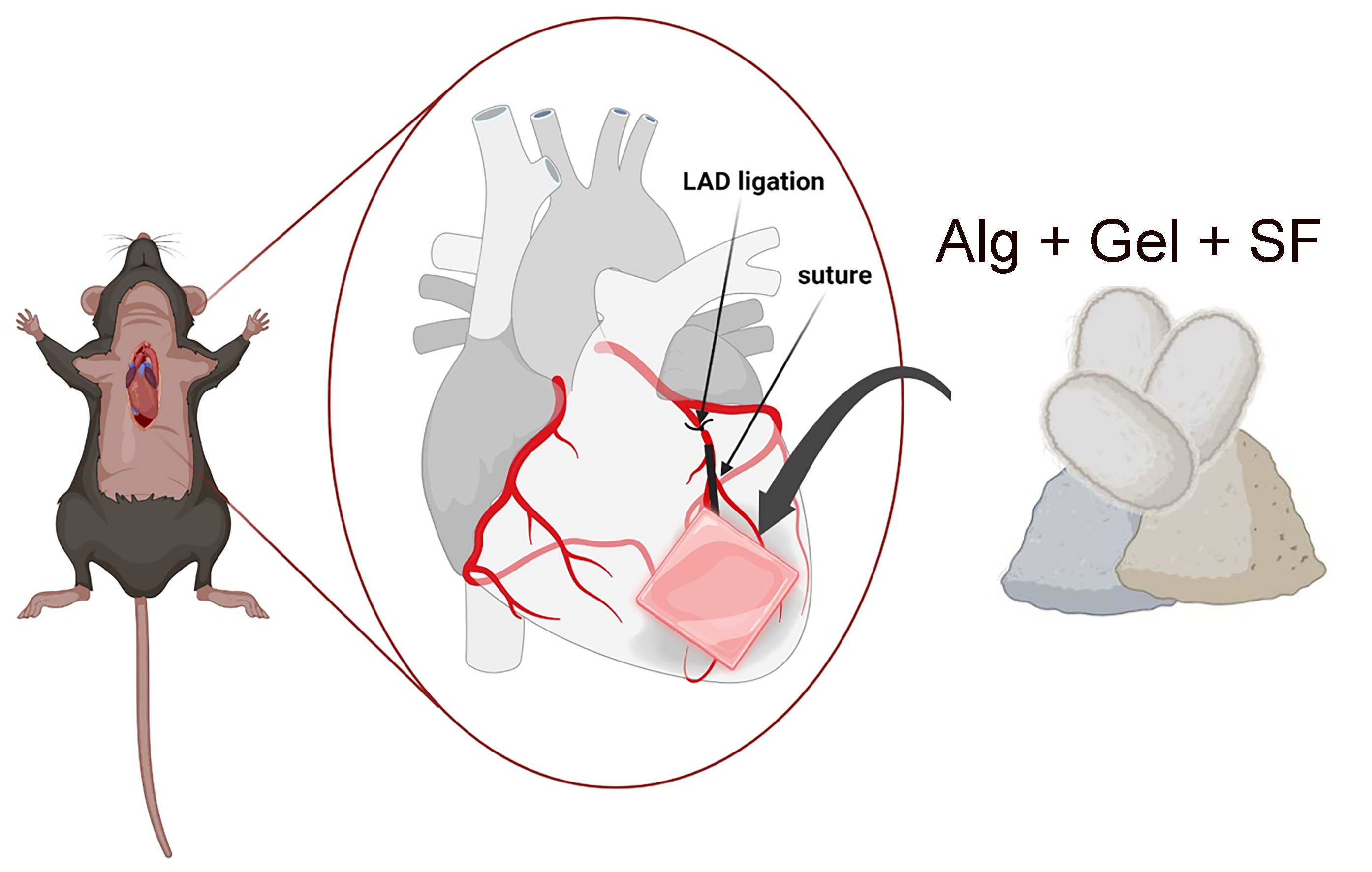
Transplantation of patches based on alginate and gelatin biomaterials represents a promising approach for improving cardiac function in vivo. To enhance the long-term durability and mechanical properties of alginate-gelatin patches, silk fibroin was added to the formulation, and 3D printed alginate-gelatin-silk fibroin patches without cells were transplanted into an in vivo myocardial infarction murine model. The focus of this study is to evaluate whether the addition of silk fibroin to alginate-gelatin hydrogels can protect against the reduction in cardiac function after myocardial infarction. Mice were divided into four experimental groups: sham, myocardial infarction, myocardial infarction with the transplanted alginate-gelatin patch and myocardial infarction with the transplanted alginate-gelatin-silk fibroin patches. The ultrasound analyses confirmed that silk fibroin-containing patches increase the left ventricular ejection fraction % by 20% in infarcted mice. Furthermore, histological and transcriptomic analyses demonstrate that alginate-gelatin-silk fibroin patches stimulate tissue-repairing processes, reduce cardiac fibrosis, increase cardiac remodeling and inflammatory-regulating gene expression, and recapitulate cardiac function and tissue homeostasis in myocardial infarction mice. Altogether, our in vivo findings support the biofabrication of advanced cardiac tissues containing alginate-gelatin-silk fibroin for tissue engineering and regenerative medicine, representing a potential therapeutic approach for patients with myocardial infarction.
- Wang Y, Wang C, Shen L, Xu D. The Role of Regulatory T Cells in Heart Repair After Myocardial Infarction. J Cardiovasc Transl Res. 2023;16(3):590-597. doi: 10.1007/s12265-022-10290-5
- Liu T, Hao Y, Zhang Z, et al. Advanced Cardiac Patches for the Treatment of Myocardial Infarction. Circulation. 2024;149(25):2002-2020. doi: 10.1161/CIRCULATIONAHA.123.067097
- Sharma P, Wang X, Ming CLC, et al. Considerations for the Bioengineering of Advanced Cardiac In Vitro Models of Myocardial Infarction. Small. 2021;17(15):e2003765. doi: 10.1002/smll.202003765
- Vettori L, Sharma P, Rnjak-Kovacina J, Gentile C. 3D Bioprinting of Cardiovascular Tissues for In Vivo and In Vitro Applications Using Hybrid Hydrogels Containing Silk Fibroin: State of the Art and Challenges. Curr Tissue Microenviron Rep. 2020;1(4):261-276. doi: 10.1007/s43152-020-00026-5
- Chen R, Zhang H, Tang B, et al. Macrophages in cardiovascular diseases: molecular mechanisms and therapeutic targets. Signal Transduct Target Ther. 2024;9(1):130. doi: 10.1038/s41392-024-01840-1
- Frangogiannis NG. Cardiac fibrosis: Cell biological mechanisms, molecular pathways and therapeutic opportunities. Mol Aspects Med. 2019;65:70-99. doi: 10.1016/j.mam.2018.07.001
- Raziyeva K, Kim Y, Zharkinbekov Z, Temirkhanova K, Saparov A. Novel Therapies for the Treatment of Cardiac Fibrosis Following Myocardial Infarction. Biomedicines. 2022;10(9):2178. doi: 10.3390/biomedicines10092178
- Yin X, Yin X, Pan X, et al. Post-myocardial infarction fibrosis: Pathophysiology, examination, and intervention. Front Pharmacol. 2023;14:1070973. doi: 10.3389/fphar.2023.1070973
- Vettori L, Tran HA, Mahmodi H, et al. Silk fibroin increases the elasticity of alginate-gelatin hydrogels and regulates cardiac cell contractile function in cardiac bioinks. Biofabrication. 2024;16(3):035025. doi: 10.1088/1758-5090/ad4f1b
- Chen J, Zhan Y, Wang Y, et al. Chitosan/silk fibroin modified nanofibrous patches with mesenchymal stem cells prevent heart remodeling post-myocardial infarction in rats. Acta Biomater. 2018;80:154-168. doi: 10.1016/j.actbio.2018.09.013
- de Winter N, Ji J, Sintou A, et al. Persistent transcriptional changes in cardiac adaptive immune cells following myocardial infarction: New evidence from the re-analysis of publicly available single cell and nuclei RNA-sequencing data sets. J Mol Cell Cardiol. 2024;192:48-64. doi: 10.1016/j.yjmcc.2024.04.016
- Polonchuk L, Surija L, Lee MH, et al. Towards engineering heart tissues from bioprinted cardiac spheroids. Biofabrication. 2021;13(4):045009. doi: 10.1088/1758-5090/ac14ca
- Roche CD, Sharma P, Ashton AW, Jackson C, Xue M, Gentile C. Printability, Durability, Contractility and Vascular Network Formation in 3D Bioprinted Cardiac Endothelial Cells Using Alginate-Gelatin Hydrogels. Front Bioeng Biotechnol. 2021;9:636257. doi: 10.3389/fbioe.2021.636257
- Das P, Ganguly S, Marvi PK, et al. Carbon Dots Infused 3D Printed Cephalopod Mimetic Bactericidal and Antioxidant Hydrogel for Uniaxial Mechano-Fluorescent Tactile Sensor. Adv Mater. 2024;36(48):e2409819. doi: 10.1002/adma.202409819
- Panico A, Corvi A, Collini L, Sciancalepore C. Multi objective optimization of FDM 3D printing parameters set via design of experiments and machine learning algorithms. Sci Rep. 2025;15(1):16753. doi: 10.1038/s41598-025-01016-z
- Wang X, Liu X, Li K, et al. A Microgel-Hydrogel Hybrid for Functional Compensation and Mechanical Stability in 3D Printed Cell-Dense Vascularized Liver Tissue. Adv Mater. 2025;37(28):e2413940. doi: 10.1002/adma.202413940
- Roche CD, Lin H, Huang Y, et al. 3D bioprinted alginate-gelatin hydrogel patches containing cardiac spheroids recover heart function in a mouse model of myocardial infarction. Bioprinting. 2023;30:e00263. doi: 10.1016/j.bprint.2023.e00263
- Huang K, Ozpinar EW, Su T, et al. An off-the-shelf artificial cardiac patch improves cardiac repair after myocardial infarction in rats and pigs. Sci Transl Med. 2020;12(538):eaat9683. doi: 10.1126/scitranslmed.aat9683
- Tran HA, Hoang TT, Maraldo A, et al. Emerging silk fibroin materials and their applications: New functionality arising from innovations in silk crosslinking. Mater Today. 2023;65:244-259. doi: 10.1016/j.mattod.2023.03.027
- Giuseppe MD, Law N, Webb B, et al. Mechanical behaviour of alginate-gelatin hydrogels for 3D bioprinting. J Mech Behav Biomed Mater. 2018;79:150-157. doi: 10.1016/j.jmbbm.2017.12.018
- Kumaraswami K, Arnholdt C, Deindl E, Lasch M. Rag1 Deficiency Impairs Arteriogenesis in Mice. Int J Mol Sci. 2023;24(16):12839. doi: 10.3390/ijms241612839
- Cui X, Soliman BG, Alcala-Orozco CR, et al. Rapid Photocrosslinking of Silk Hydrogels with High Cell Density and Enhanced Shape Fidelity. Adv Healthc Mater. 2020;9(4):e1901667. doi: 10.1002/adhm.201901667
- Karimi F, Lau K, Kim HN, et al. Surface Biofunctionalization of Silk Biomaterials Using Dityrosine Cross-Linking. ACS Appl Mater Interfaces. 2022;14(28):31551-31566. doi: 10.1021/acsami.2c03345
- Roche CD, Gentile C. Transplantation of a 3D Bioprinted Patch in a Murine Model of Myocardial Infarction. J Vis Exp. 2020;(163). doi: 10.3791/61675
- O’Riordan CE, Trochet P, Steiner M, Fuchs D. Standardisation and future of preclinical echocardiography. Mamm Genome. 2023;34(2):123-155. doi: 10.1007/s00335-023-09981-4
- Mancuso A, Cianflone E, Cristiano MC, et al. Lyotropic Liquid Crystals: A Biocompatible and Safe Material for Local Cardiac Application. Pharmaceutics. 2022;14(2):452. doi: 10.3390/pharmaceutics14020452
- Feldman AT, Wolfe D. Tissue processing and hematoxylin and eosin staining. Methods Mol Biol. 2014;1180:31-43. doi: 10.1007/978-1-4939-1050-2_3
- Kim J, Choi W, Yoo D, et al. Solution-free and simplified H&E staining using a hydrogel-based stamping technology. Front Bioeng Biotechnol. 2023;11:1292785. doi: 10.3389/fbioe.2023.1292785
- Lattouf R, Younes R, Lutomski D, et al. Picrosirius red staining: a useful tool to appraise collagen networks in normal and pathological tissues. J Histochem Cytochem. 2014;62(10):751-758. doi: 10.1369/0022155414545787
- Li WJ, Yan H, Zhou ZY, et al. Cryptotanshinone Attenuated Pathological Cardiac Remodeling In Vivo and In Vitro Experiments. Oxid Med Cell Longev. 2023;2023:4015199. doi: 10.1155/2023/4015199
- Sridharan D, Pracha N, Dougherty JA, Akhtar A, Alvi SB, Khan M. A One-Stop Protocol to Assess Myocardial Fibrosis in Frozen and Paraffin Sections. Methods Protoc. 2022;5(1):13. doi: 10.3390/mps5010013
- Van De Vlekkert D, Machado E, d’Azzo A. Analysis of Generalized Fibrosis in Mouse Tissue Sections with Masson’s Trichrome Staining. Bio Protoc. 2020;10(10):e3629. doi: 10.21769/BioProtoc.3629
- Dobin A, Davis CA, Schlesinger F, et al. STAR: ultrafast universal RNA-seq aligner. Bioinformatics. 2013;29(1):15-21. doi: 10.1093/bioinformatics/bts635
- Liao Y, Smyth GK, Shi W. featureCounts: an efficient general purpose program for assigning sequence reads to genomic features. Bioinformatics. 2014;30(7):923-930. doi: 10.1093/bioinformatics/btt656
- Love MI, Huber W, Anders S. Moderated estimation of fold change and dispersion for RNA-seq data with DESeq2. Genome Biol. 2014;15(12):550. doi: 10.1186/s13059-014-0550-8
- R Core Team. R: A Language and Environment for Statistical Computing. R Foundation for Statistical Computing. Accessed June 9, 2023. http://www.R-project.org/
- Corchete LA, Rojas EA, Alonso-López D, De Las Rivas J, Gutiérrez NC, Burguillo FJ. Systematic comparison and assessment of RNA-seq procedures for gene expression quantitative analysis. Sci Rep. 2020;10(1):19737. doi: 10.1038/s41598-020-76881-x
- Deshpande D, Chhugani K, Chang Y, et al. RNA-seq data science: From raw data to effective interpretation. Front Genet. 2023;14:997383. doi: 10.3389/fgene.2023.997383
- Koch CM, Chiu SF, Akbarpour M, et al. A Beginner’s Guide to Analysis of RNA Sequencing Data. Am J Respir Cell Mol Biol. 2018;59(2):145-157. doi: 10.1165/rcmb.2017-0430TR
- Arif M, Klevstig M, Benfeitas R, et al. Integrative transcriptomic analysis of tissue-specific metabolic crosstalk after myocardial infarction. eLife. 2021;10:e66921. doi: 10.7554/eLife.66921
- Gehlenborg N, Wong B. Points of View: Heat maps. Nat Methods. 2012;9(3):213. doi: 10.1038/nmeth.1902
- Li X, Li B, Jiang H. Identification of time‑series differentially expressed genes and pathways associated with heart failure post‑myocardial infarction using integrated bioinformatics analysis. Mol Med Rep. 2019;19(6):5281-5290. doi: 10.3892/mmr.2019.10190
- Zhang Q, Guo Y, Zhang B, et al. Identification of hub biomarkers of myocardial infarction by single-cell sequencing, bioinformatics, and machine learning. Front Cardiovasc Med. 2022;9:939972. doi: 10.3389/fcvm.2022.939972
- Majid QA, Fricker ATR, Gregory DA, et al. Natural Biomaterials for Cardiac Tissue Engineering: A Highly Biocompatible Solution. Front Cardiovasc Med. 2020;7:554597. doi: 10.3389/fcvm.2020.554597
- Settembrini A, Buongiovanni G, Settembrini P, et al. In-vivo evaluation of silk fibroin small-diameter vascular grafts: state of art of preclinical studies and animal models. Front Surg. 2023;10:1090565. doi: 10.3389/fsurg.2023.1090565
- Schneider KH, Goldberg BJ, Hasturk O, et al. Silk fibroin, gelatin, and human placenta extracellular matrix-based composite hydrogels for 3D bioprinting and soft tissue engineering. Biomater Res. 2023;27(1):117. doi: 10.1186/s40824-023-00431-5
- Johnston ER, Miyagi Y, Chuah JA, Numata K, Serban MA. The interplay between silk fibroin’s structure and its adhesive properties. ACS Biomater Sci Eng. 2018;4(8):2815-2824. doi: 10.1021/acsbiomaterials.8b00544
- Lyu Y, Liu Y, He H, Wang H. Application of Silk-Fibroin-Based Hydrogels in Tissue Engineering. Gels. 2023;9(5):431. doi: 10.3390/gels9050431
- Pan X, Li R, Li W, et al. Silk fibroin hydrogel adhesive enables sealed-tight reconstruction of meniscus tears. Nat Commun. 2024;15(1):2651. doi: 10.1038/s41467-024-47029-6
- Patra C, Talukdar S, Novoyatleva T, et al. Silk protein fibroin from Antheraea mylitta for cardiac tissue engineering. Biomaterials. 2012;33(9):2673-2680. doi: 10.1016/j.biomaterials.2011.12.036
- Bazgir F, Nau J, Nakhaei-Rad S, et al. The Microenvironment of the Pathogenesis of Cardiac Hypertrophy. Cells. 2023;12(13):1780. doi: 10.3390/cells12131780
- Feng Y, Zhao G, Xu M, et al. rGO/Silk Fibroin-Modified Nanofibrous Patches Prevent Ventricular Remodeling via Yap/Taz-TGFβ1/Smads Signaling After Myocardial Infarction in Rats. Front Cardiovasc Med. 2021;8:718055. doi: 10.3389/fcvm.2021.718055
- Leite FG, Marana JF, de Sá LFT, et al. Effects of a collagen hyaluronic acid silk-fibroin patch with the electroconductive element polyaniline on left ventricular remodeling in an infarct heart model. J Biomed Mater Res B Appl Biomater. 2022;110(7):1651-1666. doi: 10.1002/jbm.b.35026
- Kopp A, Schunck L, Gosau M, et al. Influence of the Casting Concentration on the Mechanical and Optical Properties of FA/CaCl2-Derived Silk Fibroin Membranes. Int J Mol Sci. 2020;21(18):6704. doi: 10.3390/ijms21186704
- Cai Y, Fu Y, Liu C, et al. Stathmin 1 is a biomarker for diagnosis of microvascular invasion to predict prognosis of early hepatocellular carcinoma. Cell Death Dis. 2022;13(2):176. doi: 10.1038/s41419-022-04625-y
- Kim MY, Seo EJ, Lee DH, et al. Gadd45beta is a novel mediator of cardiomyocyte apoptosis induced by ischaemia/hypoxia. Cardiovasc Res. 2010;87(1):119-126. doi: 10.1093/cvr/cvq048
- Kim YA, Kim MY, Yu HY, et al. Gadd45β is transcriptionally activated by p53 via p38α-mediated phosphorylation during myocardial ischemic injury. J Mol Med. 2013;91(11):1303-1313. doi: 10.1007/s00109-013-1070-9
- Wang J, Zhang G, Sui Y, et al. CD52 Is a Prognostic Biomarker and Associated With Tumor Microenvironment in Breast Cancer. Front Genet. 2020;11:578002. doi: 10.3389/fgene.2020.578002
- Zhang YF, Meng LB, Hao ML, Li XY, Zou T. CXCR4 and TYROBP mediate the development of atrial fibrillation via inflammation. J Cell Mol Med. 2022;26(12):3557-3567. doi: 10.1111/jcmm.17405
- Hedberg-Oldfors C, Abramsson A, Osborn DPS, et al. Cardiomyopathy with lethal arrhythmias associated with inactivation of KLHL24. Hum Mol Genet. 2019;28(11):1919-1929. doi: 10.1093/hmg/ddz032