

Development and characterization of a novel anti-reflux artificial lacrimal duct via 3D printing for lacrimal system reconstruction

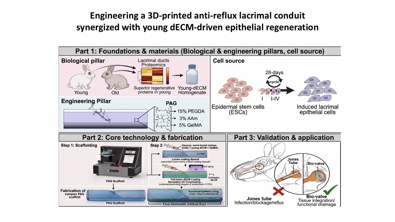
Severe lacrimal duct defects, including congenital absence and traumatic obliteration, remain formidable clinical challenges. Current therapeutic strategies, such as Jones tube prosthesis implantation, are severely limited by poor biocompatibility, susceptibility to displacement, and retrograde infections caused by fluid reflux. To address these limitations, we developed a novel anti-reflux artificial lacrimal duct using three-dimensional (3D) printing that integrates biomimetic hydrodynamic design with a biochemically optimized microenvironment. A meticulously engineered photopolymerizable composite hydrogel—comprising 15% polyethylene glycol diacrylate (PEGDA), 3% acrylamide, and 5% gelatin methacryloyl (GelMA) (60% degree of substitution)—was formulated as a high-strength hydrogel ink. Using 3D printing, we precisely fabricated an artificial lacrimal duct with a biomimetic unidirectional valve that simulates the natural Hasner valve. Guided by proteomic screening, the luminal surface of the construct was subsequently functionalized with a young donor-derived decellularized extracellular matrix (dECM) coating, onto which epidermal stem cells (ESCs) were seeded. The structural PEGDA/polyacrylamide network endowed the construct with superior printability and the requisite mechanical resilience to withstand valve fatigue. Hydrodynamic characterizations demonstrated that the 3D-printed valve effectively prevented fluid reflux while maintaining normal drainage, providing a robust physical barrier against retrograde infections. Biologically, the incorporation of GelMA-60 into the bulk scaffold and the young dECM coating at the luminal interface established a rejuvenated biochemical niche that exhibited excellent biocompatibility and successfully induced the targeted differentiation of ESCs into the lacrimal epithelial lineage. By synergizing physical anti-reflux hydrodynamics with omics-driven microenvironment rejuvenation, this composite 3D-printed construct enables superior tissue integration and infection resistance, presenting a highly promising tissue-engineered alternative for lacrimal system reconstruction.
- Fiorino MG, Quaranta‐Leoni C, Quaranta‐Leoni FM. Proximal lacrimal obstructions: a review. Acta Ophthalmol. 2021;99(7):701-711. doi: 10.1111/aos.14762
- Ali MJ, Paulsen F. Ultrastructure of the lacrimal drainage system in health and disease: A major review. Ann Anat. 2019;224:1-7. doi: 10.1016/j.aanat.2019.02.003
- Ali MJ. Updates on congenital lacrimal drainage anomalies and their association with syndromes and systemic disorders: A major review. Ann Anat. 2021;233:151613. doi: 10.1016/j.aanat.2020.151613
- Iandelli A, Carobbio ALC, Migliardi R, et al. Application of a symptoms score questionnaire after conjunctivodacryocystorhinostomy: outcomes. Acta Otorhinolaryngol Ital. 2021;41(3):248-254. doi: 10.14639/0392-100X-N0881
- Nowak R. Management of inferior dislocation of a StopLoss Jones tube after conjunctivodacryocystorhinostomy. BMJ Case Rep. 2020;13(11):e236003. doi: 10.1136/bcr-2020-236003
- Jamshidian-Tehrani M, Kashkouli MB, Ghahvehchian H, Amini M. Lacrimal Canalicular Bypass Surgery with Autologous Superficial Temporal Artery Graft. J Curr Ophthalmol. 2023;34(4):486-488. doi: 10.4103/joco.joco_98_22
- Park MS, Chi MJ, Baek SH. Clinical study of endoscopic endonasal conjunctivodacryocystorhinostomy with Jones tube placement. Ophthalmologica. 2007;221(1):36-40. doi: 10.1159/000096520
- Ullrich K, Malhotra R, Patel BC. Dacryocystorhinostomy. In: StatPearls. Treasure Island (FL): StatPearls Publishing; 2026. Available March 15, 2026. https://www.ncbi.nlm.nih.gov/books/NBK557851/
- Steele EA. Conjunctivodacryocystorhinostomy with Jones tube: a history and update. Curr Opin Ophthalmol. 2016;27(5):439-442. doi: 10.1097/ICU.0000000000000287
- Ahn ES, Dailey RA, Radmall B. The Effectiveness and Long-Term Outcome of Conjunctivodacryocystorhinostomy With Frosted Jones Tubes. Ophthalmic Plast Reconstr Surg. 2017;33(4):294-299. doi: 10.1097/IOP.0000000000000764
- Dondossola E, Holzapfel BM, Alexander S, Filippini S, Hutmacher DW, Friedl P. Examination of the foreign body response to biomaterials by nonlinear intravital microscopy. Nat Biomed Eng. 2016;1(1):0007. doi: 10.1038/s41551-016-0007
- Abel AD, Meyer DR. Refractory medial conjunctival inflammation associated with Jones tubes. Ophthalmic Plast Reconstr Surg. 2003;19(4):309-312. doi: 10.1097/01.IOP.0000075017.89210.1E
- Shah SS, Ashurst JV. Dacryocystitis. In: StatPearls. Treasure Island (FL): StatPearls Publishing; 2026. Available March 15, 2026. https://www.ncbi.nlm.nih.gov/books/NBK470565/
- Can I, Aribal E, Yarangümeli A, Ataoğlu H, Kural G. Changes in the conjunctival flora after conjunctivodacryocystorhinostomy (CDCR): a preliminary report. Eur J Ophthalmol. 1998;8(3):142-147. doi: 10.1177/112067219800800304
- Epling J. Bacterial conjunctivitis. Am Fam Physician. 2010;82(6):665.
- MacNeil S. Progress and opportunities for tissue-engineered skin. Nature. 2007;445(7130):874-880. doi: 10.1038/nature05664
- Fuchs E, Blau HM. Tissue Stem Cells: Architects of Their Niches. Cell Stem Cell. 2020;27(4):532-556. doi: 10.1016/j.stem.2020.09.011
- Watt FM. Mammalian skin cell biology: at the interface between laboratory and clinic. Science. 2014;346(6212):937-940. doi: 10.1126/science.1253734
- Jackson CJ, Tønseth KA, Utheim TP. Cultured epidermal stem cells in regenerative medicine. Stem Cell Res Ther. 2017;8(1):155. doi: 10.1186/s13287-017-0587-1
- Raja E, Changarathil G, Oinam L, et al. The extracellular matrix fibulin 7 maintains epidermal stem cell heterogeneity during skin aging. EMBO Rep. 2022;23(12):e55478. doi: 10.15252/embr.202255478
- Statzer C, Park JYC, Ewald CY. Extracellular Matrix Dynamics as an Emerging yet Understudied Hallmark of Aging and Longevity. Aging Dis. 2023;14(3):670-693. doi: 10.14336/AD.2022.1116
- Schultz GS, Wysocki A. Interactions between extracellular matrix and growth factors in wound healing. Wound Repair Regen. 2009;17(2):153-162. doi: 10.1111/j.1524-475X.2009.00466.x
- Sun Y, Li W, Lu Z, et al. Rescuing replication and osteogenesis of aged mesenchymal stem cells by exposure to a young extracellular matrix. FASEB J. 2011;25(5):1474-1485. doi: 10.1096/fj.10-161497
- Jiang D, Xu B, Gao P. Effects of young extracellular matrix on the biological characteristics of aged tendon stem cells. Adv Clin Exp Med. 2018;27(12):1625-1630. doi: 10.17219/acem/75503
- Laude M, Kolliopoulos V, Mikos AG, White LJ, Cosgriff-Hernandez E. Extracellular-Matrix-Based Materials from Decellularized Tissue: Opportunities, Challenges, and Future Directions in Regenerative Medicine. Adv Healthc Mater. 2026;15(1):e02107. doi: 10.1002/adhm.202502107
- Taylor DA, Sampaio LC, Ferdous Z, Gobin AS, Taite LJ. Decellularized matrices in regenerative medicine. Acta Biomater. 2018;74:74-89. doi: 10.1016/j.actbio.2018.04.044
- Yan X, Huang H, Bakry AM, Wu W, Liu X, Liu F. Advances in enhancing the mechanical properties of biopolymer hydrogels via multi-strategic approaches. Int J Biol Macromol. 2024;272(Pt 2):132583. doi: 10.1016/j.ijbiomac.2024.132583
- Zhao X. Multi-scale multi-mechanism design of tough hydrogels: building dissipation into stretchy networks. Soft Matter. 2014;10(5):672-87. doi: 10.1039/c3sm52272e
- Liu Y, He W, Zhang Z, Lee BP. Recent Developments in Tough Hydrogels for Biomedical Applications. Gels. 2018;4(2):46. doi: 10.3390/gels4020046
- Grigoryan B, Paulsen SJ, Corbett DC, et al. Multivascular networks and functional intravascular topologies within biocompatible hydrogels. Science. 2019;364(6439):458-464. doi: 10.1126/science.aav9750
- Xu R, Ooi HS, Bian L, Ouyang L, Sun W. Dynamic hydrogels for biofabrication: A review. Biomaterials. 2025;320:123266. doi: 10.1016/j.biomaterials.2025.123266
- Patrocinio D, Galván-Chacón V, Gómez-Blanco JC, et al. Biopolymers for Tissue Engineering: Crosslinking, Printing Techniques, and Applications. Gels. 2023;9(11):890. doi: 10.3390/gels9110890.
- Alparslan C, Bayraktar Ş. Advances in Digital Light Processing (DLP) Bioprinting: A Review of Biomaterials and Its Applications, Innovations, Challenges, and Future Perspectives. Polymers. 2025;17(9):1287. doi: 10.3390/polym17091287
- Kunwar P, Andrada BL, Poudel A, et al. Printing Double-Network Tough Hydrogels Using Temperature-Controlled Projection Stereolithography (TOPS). ACS Appl Mater Interfaces. 2023;15(25):30780-30792. doi: 10.1021/acsami.3c04661
- Li W, Hu X, Liu H, et al. 3D light-curing printing to construct versatile octopus-bionic patches. J Mater Chem B. 2023;11(22):5010-5020. doi: 10.1039/D3TB00590A
- Zhang D, Yang J, Le X, Song D. Editorial for Special Issue: Advances in Smart and Tough Hydrogels. Gels. 2023;9(10):789. doi: 10.3390/gels9100789
- Ban Y, Yoshida Y, Aziza Y, Kinoshita S, Sotozono C. Strengthening of the barrier function in human telomerase reverse transcription (hTERT) immortalized corneal and conjunctival epithelium by double-stranded RNA. Exp Eye Res. 2023;227:109357. doi: 10.1016/j.exer.2022.109357
- Mohamed YH, Uematsu M, Kusano M, Inoue D, Tang D, Suzuki K, Kitaoka T. A Novel Technique for Corneal Transepithelial Electrical Resistance Measurement in Mice. Life. 2024;14(8):1046. doi: 10.3390/life14081046
- Gwaltney JM Jr, Hendley JO, Phillips CD, Bass CR, Mygind N, Winther B. Nose blowing propels nasal fluid into the paranasal sinuses. Clin Infect Dis. 2000;30(2):387-391. doi: 10.1086/313661
- Kudo A. Periostin in fibrillogenesis for tissue regeneration: periostin actions inside and outside the cell. Cell Mol Life Sci. 2011;68(19):3201-3207. doi: 10.1007/s00018-011-0784-5
- Zihni C, Mills C, Matter K, Balda MS. Tight junctions: from simple barriers to multifunctional molecular gates. Nat Rev Mol Cell Biol. 2016;17(9):564-580. doi: 10.1038/nrm.2016.80