

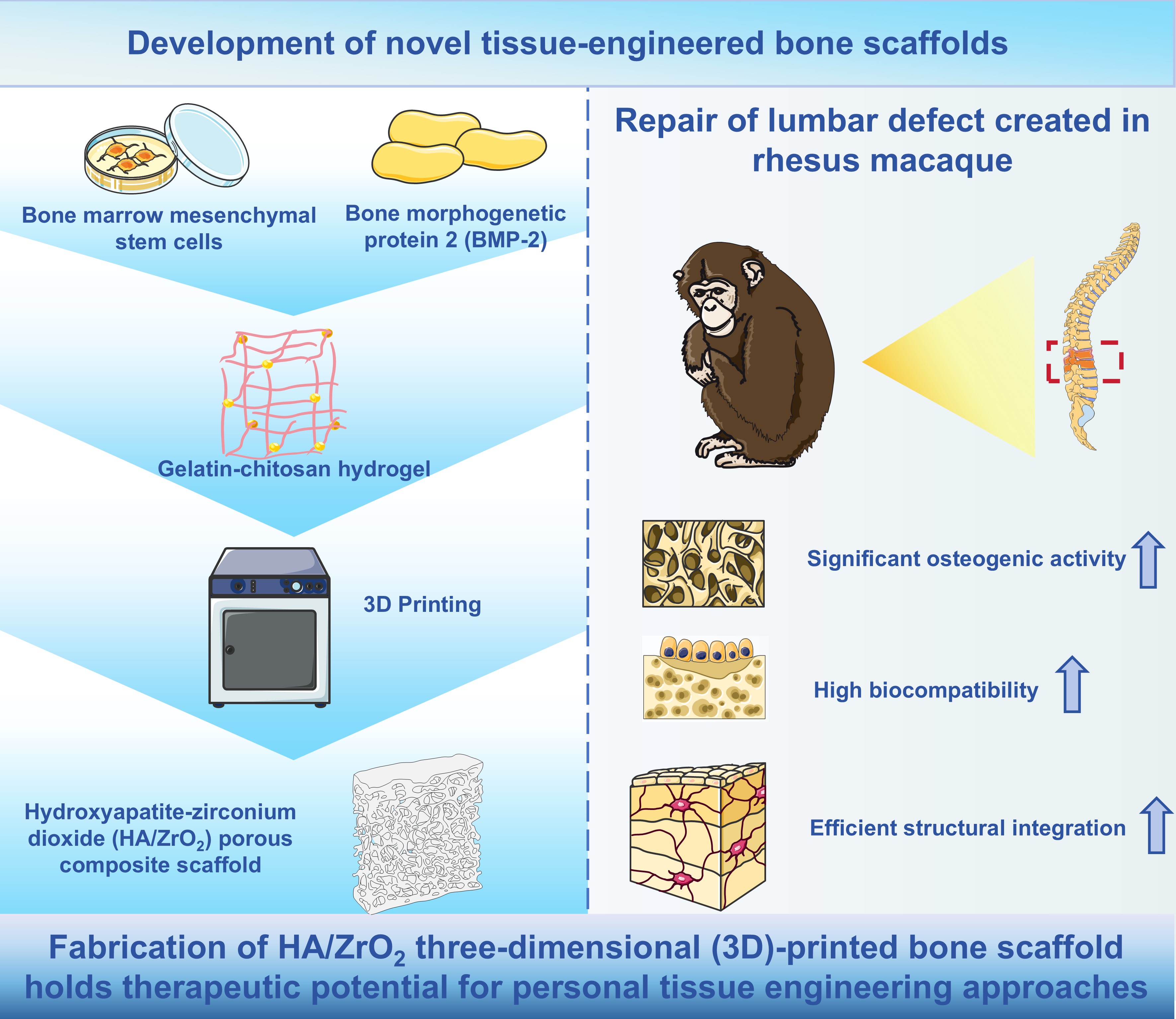
Three-dimensional-printed hydroxyapatite/ zirconium dioxide composite scaffold incorporating bone morphogenetic protein-2 for lumbar vertebral bone defect repair in rhesus macaques

Bone defects caused by various factors have become a persistent challenge in orthopedic clinics, and traditional treatment methods mainly involve the use of artificial bone or autologous bone transplantation. However, these methods have considerable limitations, such as donor site bone loss, immune rejection, and the risk of secondary infection at the donor site. Therefore, considering these limitations and the rapid development of the field of bone tissue engineering, this study adopted light-curing stereolithography three-dimensional (3D) printing technology to design bone scaffold materials. The technology was used to prepare hydroxyapatite (HA)/zirconium dioxide (ZrO2) porous composites with satisfactory mechanical properties as tissue-engineering bone scaffolds. A bone morphogenetic protein- 2-loaded gelatin/chitosan hydrogel sustained-release system was prepared via an emulsification and cross-linking process. Subsequently, rhesus macaque bone marrow mesenchymal stem cells, acting as osteogenic progenitors, were seeded into the system. Novel HA/ZrO2 scaffolds were fabricated using stereolithography 3D printing technology to serve as bone graft substitutes. The resulting scaffold exhibited a 3D interconnected porous structure and showed good biocompatibility and osteoinductive ability in a rhesus macaque lumbar vertebral bone defect model. The results confirmed that the scaffold achieved osteogenic efficiency comparable to that of autologous bone grafting in rhesus macaques. Therefore, the developed scaffold material has promising potential in bone defect repair.
- Dec P, Modrzejewski A, Pawlik A, Existing and novel biomaterials for bone tissue engineering. Int J Mol Sci. 2022:24:529. doi: 10.3390/ijms24010529
- Jagadale S, Damle M, Joshi MG. Bone tissue engineering: from biomaterials to clinical trials. Adv Exp Med Biol. 2025;1479:73-115. doi: 10.1007/5584_2024_841
- Brachet A, Bełżek A, Furtak D. et al. Application of 3D printing in bone grafts. Cells. 2023;12:859. doi: 10.3390/cells12060859
- Maresca JA, DeMel DC, Wagner GA, et al. Three-dimensional bioprinting applications for bone tissue engineering. Cells. 2023;12:1230.
- Chatzipetros E, Damaskos S, Tosios KI, et al. The effect of nano-hydroxyapatite/chitosan scaffolds on rat calvarial defects for bone regeneration. Int J Implant Dent. 2021;7:40. doi: 10.1186/s40729-021-00327-w
- Quan RF, Tang YH, Huang ZM, et al. Difference of adherence, proliferation and osteogenesis of mesenchymal stem cells cultured on different HA/ZrO2 composites. Chin J Traumatol. 2012;15:131-139.
- Ingwersen LC, Frank M, Naujokat H, et al. BMP-2 long-term stimulation of human pre-osteoblasts induces osteogenic differentiation and promotes transdifferentiation and bone remodeling processes. Int J Mol Sci. 2022;23:3077. doi: 10.3390/ijms23063077
- Grosso A, Lunger A, Burger MG, et al. VEGF dose controls the coupling of angiogenesis and osteogenesis in engineered bone. NPJ Regen Med. 2023;8:15. doi: 10.1038/s41536-023-00288-1
- Gorskaya YF, Danilova TA, Karyagina AS, et al. Effects of combined treatment with complex S. typhimurium antigens and factors stimulating osteogenesis (curettage, BMP- 2) on multipotent bone marrow stromal cells and serum concentration of cytokines in CBA mice. Bull Exp Biol Med. 2015;158:465-470. doi: 10.1007/s10517-015-2786-z
- Kong D, Shi Y, Gao Y, et al. Preparation of BMP-2 loaded MPEG-PCL microspheres and evaluation of their bone repair properties. Biomed Pharmacother. 2020;130: 110516. doi: 10.1016/j.biopha.2020.110516
- Arthur A, Gronthos S. Clinical application of bone marrow mesenchymal stem/stromal cells to repair skeletal tissue. Int J Mol Sci. 2020;21:9759. doi: 10.3390/ijms21249759
- Gholami Farashah MS, Javadi M, Mohammadi A, et al. Bone marrow mesenchymal stem cell’s exosomes as key nanoparticles in osteogenesis and bone regeneration: specific capacity based on cell type. Mol Biol Rep. 2022;49:12203-12218. doi: 10.1007/s11033-022-07807-1
- Yazdanpanah Z, Sharma NK, Raquin A, et al. Printing tissue-engineered scaffolds made of polycaprolactone and nano-hydroxyapatite with mechanical properties appropriate for trabecular bone substitutes. Biomed Eng Online. 2023; 22:73. doi: 10.1186/s12938-023-01135-6
- Rajabi M, Cabral JD, Saunderson S, et al. Development and optimisation of hydroxyapatite-polyethylene glycol diacrylate hydrogel inks for 3D printing of bone tissue engineered scaffolds. Biomed Mater. 2023;18(6). doi: 10.1088/1748-605X/acf90a
- Bahraminasab M, Talebi A, Doostmohammadi N, et al. The healing of bone defects by cell-free and stem cell-seeded 3D-printed PLA tissue-engineered scaffolds. J Orthop Surg Res. 2022;17:320. doi: 10.1186/s13018-022-03213-2
- Zalewska J, Przekora A, Pałka K, et al. Gypsum-related compensation of ions uptake by highly porous hydroxyapatite ceramics: – consequences for osteoblasts growth and proliferation. Biomater Adv. 2022;133:112665. doi: 10.1016/j.msec.2022.112665
- Ostrowska B, Di Luca A, Szlazak K, et al. Influence of internal pore architecture on biological and mechanical properties of three-dimensional fiber deposited scaffolds for bone regeneration. J Biomed Mater Res A. 2016;104:991-1001. doi: 10.1002/jbm.a.35637
- Shao RX, Quan RF, Huang XL, et al. Evaluation of porous gradient hydroxyapatite/zirconia composites for repair of lumbar vertebra defect in dogs. J Biomater Appl. 2016;30:1312-1321. doi: 10.1177/0885328215627616
- Julien A, Perrin S, Martínez-Sarrà E, et al. Skeletal stem/ progenitor cells in periosteum and skeletal muscle share a common molecular response to bone injury. J Bone Miner Res. 2022;37:1545-1561. doi: 10.1002/jbmr.4616
- Salamanca E, Wu YF, Aung LM, et al. Allylamine coating on zirconia dental implant surface promotes osteogenic differentiation in vitro and accelerates osseointegration in vivo. Clin Oral Implants Res. 2024;35:1101-1113. doi: 10.1111/clr.14300
- Kim J, Kang IG, Cheon KH, et al. Stable sol-gel hydroxyapatite coating on zirconia dental implant for improved osseointegration. J Mater Sci Mater Med. 2021;32:81. doi: 10.1007/s10856-021-06550-6
- Kusuyama J, Amir MS, Albertson BG, et al. JNK inactivation suppresses osteogenic differentiation, but robustly induces osteopontin expression in osteoblasts through the induction of inhibitor of DNA binding 4 (Id4). FASEB J. 2019;33:7331-7347. doi: 10.1096/fj.201802465R
- Mardiyantoro F, Chiba N, Seong CH, et al. Two-sided function of osteopontin during osteoblast differentiation. J Biochem. 2025;177:121-131. doi: 10.1093/jb/mvae080
- Selvaraj V, Sekaran S, Dhanasekaran A, et al. Type 1 collagen: synthesis, structure and key functions in bone mineralization. Differentiation. 2024;136:100757. doi: 10.1016/j.diff.2024.100757
- Asakura T, Diep TTT, Ueda Y, et al. Analysis of the effect of human type I collagen-derived peptide on bone regenerative capacity and comparison with various collagen materials in vivo. Medicina (Kaunas). 2025;61:57. doi: 10.3390/medicina61010057
- Tateno A, Asano M, Akita D, et al. Transplantation of dedifferentiated fat cells combined with a biodegradable type I collagen-recombinant peptide scaffold for critical-size bone defects in rats. J Oral Sci. 2019;61:534-538. doi: 10.2334/josnusd.18-0458
- Vimalraj S. Alkaline phosphatase: structure, expression and its function in bone mineralization. Gene. 2020;754:144855.
- Wu H, Yin G, Pu X, et al. Inhibitory effects of combined bone morphogenetic protein 2, vascular endothelial growth factor, and basic fibroblast growth factor on osteoclast differentiation and activity. Tissue Eng Part A. 2021;27:1387-1398. doi: 10.1089/ten.TEA.2020.0325
- Nakamichi M, Akishima-Fukasawa Y, Fujisawa C, et al. Basic fibroblast growth factor induces angiogenic properties of fibrocytes to stimulate vascular formation during wound healing. Am J Pathol. 2016;186:3203-3216. doi: 10.1016/j.ajpath.2016.08.015
- Pasanen S, Mootha A, Hirata I, et al. Enhanced growth of bone marrow-derived mesenchymal stem cells on the microcarriers tethered with engineered basic fibroblast growth factor. Biotechnol J. 2025;20:e70057. doi: 10.1002/biot.70057
- Hu K, Olsen BR. Osteoblast-derived VEGF regulates osteoblast differentiation and bone formation during bone repair. J Clin Invest. 2016;126:509-526. doi: 10.1172/JCI82585
- Veraitch O, Mabuchi Y, Matsuzaki Y, et al. Induction of hair follicle dermal papilla cell properties in human induced pluripotent stem cell-derived multipotent LNGFR(+)THY- 1(+) mesenchymal cells. Sci Rep. 2017;7:42777. doi: 10.1038/srep42777