

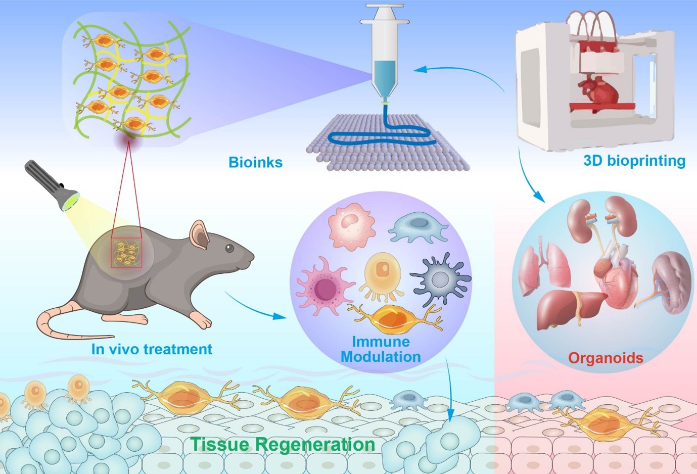
3D bioprinting-driven strategies for tissue regeneration and controlled immune modulation

Tissue loss, fibrosis-prone repair, and immune-mediated graft failure remain persistent obstacles in regenerative medicine. Within this context, 3D bioprinting is shifting from structure-centric fabrication to a platform for programmed immune modulation. This review synthesizes evidence across materials, architecture, and living components to delineate how bioprinted constructs can steer host responses toward resolution and durable function. We first examine events at the blood–biomaterial interface, including protein corona formation, complement–coagulation crosstalk, and leukocyte recruitment, and map them to tunable parameters, such as chemistry, stiffness, degradability, topography, and pore geometry that direct macrophage and dendritic-cell programs. We compare natural and synthetic bioinks, emphasizing printability windows, batch control, and impurity management as prerequisites for interpretable immunological readouts. We survey stimuli-responsive inks triggered by pH, reactive oxygen species, enzymes, light, or magnetic fields to deliver cytokines, chemokines, and metabolites with temporal precision, and highlight architected lattices and gradients that guide cell trafficking, vascular and lymphatic integration, and mechano-immune conditioning. Cell- and signal-centric strategies include immune–stromal coprinting, extracellular vesicle embedding, membrane cloaking for immune stealth or targeting, and synthetic circuits that sense inflammation and secrete immunoregulatory payloads. Finally, we identify translational bottlenecks and outline opportunities in 4D bioprinting, AI-assisted design, digital twins, and in situ printing. Treating immunity as a primary design variable is essential for predictable, durable, and clinically credible bioprinted therapies.
1 Anderson JM, Rodriguez A, Chang DT. Foreign body reaction to biomaterials. Semin Immunol. 2008;20(2):86-100. doi: 10.1016/j.smim.2007.11.004
2 Carnicer-Lombarte A, Chen ST, Malliaras GG, Barone DG. Foreign body reaction to implanted biomaterials and its impact in nerve neuroprosthetics. Front Bioeng Biotechnol. 2021;9:622524. doi: 10.3389/fbioe.2021.622524
3 Gomes YVR, Tavares AA, Barbosa RC, et al. Biological responses to biomaterials: a review. Braz J Med Biol Res. 2025;58:e14599. doi: 10.1590/1414-431X2025e14599
4 Nilsson B, Ekdahl KN, Mollnes TE, Lambris JD. The role of complement in biomaterial-induced inflammation. Mol Immunol. 2007;44(1-3):82-94. doi: 10.1016/j.molimm.2006.06.020
5 Chenoweth DE. Complement activation produced by biomaterials. Artif Organs. 1988;12(6):508-510. doi: 10.1111/j.1525-1594.1988.tb02815.x
6 Thakur KK, Lekurwale R, Bansode S, Pansare R. 3D bioprinting: a systematic review for future research direction. Indian J Orthop. 2023;57(12):1949-1967. doi: 10.1007/s43465-023-01000-7
7 Agarwal T, Onesto V, Banerjee D, et al. 3D bioprinting in tissue engineering: current state-of-the-art and challenges towards system standardization and clinical translation. Biofabrication. 2025;17:4. doi: 10.1088/1758-5090/ade47a
8 Raees S, Ullah F, Javed F, et al. Classification, processing, and applications of bioink and 3D bioprinting: a detailed review. Int J Biol Macromol. 2023;232:123476. doi: 10.1016/j.ijbiomac.2023.123476
9 Lee J, Byun H, Madhurakkat Perikamana SK, Lee S, Shin H. Current advances in immunomodulatory biomaterials for bone regeneration. Adv Healthc Mater. 2019;8(4): e1801106. doi: 10.1002/adhm.201801106
10 Yang T, Fang Z, Zhang J, Zheng S. Physical cues in biomaterials modulate macrophage polarization for bone regeneration: a review. Front Bioeng Biotechnol. 2025;13:1640560. doi: 10.3389/fbioe.2025.1640560
11 Yang HC, Park HC, Quan H, Kim Y. Immunomodulation of biomaterials by controlling macrophage polarization. Adv Exp Med Biol. 2018;1064:197-206. doi: 10.1007/978-981-13-0445-3_12
12 Zarubova J, Hasani-Sadrabadi MM, Ardehali R, Li S. Immunoengineering strategies to enhance vascularization and tissue regeneration. Adv Drug Deliv Rev. 2022;184:114233. doi: 10.1016/j.addr.2022.114233
13 Est-Witte S, Jain M, Ben-Akiva E, et al. Immunomodulatory delivery materials for tissue repair. Adv Healthc Mater. 2025;14(23):e2501400. doi: 10.1002/adhm.202501400
14 Modinger Y, Teixeira GQ, Neidlinger-Wilke C, Ignatius A. Role of the complement system in the response to orthopedic biomaterials. Int J Mol Sci. 2018;19:11. doi: 10.3390/ijms19113367
15 Trindade R, Albrektsson T, Tengvall P, Wennerberg A. Foreign body reaction to biomaterials: on mechanisms for buildup and breakdown of osseointegration. Clin Implant Dent Relat Res. 2016;18(1):192-203. doi: 10.1111/cid.12274
16 Durieux N, Vandenput S, Pasleau F. OCEBM levels of evidence system. Rev Med Liege. 2013;68(12):644-649.
17 Page MJ, McKenzie JE, Bossuyt PM, et al. The PRISMA 2020 statement: an updated guideline for reporting systematic reviews. BMJ. 2021;372:n71. doi: 10.1136/bmj.n71
18 Page MJ, McKenzie JE, Bossuyt PM, et al. The PRISMA 2020 statement: an updated guideline for reporting systematic reviews. PLoS Med. 2021;18(3):e1003583. doi: 10.1371/journal.pmed.1003583
19 Gurtner GC, Werner S, Barrandon Y, Longaker MT. Wound repair and regeneration. Nature. 2008;453(7193):314-321. doi: 10.1038/nature07039
20 Landen NX, Li D, Stahle M. Transition from inflammation to proliferation: a critical step during wound healing. Cell Mol Life Sci. 2016;73(20):3861-3885. doi: 10.1007/s00018-016-2268-0
21 Reinke JM, Sorg H. Wound repair and regeneration. Eur Surg Res. 2012;49(1):35-43. doi: 10.1159/000339613
22 Ekdahl KN, Huang S, Nilsson B, Teramura Y. Complement inhibition in biomaterial- and biosurface-induced thromboinflammation. Semin Immunol. 2016;28(3):268-277. doi: 10.1016/j.smim.2016.04.006
23 Netea MG, Dominguez-Andres J, Barreiro LB, et al. Defining trained immunity and its role in health and disease. Nat Rev Immunol. 2020;20(6):375-388. doi: 10.1038/s41577-020-0285-6
24 Dagenais A, Villalba-Guerrero C, Olivier M. Trained immunity: a “new” weapon in the fight against infectious diseases. Front Immunol. 2023;14:1147476. doi: 10.3389/fimmu.2023.1147476
25 Mestres G, Carter SD, Hailer NP, Diez-Escudero A. A practical guide for evaluating the osteoimmunomodulatory properties of biomaterials. Acta Biomater. 2021;130:115-137. doi: 10.1016/j.actbio.2021.05.038
26 Martin KE, Garcia AJ. Macrophage phenotypes in tissue repair and the foreign body response: implications for biomaterial-based regenerative medicine strategies. Acta Biomater. 2021;133:4-16. doi: 10.1016/j.actbio.2021.03.038
27 Piatnitskaia S, Rafikova G, Bilyalov A, et al. Modelling of macrophage responses to biomaterials in vitro: state-of-the-art and the need for the improvement. Front Immunol. 2024;15:1349461. doi: 10.3389/fimmu.2024.1349461
28 Holzl K, Lin S, Tytgat L, Van Vlierberghe S, Gu L, Ovsianikov A. Bioink properties before, during and after 3D bioprinting. Biofabrication. 2016;8(3):032002. doi: 10.1088/1758-5090/8/3/032002
29 Gungor-Ozkerim PS, Inci I, Zhang YS, Khademhosseini A, Dokmeci MR. Bioinks for 3D bioprinting: an overview. Biomater Sci. 2018;6(5):915-946. doi: 10.1039/c7bm00765e
30 Crapo PM, Gilbert TW, Badylak SF. An overview of tissue and whole organ decellularization processes. Biomaterials. 2011;32(12):3233-3243. doi: 10.1016/j.biomaterials.2011.01.057
31 Lieder R, Petersen PH, Sigurjonsson OE. Endotoxins-the invisible companion in biomaterials research. Tissue Eng Part B Rev. 2013;19(5):391-402. doi: 10.1089/ten.TEB.2012.0636
32 Lin CC, Anseth KS. PEG hydrogels for the controlled release of biomolecules in regenerative medicine. Pharm Res. 2009;26(3):631-643. doi: 10.1007/s11095-008-9801-2
33 Fernandez-Yague MA, Hymel LA, Olingy CE, et al. Analyzing immune response to engineered hydrogels by hierarchical clustering of inflammatory cell subsets. Sci Adv. 2022;8(8):eabd8056. doi: 10.1126/sciadv.abd8056
34 Yang D, Jones KS. Effect of alginate on innate immune activation of macrophages. J Biomed Mater Res A. 2009;90(2):411-418. doi: 10.1002/jbm.a.32096
35 Choudhury D, Tun HW, Wang T, Naing MW. Organ-derived decellularized extracellular matrix: a game changer for bioink manufacturing? Trends Biotechnol. 2018;36(8):787-805. doi: 10.1016/j.tibtech.2018.03.003
36 Abaci A, Guvendiren M. Designing decellularized extracellular matrix-based bioinks for 3D bioprinting. Adv Healthc Mater. 2020;9(24):e2000734. doi: 10.1002/adhm.202000734
37 Ma S, Feng X, Liu F, Wang B, Zhang H, Niu X. The pro-inflammatory response of macrophages regulated by acid degradation products of poly(lactide-co-glycolide) nanoparticles. Eng Life Sci. 2021;21(10):709-720. doi: 10.1002/elsc.202100040
38 Zhao Y, Feng X, Zhao Z, Song Z, Wang W, Zhao H. Interleukin-4-loaded heparin hydrogel regulates macrophage polarization to promote osteogenic differentiation. ACS Biomater Sci Eng. 2024;10(9):5774-5783. doi: 10.1021/acsbiomaterials.4c00589
39 Pacifici N, Bolandparvaz A, Lewis JS. Stimuli-responsive biomaterials for vaccines and immunotherapeutic applications. Adv Ther (Weinh). 2020;3(11):2000129. doi: 10.1002/adtp.202000129
40 Yu H, Gao R, Liu Y, Fu L, Zhou J, Li L. Stimulus-responsive hydrogels as drug delivery systems for inflammation targeted therapy. Adv Sci (Weinh). 2024;11(1):e2306152. doi: 10.1002/advs.202306152
41 Pranantyo D, Yeo CK, Wu Y, et al. Hydrogel dressings with intrinsic antibiofilm and antioxidative dual functionalities accelerate infected diabetic wound healing. Nat Commun. 2024;15(1):954. doi: 10.1038/s41467-024-44968-y
42 Huang Q, Qu Y, Tang M, et al. ROS-responsive hydrogel for bone regeneration: controlled dimethyl fumarate release to reduce inflammation and enhance osteogenesis. Acta Biomater. 2025;195:183-200. doi: 10.1016/j.actbio.2025.02.026
43 Chen H, Wang W, Yang Y, et al. A sequential stimuli-responsive hydrogel promotes structural and functional recovery of severe spinal cord injury. Biomaterials. 2025;316:122995. doi: 10.1016/j.biomaterials.2024.122995
44 Lai Y, Xiao X, Huang Z, et al. Photocrosslinkable biomaterials for 3D bioprinting: mechanisms, recent advances, and future prospects. Int J Mol Sci. 2024;25:23. doi: 10.3390/ijms252312567
45 Li L, Alsema E, Beijer NRM, Gumuscu B. Magnetically driven hydrogel surfaces for modulating macrophage behavior. ACS Biomater Sci Eng. 2024;10(11):6974-6983. doi: 10.1021/acsbiomaterials.4c01624
46 Gorronogoitia I, Urtaza U, Zubiarrain-Laserna A, Alonso- Varona A, Zaldua AM. A study of the printability of alginate-based bioinks by 3D bioprinting for articular cartilage tissue engineering. Polymers (Basel). 2022;14:2. doi: 10.3390/polym14020354
47 Cruz EM, Machado LS, Zamproni LN, et al. A gelatin methacrylate-based hydrogel as a potential bioink for 3D bioprinting and neuronal differentiation. Pharmaceutics. 2023;15:2. doi: 10.3390/pharmaceutics15020627
48 Wang D, Guo Y, Zhu J, et al. Hyaluronic acid methacrylate/ pancreatic extracellular matrix as a potential 3D printing bioink for constructing islet organoids. Acta Biomater. 2023;165:86-101. doi: 10.1016/j.actbio.2022.06.036
49 Osidak EO, Kozhukhov VI, Osidak MS, Domogatsky SP. Collagen as bioink for bioprinting: a comprehensive review. Int J Bioprint. 2020;6(3):270. doi: 10.18063/ijb.v6i3.270
50 de Melo BAG, Jodat YA, Cruz EM, Benincasa JC, Shin SR, Porcionatto MA. Strategies to use fibrinogen as bioink for 3D bioprinting fibrin-based soft and hard tissues. Acta Biomater. 2020;117:60-76. doi: 10.1016/j.actbio.2020.09.024
51 Hidaka M, Kojima M, Nakahata M, Sakai S. Visible light-curable chitosan ink for extrusion-based and vat polymerization-based 3D bioprintings. Polymers (Basel). 2021;13:9. doi: 10.3390/polym13091382
52 Markstedt K, Mantas A, Tournier I, Martinez Avila H, Hagg D, Gatenholm P. 3D bioprinting human chondrocytes with nanocellulose-alginate bioink for cartilage tissue engineering applications. Biomacromolecules. 2015;16(5): 1489-1496. doi: 10.1021/acs.biomac.5b00188
53 Alheib O, da Silva LP, Youn YH, Kwon IK, Reis RL, Correlo VM. 3D bioprinting of gellan gum-based hydrogels tethered with laminin-derived peptides for improved cellular behavior. J Biomed Mater Res A. 2022;110(10):1655-1668. doi: 10.1002/jbm.a.37415
54 Marques DMC, Silva JC, Serro AP, Cabral JMS, Sanjuan- Alberte P, Ferreira FC. 3D bioprinting of novel kappa-carrageenan bioinks: an algae-derived polysaccharide. Bioengineering (Basel). 2022;9:3. doi: 10.3390/bioengineering9030109
55 Gu Y, Schwarz B, Forget A, Barbero A, Martin I, Shastri VP. Advanced bioink for 3D bioprinting of complex free-standing structures with high stiffness. Bioengineering (Basel). 2020;7:4. doi: 10.3390/bioengineering7040141
56 Piluso S, Skvortsov GA, Altunbek M, et al. 3D bioprinting of molecularly engineered PEG-based hydrogels utilizing gelatin fragments. Biofabrication. 2021;13:4. doi: 10.1088/1758-5090/ac0ff0
57 Rossi A, Pescara T, Gambelli AM, et al. Biomaterials for extrusion-based bioprinting and biomedical applications. Front Bioeng Biotechnol. 2024;12:1393641. doi: 10.3389/fbioe.2024.1393641
58 Li X, Liu B, Pei B, et al. Inkjet bioprinting of biomaterials. Chem Rev. 2020;120(19):10793-10833. doi: 10.1021/acs.chemrev.0c00008
59 Jeong YG, Yoo JJ, Lee SJ, Kim MS. 3D digital light process bioprinting: cutting-edge platforms for resolution of organ fabrication. Mater Today Bio. 2024;29:101284. doi: 10.1016/j.mtbio.2024.101284
60 Jing X, Fu H, Yu B, Sun M, Wang L. Two-photon polymerization for 3D biomedical scaffolds: overview and updates. Front Bioeng Biotechnol. 2022;10:994355. doi: 10.3389/fbioe.2022.994355
61 Lee SJ, Jeong W, Atala A. 3D bioprinting for engineered tissue constructs and patient-specific models: current progress and prospects in clinical applications. Adv Mater. 2024;36(49):e2408032. doi: 10.1002/adma.202408032
62 Hady TF, Hwang B, Pusic AD, et al. Uniform 40-microm-pore diameter precision templated scaffolds promote a pro-healing host response by extracellular vesicle immune communication. J Tissue Eng Regen Med. 2021; 15(1):24-36. doi: 10.1002/term.3160
63 Yang X, Gao J, Yang S, et al. Pore size-mediated macrophage M1 to M2 transition affects osseointegration of 3D-printed PEEK scaffolds. Int J Bioprint. 2023;9(5):755. doi: 10.18063/ijb.755
64 Liao C, Li Y, Tjong SC. Polyetheretherketone and its composites for bone replacement and regeneration. Polymers (Basel). 2020;12:12. doi: 10.3390/polym12122858
65 Adamson M, Eslami B. Post-processing PEEK 3D-printed parts: experimental investigation of annealing on microscale and macroscale properties. Polymers (Basel). 2025;17:6. doi: 10.3390/polym17060744
66 Sun Y, Zhang Y, Guo Y, et al. Electrical aligned polyurethane nerve guidance conduit modulates macrophage polarization and facilitates immunoregulatory peripheral nerve regeneration. J Nanobiotechnol. 2024;22(1):244. doi: 10.1186/s12951-024-02507-3
67 McWhorter FY, Wang T, Nguyen P, Chung T, Liu WF. Modulation of macrophage phenotype by cell shape. Proc Natl Acad Sci USA. 2013;110(43):17253-17258. doi: 10.1073/pnas.1308887110
68 Wu J, Mao Z, Tan H, Han L, Ren T, Gao C. Gradient biomaterials and their influences on cell migration. Interface Focus. 2012;2(3):337-355. doi: 10.1098/rsfs.2011.0124
69 Miri AK, Mirzaee I, Hassan S, et al. Effective bioprinting resolution in tissue model fabrication. Lab Chip. 2019;19(11):2019-2037. doi: 10.1039/c8lc01037d
70 Miri AK, Nieto D, Iglesias L, et al. Microfluidics-enabled multimaterial maskless stereolithographic bioprinting. Adv Mater. 2018;30(27):e1800242. doi: 10.1002/adma.201800242
71 Kim YK, Park JA, Yoon WH, Kim J, Jung S. Drop-on-demand inkjet-based cell printing with 30-mum nozzle diameter for cell-level accuracy. Biomicrofluidics. 2016;10(6):064110. doi: 10.1063/1.4968845
72 Jia W, Gungor-Ozkerim PS, Zhang YS, et al. Direct 3D bioprinting of perfusable vascular constructs using a blend bioink. Biomaterials. 2016;106:58-68. doi: 10.1016/j.biomaterials.2016.07.038
73 Zhu W, Qu X, Zhu J, et al. Direct 3D bioprinting of prevascularized tissue constructs with complex microarchitecture. Biomaterials. 2017;124:106-115. doi: 10.1016/j.biomaterials.2017.01.042
74 Dellaquila A, Le Bao C, Letourneur D, Simon-Yarza T. In vitro strategies to vascularize 3D physiologically relevant models. Adv Sci (Weinh). 2021;8(19):e2100798. doi: 10.1002/advs.202100798
75 Cao X, Ashfaq R, Cheng F, et al. A tumor-on-a-chip system with bioprinted blood and lymphatic vessel pair. Adv Funct Mater. 2019;29:31. doi: 10.1002/adfm.201807173
76 Lu R, Lee BJ, Lee E. Three-dimensional lymphatics-on-a-chip reveals distinct, size-dependent nanoparticle transport mechanisms in lymphatic drug delivery. ACS Biomater Sci Eng. 2024;10(9):5752-5763. doi: 10.1021/acsbiomaterials.4c01005
77 Sergis V, Kelly D, Pramanick A, Britchfield G, Mason K, Daly AC. In-situquality monitoring during embedded bioprinting using integrated microscopy and classical computer vision. Biofabrication. 2025;17:2. doi: 10.1088/1758-5090/adaa22
78 Quirk EL, Burroughs MC, Mai DJ. In situ photo-rheology monitors viscoelastic changes in photo-responsive polymer networks. J Vis Exp. 2025:220. doi: 10.3791/68394
79 Malekpour A, Chen X. Printability and cell viability in extrusion-based bioprinting from experimental, computational, and machine learning views. J Funct Biomater. 2022;13:2. doi: 10.3390/jfb13020040
80 Nair K, Gandhi M, Khalil S, et al. Characterization of cell viability during bioprinting processes. Biotechnol J. 2009;4(8):1168-1177. doi: 10.1002/biot.200900004
81 Jui E, Kingsley G, Phan HKT, et al. Shear stress induces a time-dependent inflammatory response in human monocyte-derived macrophages. Ann Biomed Eng. 2024;52(11):2932-2947. doi: 10.1007/s10439-024-03546-5
82 Son H, Choi HS, Baek SE, et al. Shear stress induces monocyte/macrophage-mediated inflammation by upregulating cell-surface expression of heat shock proteins. Biomed Pharmacother. 2023;161:114566. doi: 10.1016/j.biopha.2023.114566
83 Blaeser A, Duarte Campos DF, Puster U, Richtering W, Stevens MM, Fischer H. Controlling shear stress in 3D bioprinting is a key factor to balance printing resolution and stem cell integrity. Adv Healthc Mater. 2016;5(3):326-333. doi: 10.1002/adhm.201500677
84 Hoque R, Farooq A, Ghani A, Gorelick F, Mehal WZ. Lactate reduces liver and pancreatic injury in toll-like receptor-and inflammasome-mediated inflammation via GPR81- mediated suppression of innate immunity. Gastroenterology. 2014;146(7):1763-1774. doi: 10.1053/j.gastro.2014.03.014
85 Ekdahl KN, Lambris JD, Elwing H, et al. Innate immunity activation on biomaterial surfaces: a mechanistic model and coping strategies. Adv Drug Deliv Rev. 2011;63(12):1042-1050. doi: 10.1016/j.addr.2011.06.012
86 Beskid NM, Kolawole EM, Coronel MM, et al. IL-10- functionalized hydrogels support immunosuppressive dendritic cell phenotype and function. ACS Biomater Sci Eng. 2022;8(10):4341-4353. doi: 10.1021/acsbiomaterials.2c00465
87 Liu JMH, Zhang J, Zhang X, et al. Transforming growth factor-beta 1 delivery from microporous scaffolds decreases inflammation post-implant and enhances function of transplanted islets. Biomaterials. 2016;80:11-19. doi: 10.1016/j.biomaterials.2015.11.065
88 McHugh MD, Park J, Uhrich R, Gao W, Horwitz DA, Fahmy TM. Paracrine co-delivery of TGF-beta and IL-2 using CD4- targeted nanoparticles for induction and maintenance of regulatory T cells. Biomaterials. 2015;59:172-181. doi: 10.1016/j.biomaterials.2015.04.003
89 Spiller KL, Nassiri S, Witherel CE, et al. Sequential delivery of immunomodulatory cytokines to facilitate the M1-to-M2 transition of macrophages and enhance vascularization of bone scaffolds. Biomaterials. 2015;37:194-207. doi: 10.1016/j.biomaterials.2014.10.017
90 Zhang D, Tang Z, Huang H, et al. Metabolic regulation of gene expression by histone lactylation. Nature. 2019;574(7779):575-580. doi: 10.1038/s41586-019-1678-1
91 Tannahill GM, Curtis AM, Adamik J, et al. Succinate is an inflammatory signal that induces IL-1beta through HIF- 1alpha. Nature. 2013;496(7444):238-242. doi: 10.1038/nature11986
92 Furusawa Y, Obata Y, Fukuda S, et al. Commensal microbe-derived butyrate induces the differentiation of colonic regulatory T cells. Nature. 2013;504(7480):446-450. doi: 10.1038/nature12721
93 Vauthier C, Persson B, Lindner P, Cabane B. Protein adsorption and complement activation for di-block copolymer nanoparticles. Biomaterials. 2011;32(6):1646-1656. doi: 10.1016/j.biomaterials.2010.10.026
94 Debnath S, Latiyan S, Jain N, et al. 3D Bioprinted Immunomodulation horizontal line the advancing landscape of next-generation immuno-oncology. Biomacromolecules. 2025;26(6):3255-3280. doi: 10.1021/acs.biomac.4c01816
95 Li C, Li C, Ma Z, et al. Regulated macrophage immune microenvironment in 3D printed scaffolds for bone tumor postoperative treatment. Bioact Mater. 2023;19:474-485. doi: 10.1016/j.bioactmat.2022.04.028
96 Xuan Y, Guo Y, Li L, et al. 3D-printed bredigite scaffolds with ordered arrangement structures promote bone regeneration by inducing macrophage polarization in onlay grafts. J Nanobiotechnology. 2024;22(1):102. doi: 10.1186/s12951-024-02362-2
97 Muller L, Tunger A, Wobus M, et al. Immunomodulatory properties of mesenchymal stromal cells: an update. Front Cell Dev Biol. 2021;9:637725. doi: 10.3389/fcell.2021.637725
98 Kou M, Huang L, Yang J, et al. Mesenchymal stem cell-derived extracellular vesicles for immunomodulation and regeneration: a next generation therapeutic tool? Cell Death Dis. 2022;13(7):580. doi: 10.1038/s41419-022-05034-x
99 Fang RH, Gao W, Zhang L. Targeting drugs to tumours using cell membrane-coated nanoparticles. Nat Rev Clin Oncol. 2023;20(1):33-48. doi: 10.1038/s41571-022-00699-x
100 Han H, Bartolo R, Li J, Shahbazi MA, Santos HA. Biomimetic platelet membrane-coated nanoparticles for targeted therapy. Eur J Pharm Biopharm. 2022;172:1-15. doi: 10.1016/j.ejpb.2022.01.004
101 Liu H, Su YY, Jiang XC, Gao JQ. Cell membrane-coated nanoparticles: a novel multifunctional biomimetic drug delivery system. Drug Deliv Transl Res. 2023;13(3):716-737. doi: 10.1007/s13346-022-01252-0
102 Cai Y, Wang Y, Hu S. Synthetic Gene circuits enable sensing in engineered living materials. Biosensors (Basel). 2025;15:9. doi: 10.3390/bios15090556
103 Teng F, Cui T, Zhou L, Gao Q, Zhou Q, Li W. Programmable synthetic receptors: the next-generation of cell and gene therapies. Signal Transduct Target Ther. 2024;9(1):7. doi: 10.1038/s41392-023-01680-5
104 Guo C, Ma X, Gao F, Guo Y. Off-target effects in CRISPR/ Cas9 gene editing. Front Bioeng Biotechnol. 2023;11:1143157. doi: 10.3389/fbioe.2023.1143157
105 Luo J, Chen J, Huang Y, You L, Dai Z. Engineering living materials by synthetic biology. Biophys Rev (Melville). 2023;4(1):011305. doi: 10.1063/5.0115645
106 Ou Y, Guo S. Safety risks and ethical governance of biomedical applications of synthetic biology. Front Bioeng Biotechnol. 2023;11:1292029. doi: 10.3389/fbioe.2023.1292029
107 Baltazar T, Merola J, Catarino C, et al. Three dimensional bioprinting of a vascularized and perfusable skin graft using human keratinocytes, fibroblasts, pericytes, and endothelial cells. Tissue Eng Part A. 2020;26(5-6):227-238. doi: 10.1089/ten.TEA.2019.0201
108 Pasierb A, Jezierska M, Karpuk A, Czuwara J, Rudnicka L. 3D skin bioprinting: future potential for skin regeneration. Postepy Dermatol Alergol. 2022;39(5):845-851. doi: 10.5114/ada.2021.109692
109 Aureal M, Machuca-Gayet I, Coury F. Rheumatoid arthritis in the view of osteoimmunology. Biomolecules. 2020;11:1. doi: 10.3390/biom11010048
110 Dutta SD, Ganguly K, Patil TV, Randhawa A, Lim KT. Unraveling the potential of 3D bioprinted immunomodulatory materials for regulating macrophage polarization: state-of-the-art in bone and associated tissue regeneration. Bioact Mater. 2023;28:284-310. doi: 10.1016/j.bioactmat.2023.05.014
111 Yao Y, Cai X, Chen Y, Zhang M, Zheng C. Estrogen deficiency-mediated osteoimmunity in postmenopausal osteoporosis. Med Res Rev. 2025;45(2):561-575. doi: 10.1002/med.22081
112 Guo Y, Shi Z, Han L, et al. Infection-sensitive SPION/PLGA scaffolds promote periodontal regeneration via antibacterial activity and macrophage-phenotype modulation. ACS Appl Mater Interfaces. 2024;16(32):41855-41868. doi: 10.1021/acsami.4c06430
113 Qiao Z, Zhang W, Jiang H, Li X, An W, Yang H. 3D-printed composite scaffold with anti-infection and osteogenesis potential against infected bone defects. RSC Adv. 2022;12(18):11008-11020. doi: 10.1039/d2ra00214k
114 Zou F, Jiang J, Lv F, Xia X, Ma X. Preparation of antibacterial and osteoconductive 3D-printed PLGA/Cu(I)@ZIF- 8 nanocomposite scaffolds for infected bone repair. J Nanobiotechnology. 2020;18(1):39. doi: 10.1186/s12951-020-00594-6
115 Dubey A, Vahabi H, Kumaravel V. Antimicrobial and biodegradable 3D printed scaffolds for orthopedic infections. ACS Biomater Sci Eng. 2023;9(7):4020-4044. doi: 10.1021/acsbiomaterials.3c00115
116 Wang Z, Wang L, Li T, et al. 3D bioprinting in cardiac tissue engineering. Theranostics. 2021;11(16):7948-7969. doi: 10.7150/thno.61621
117 Elalouf A. Immune response against the biomaterials used in 3D bioprinting of organs. Transpl Immunol. 2021;69:101446. doi: 10.1016/j.trim.2021.101446
118 Ma Y, He R, Deng B, et al. Advanced 3D bioprinted liver models with human-induced hepatocytes for personalized toxicity screening. J Tissue Eng. 2025;16:20417314241313341. doi: 10.1177/20417314241313341
119 Ali ASM, Berg J, Roehrs V, et al. Xeno-free 3D bioprinted liver model for hepatotoxicity assessment. Int J Mol Sci. 2024;25:3. doi: 10.3390/ijms25031811
120 King SM, Higgins JW, Nino CR, et al. 3D proximal tubule tissues recapitulate key aspects of renal physiology to enable nephrotoxicity testing. Front Physiol. 2017;8:123. doi: 10.3389/fphys.2017.00123
121 Andreev AL, Andreeva TB, Kompanets IN, Zalyapin NV. Space-inhomogeneous phase modulation of laser radiation in an electro-optical ferroelectric liquid crystal cell for suppressing speckle noise. Appl Opt. 2018;57(6):1331-1337. doi: 10.1364/AO.57.001331
122 Feile A, Wegner VD, Raasch M, Mosig AS. Immunocompetent intestine-on-chip model for analyzing gut mucosal immune responses. J Vis Exp. 2024:207. doi: 10.3791/66603
123 Ji W, Hou B, Lin W, et al. 3D bioprinting a human iPSC-derived MSC-loaded scaffold for repair of the uterine endometrium. Acta Biomater. 2020;116:268-284. doi: 10.1016/j.actbio.2020.09.012
124 Tinggaard Pedersen N, Cohn J. Intact megakaryocytes in the venous blood as a marker for thrombopoiesis. Scand J Haematol. 1981;27(1):57-63. doi: 10.1111/j.1600-0609.1981.tb00452.x
125 Richards C, Chen H, O’Rourke M, et al. Matrix directs trophoblast differentiation in a bioprinted organoid model of early placental development. Nat Commun. 2025;16(1):8267. doi: 10.1038/s41467-025-62996-0
126 Luo X, Liu Z, Fang S, et al. Implantable hydrogel-based scaffold integrating cascaded chemotherapy and self-amplified immunotherapy to suppress postsurgical tumor recurrence. J Colloid Interface Sci. 2025;700(Pt 2):138475. doi: 10.1016/j.jcis.2025.138475
127 Fang L, Liu Y, Qiu J, Wan W. Bioprinting and its use in tumor-on-a-chip technology for cancer drug screening: a review. Int J Bioprint. 2022;8(4):603. doi: 10.18063/ijb.v8i4.603
128 Neal JT, Li X, Zhu J, et al. Organoid modeling of the tumor immune microenvironment. Cell. 2018;175(7):1972-1988 e1916. doi: 10.1016/j.cell.2018.11.021
129 Zhao Z, Zhang S, Jiang N, et al. Patient-derived Immunocompetent tumor organoids: a platform for chemotherapy evaluation in the context of t-cell recognition. Angew Chem Int Ed Engl. 2024;63(9):e202317613. doi: 10.1002/anie.202317613
130 Logun M, Wang X, Sun Y, et al. Patient-derived glioblastoma organoids as real-time avatars for assessing responses to clinical CAR-T cell therapy. Cell Stem Cell. 2025;32(2):181- 190 e184. doi: 10.1016/j.stem.2024.11.010
131 Dekkers JF, Alieva M, Cleven A, et al. Uncovering the mode of action of engineered T cells in patient cancer organoids. Nat Biotechnol. 2023;41(1):60-69. doi: 10.1038/s41587-022-01397-w
132 Alieva M, Barrera Roman M, de Blank S, et al. BEHAV3D: a 3D live imaging platform for comprehensive analysis of engineered T cell behavior and tumor response. Nat Protoc. 2024;19(7):2052-2084. doi: 10.1038/s41596-024-00972-6
133 Abdulrahman Z, Slieker RC, McGuire D, Welters MJP, van Poelgeest MIE, van der Burg SH. Single-cell spatial transcriptomics unravels cell states and ecosystems associated with clinical response to immunotherapy. J Immunother Cancer. 2025;13:3. doi: 10.1136/jitc-2024-011308
134 Renauer P, Park JJ, Bai M, et al. Immunogenetic metabolomics reveals key enzymes that modulate CAR T-cell metabolism and function. Cancer Immunol Res. 2023;11(8):1068-1084. doi: 10.1158/2326-6066.CIR-22-0565
135 Shi H, Chen S, Chi H. Immunometabolism of CD8(+) T cell differentiation in cancer. Trends Cancer. 2024;10(7): 610-626. doi: 10.1016/j.trecan.2024.03.010
136 Ingber DE. Human organs-on-chips for disease modelling, drug development and personalized medicine. Nat Rev Genet. 2022;23(8):467-491. doi: 10.1038/s41576-022-00466-9
137 Wang H, Ning X, Zhao F, Zhao H, Li D. Human organoids-on-chips for biomedical research and applications. Theranostics. 2024;14(2):788-818. doi: 10.7150/thno.90492
138 Man Y, Liu Y, Chen Q, et al. Organoids-on-a-chip for personalized precision medicine. Adv Healthc Mater. 2024;13(30):e2401843. doi: 10.1002/adhm.202401843
139 Yang Y, Chen Y, Wang L, et al. PBPK modeling on organs-on-chips: an overview of recent advancements. Front Bioeng Biotechnol. 2022;10:900481. doi: 10.3389/fbioe.2022.900481
140 Li T, Yang Y, Yang F, et al. Organoids and organoids-on-chip in traditional chinese medicine research: applications, advantages, and future prospects. Cell Biol Int. 2025;49(10):1233-1244. doi: 10.1002/cbin.70067
141 Liu J, Li T, Li R, et al. Hepatic organoid-based high-content imaging boosts evaluation of stereoisomerism-dependent hepatotoxicity of stilbenes in herbal medicines. Front Pharmacol. 2022;13:862830. doi: 10.3389/fphar.2022.862830
142 Huang C, Jiang Y, Bao Q, et al. Study on the differential hepatotoxicity of raw polygonum multiflorum and polygonum multiflorum praeparata and its mechanism. BMC Complement Med Ther. 2024;24(1):161. doi: 10.1186/s12906-024-04463-9
143 Li CG, Yan L, Mai FY, et al. Baicalin inhibits NOD-like receptor family, pyrin containing domain 3 inflammasome activation in murine macrophages by augmenting protein kinase A signaling. Front Immunol. 2017;8:1409. doi: 10.3389/fimmu.2017.01409
144 Ning B, Shen J, Liu F, Zhang H, Jiang X. Baicalein suppresses NLRP3 and AIM2 inflammasome-mediated pyroptosis in macrophages infected by mycobacterium tuberculosis via induced autophagy. Microbiol Spectr. 2023;11(3): e0471122. doi: 10.1128/spectrum.04711-22
145 Xu F, Cui WQ, Wei Y, et al. Astragaloside IV inhibits lung cancer progression and metastasis by modulating macrophage polarization through AMPK signaling. J Exp Clin Cancer Res. 2018;37(1):207. doi: 10.1186/s13046-018-0878-0
146 Kim A, Downer MA, Berry CE, Valencia C, Fazilat AZ, Griffin M. Investigating immunomodulatory biomaterials for preventing the foreign body response. Bioengineering (Basel). 2023;10:12. doi: 10.3390/bioengineering10121411
147 Dzobo K, Motaung K, Adesida A. Recent trends in decellularized extracellular matrix bioinks for 3d printing: an updated review. Int J Mol Sci. 2019;20:18. doi: 10.3390/ijms20184628
148 Rizwan M, Chan SW, Comeau PA, Willett TL, Yim EKF. Effect of sterilization treatment on mechanical properties, biodegradation, bioactivity and printability of GelMA hydrogels. Biomed Mater. 2020;15(6):065017. doi: 10.1088/1748-605X/aba40c
149 Pohan G, Mattiassi S, Yao Y, et al. Effect of ethylene oxide sterilization on polyvinyl alcohol hydrogel compared with gamma radiation. Tissue Eng Part A. 2020;26(19-20):1077-1090. doi: 10.1089/ten.TEA.2020.0002
150 Zhang F, Scull G, Gluck JM, Brown AC, King MW. Effects of sterilization methods on gelatin methacryloyl hydrogel properties and macrophage gene expressionin vitro. Biomed Mater. 2022;18:1. doi: 10.1088/1748-605X/aca4b2
151 Xu H, Casillas J, Krishnamoorthy S, Xu C. Effects of Irgacure 2959 and lithium phenyl-2,4,6-trimethylbenzoylphosphinate on cell viability, physical properties, and microstructure in 3D bioprinting of vascular-like constructs. Biomed Mater. 2020;15(5):055021. doi: 10.1088/1748-605X/ab954e
152 Godar DE, Gurunathan C, Ilev I. 3D Bioprinting with UVA1 radiation and photoinitiator Irgacure 2959: can the ASTM Standard L929 cells predict human stem cell cytotoxicity? Photochem Photobiol. 2019;95(2):581-586. doi: 10.1111/php.13028
153 Schwab A, Levato R, D’Este M, Piluso S, Eglin D, Malda J. Printability and shape fidelity of bioinks in 3D bioprinting. Chem Rev. 2020;120(19):11028-11055. doi: 10.1021/acs.chemrev.0c00084
154 Chaudhuri O, Cooper-White J, Janmey PA, Mooney DJ, Shenoy VB. Effects of extracellular matrix viscoelasticity on cellular behaviour. Nature. 2020;584(7822):535-546. doi: 10.1038/s41586-020-2612-2
155 Wang H, Yu H, Zhou X, et al. An overview of extracellular matrix-based bioinks for 3D bioprinting. Front Bioeng Biotechnol. 2022;10:905438. doi: 10.3389/fbioe.2022.905438
156 Zhe M, Wu X, Yu P, et al. Recent advances in decellularized extracellular matrix-based bioinks for 3D bioprinting in tissue engineering. Materials (Basel). 2023;16:8. doi: 10.3390/ma16083197
157 Zhang H, Wang Y, Zheng Z, et al. Strategies for improving the 3D printability of decellularized extracellular matrix bioink. Theranostics. 2023;13(8):2562-2587. doi: 10.7150/thno.81785
158 Xin S, Deo KA, Dai J, et al. Generalizing hydrogel microparticles into a new class of bioinks for extrusion bioprinting. Sci Adv. 2021;7(42):eabk3087. doi: 10.1126/sciadv.abk3087
159 Schirmer L, Atallah P, Werner C, Freudenberg U. StarPEG-Heparin hydrogels to protect and sustainably deliver IL-4. Adv Healthc Mater. 2016;5(24):3157-3164. doi: 10.1002/adhm.201600797
160 Jui E, Kingsley G, Jimenez S, Birla RK, Keswani SG, Grande- Allen KJ. Shear-induced macrophage secretome promotes endothelial permeability. bioRxiv. 2025;660831. doi: 10.1101/2025.06.20.660831
161 Ding T, Sun J. Formation of protein corona on nanoparticle affects different complement activation pathways mediated by C1q. Pharm Res. 2019;37(1):10. doi: 10.1007/s11095-019-2747-8
162 Wiegner R, Chakraborty S, Huber-Lang M. Complement-coagulation crosstalk on cellular and artificial surfaces. Immunobiology. 2016;221(10):1073-1079. doi: 10.1016/j.imbio.2016.06.005
163 Brandwijk R, Michels M, van Rossum M, et al. Pitfalls in complement analysis: a systematic literature review of assessing complement activation. Front Immunol. 2022;13:1007102. doi: 10.3389/fimmu.2022.1007102
164 Seok J, Warren HS, Cuenca AG, et al. Genomic responses in mouse models poorly mimic human inflammatory diseases. Proc Natl Acad Sci USA. 2013;110(9):3507-3512. doi: 10.1073/pnas.1222878110
165 Takao K, Miyakawa T. Genomic responses in mouse models greatly mimic human inflammatory diseases. Proc Natl Acad Sci USA. 2015;112(4):1167-1172. doi: 10.1073/pnas.1401965111
166 Mladenovska T, Choong PF, Wallace GG, O’Connell CD. The regulatory challenge of 3D bioprinting. Regen Med. 2023;18(8):659-674. doi: 10.2217/rme-2022-0194
167 Taylor S, Mueller E, Jones LR, Makela AV, Ashammakhi N. Translational aspects of 3D and 4D printing and bioprinting. Adv Healthc Mater. 2024;13(27): e2400463. doi: 10.1002/adhm.202400463
168 Yu LX, Amidon G, Khan MA, et al. Understanding pharmaceutical quality by design. AAPS J. 2014;16(4):771-783. doi: 10.1208/s12248-014-9598-3
169 Kim EJ, Kim JH, Kim MS, Jeong SH, Choi DH. Process analytical technology tools for monitoring pharmaceutical unit operations: a control strategy for continuous process verification. Pharmaceutics. 2021;13:6. doi: 10.3390/pharmaceutics13060919
170 McCorry MC, Reardon KF, Black M, et al. Sensor technologies for quality control in engineered tissue manufacturing. Biofabrication. 2022;15:1. doi: 10.1088/1758-5090/ac94a1
171 Decoene I, Nasello G, Madeiro de Costa RF, et al. Robotics-driven manufacturing of cartilaginous microtissues for skeletal tissue engineering applications. Stem Cells Transl Med. 2024;13(3):278-292. doi: 10.1093/stcltm/szad091
172 Kandarova H, Pobis P. The “Big Three” in biocompatibility testing of medical devices: implementation of alternatives to animal experimentation-are we there yet? Front Toxicol. 2023;5:1337468. doi: 10.3389/ftox.2023.1337468
173 Lee NK, Chang JW. Manufacturing cell and gene therapies: challenges in clinical translation. Ann Lab Med. 2024;44(4):314-323. doi: 10.3343/alm.2023.0382
174 Shah NN, Qin H, Yates B, et al. Clonal expansion of CAR T cells harboring lentivector integration in the CBL gene following anti-CD22 CAR T-cell therapy. Blood Adv. 2019;3(15):2317-2322. doi: 10.1182/bloodadvances.2019000219
175 Nobles CL, Sherrill-Mix S, Everett JK, et al. CD19-targeting CAR T cell immunotherapy outcomes correlate with genomic modification by vector integration. J Clin Invest. 2020;130(2):673-685. doi: 10.1172/JCI130144
176 Wagner DL, Fritsche E, Pulsipher MA, et al. Immunogenicity of CAR T cells in cancer therapy. Nat Rev Clin Oncol. 2021;18(6):379-393. doi: 10.1038/s41571-021-00476-2
177 Wielscher M, Mandaviya PR, Kuehnel B, et al. DNA methylation signature of chronic low-grade inflammation and its role in cardio-respiratory diseases. Nat Commun. 2022;13(1):2408. doi: 10.1038/s41467-022-29792-6
178 Ashammakhi N, Ahadian S, Zengjie F, et al. Advances and future perspectives in 4D bioprinting. Biotechnol J. 2018;13(12):e1800148. doi: 10.1002/biot.201800148
179 Noroozi R, Arif ZU, Taghvaei H, et al. 3D and 4D bioprinting Technologies: a game changer for the biomedical sector? Ann Biomed Eng. 2023;51(8):1683-1712. doi: 10.1007/s10439-023-03243-9
180 Lai J, Liu Y, Lu G, et al. 4D bioprinting of programmed dynamic tissues. Bioact Mater. 2024;37:348-377. doi: 10.1016/j.bioactmat.2024.03.033
181 Rahimnejad M, Jahangiri S, Zirak Hassan Kiadeh S, et al. Stimuli-responsive biomaterials: smart avenue toward 4D bioprinting. Crit Rev Biotechnol. 2024;44(5):860-891. doi: 10.1080/07388551.2023.2213398
182 Freeman S, Calabro S, Williams R, Jin S, Ye K. Bioink formulation and machine learning-empowered bioprinting optimization. Front Bioeng Biotechnol. 2022;10:913579. doi: 10.3389/fbioe.2022.913579
183 An J, Chua CK, Mironov V. Application of machine learning in 3d bioprinting: focus on development of big data and digital twin. Int J Bioprint. 2021;7(1):342. doi: 10.18063/ijb.v7i1.342
184 Zhang Z, Zhou X, Fang Y, Xiong Z, Zhang T. AI-driven 3D bioprinting for regenerative medicine: from bench to bedside. Bioact Mater. 2025;45:201-230. doi: 10.1016/j.bioactmat.2024.11.021
185 Chen C, Xie Z, Yang S, et al. Machine learning approach to investigating macrophage polarization on various titanium surface characteristics. BME Front. 2025;6:0100. doi: 10.34133/bmef.0100
186 Samandari M, Mostafavi A, Quint J, Memic A, Tamayol A. In situ bioprinting: intraoperative implementation of regenerative medicine. Trends Biotechnol. 2022;40(10):1229-1247. doi: 10.1016/j.tibtech.2022.03.009
187 Cicha I, Priefer R, Severino P, Souto EB, Jain S. Biosensor-integrated drug delivery systems as new materials for biomedical applications. Biomolecules. 2022;12:9. doi: 10.3390/biom12091198
188 Scholten K, Meng E. A review of implantable biosensors for closed-loop glucose control and other drug delivery applications. Int J Pharm. 2018;544(2):319-334. doi: 10.1016/j.ijpharm.2018.02.022
189 Correa-Oliveira R, Fachi JL, Vieira A, Sato FT, Vinolo MA. Regulation of immune cell function by short-chain fatty acids. Clin Transl Immunol. 2016;5(4):e73. doi: 10.1038/cti.2016.17
190 Nogal A, Valdes AM, Menni C. The role of short-chain fatty acids in the interplay between gut microbiota and diet in cardio-metabolic health. Gut Microbes. 2021;13(1):1-24. doi: 10.1080/19490976.2021.1897212
191 Biagioli M, Marchiano S, Carino A, et al. Bile acids activated receptors in inflammatory bowel disease. Cells. 2021;10:6. doi: 10.3390/cells10061281
192 Roybal KT, Lim WA. Synthetic immunology: hacking immune cells to expand their therapeutic capabilities. Annu Rev Immunol. 2017;35:229-253. doi: 10.1146/annurev-immunol-051116-052302
193 Yu S, Sun H, Li Y, Wei S, Xu J, Liu J. Hydrogels as promising platforms for engineered living bacteria-mediated therapeutic systems. Mater Today Bio. 2022;16:100435. doi: 10.1016/j.mtbio.2022.100435
194 Adamo JE, Grayson WL, Hatcher H et al. Regulatory interfaces surrounding the growing field of additive manufacturing of medical devices and biologic products. J Clin Transl Sci. 2018;2(5):301-304. doi: 10.1017/cts.2018.331
195 Laubenbacher R, Mehrad B, Shmulevich I, Trayanova N. Digital twins in medicine. Nat Comput Sci. 2024;4(3):184-191. doi: 10.1038/s43588-024-00607-6
196 Niarakis A, Laubenbacher R, An G, et al. Immune digital twins for complex human pathologies: applications, limitations, and challenges. NPJ Syst Biol Appl. 2024;10(1):141. doi: 10.1038/s41540-024-00450-5