

A pilot evaluation of a 3D bioprinted tumor model for assessment of electroporation-based therapies

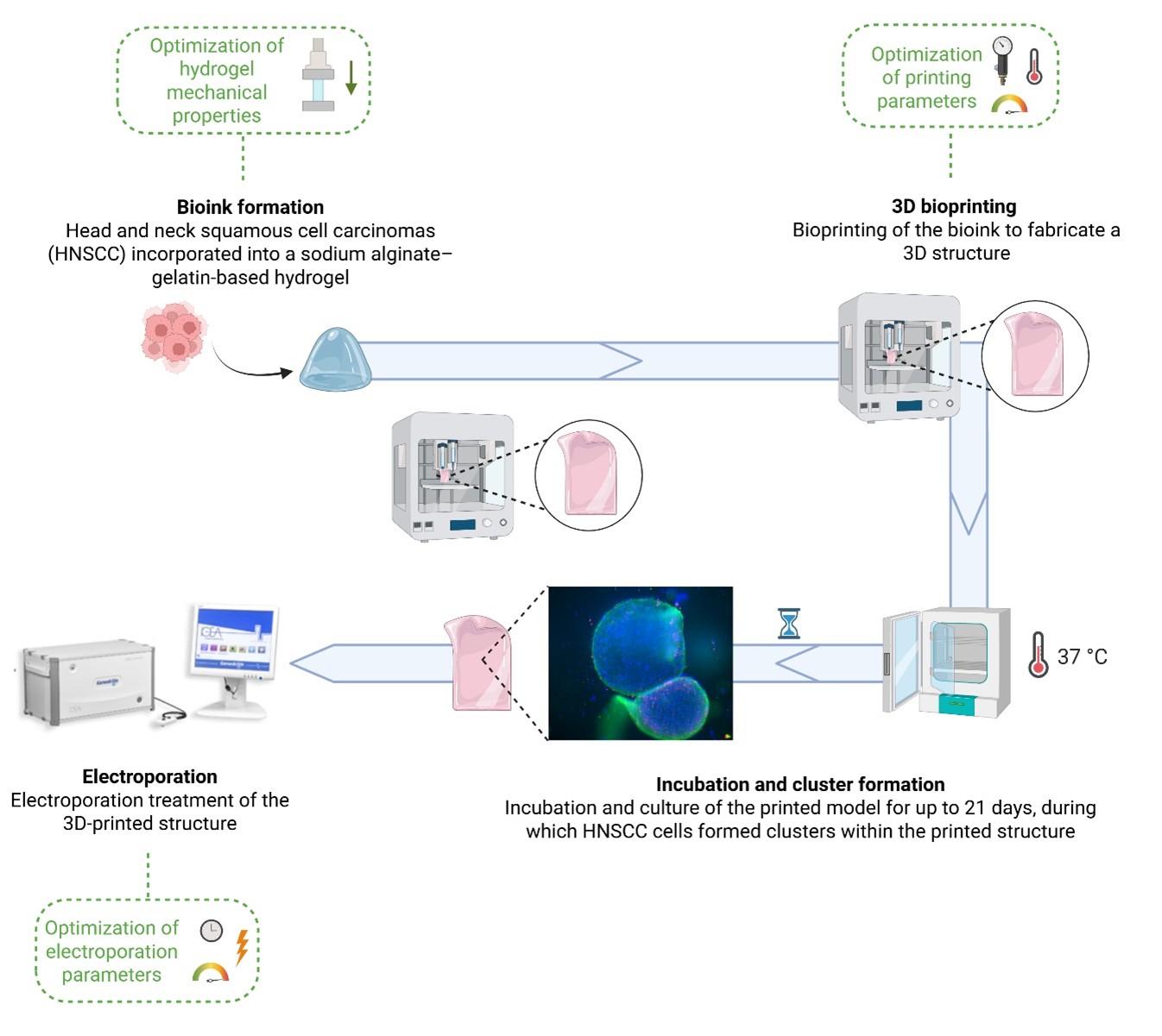
Head and neck squamous cell carcinomas (HNSCCs) are aggressive malignancies with poor prognosis and limited therapeutic options. Electrochemotherapy (ECT), combining short electric pulses with chemotherapeutic agents to enhance intracellular drug uptake, has shown clinical potential but still requires physiologically relevant in vitro models for protocol optimization and mechanistic studies. Here, we introduce a three-dimensional 3D bioprinted in vitro HNSCC model specifically designed for the assessment of electroporation. Structures were fabricated using a composite hydrogel composed of 8% sodium alginate and 4% gelatin (w/w), crosslinked with calcium chloride at concentrations of 0.5%, 1%, and 2%. Uniaxial compression testing confirmed elastic moduli spanning the physiological tumor stiffness range, with the 1% calcium chloride formulation providing optimal mechanical and handling characteristics (42.96 ± 19.89 kPa). Hypopharyngeal carcinoma FaDu cells (5×10⁶/mL) embedded in three-layer structures (thickness: 1.05 mm) maintained 75–80% viability for up to 21 days and formed tumor-like spheroids (mean diameter: 303 ± 113 μm), reflecting native tumor architecture. Electroporation with eight pulses at 200 V for 100 μs efficiently permeabilized the cell membrane, as evidenced by the internalization of propidium iodide, while maintaining high cell viability as confirmed by live/dead analysis. Programmed death-ligand 1 expression was preserved and upregulated in 3D spheroids compared to two-dimensional (2D) controls, supporting the platform’s relevance for immuno-oncology studies. Compared to other 3D HNSCC models, our system integrates mechanical tuning, electroporation compatibility, and immune-related biomarker expression, enabling functional validation of electric field-mediated intracellular delivery. This proof-of-concept platform demonstrates structural fidelity, long-term cell viability, and high reproducibility, offering a scalable, human-relevant tool for preclinical optimization of ECT and other electrically based therapies, bridging the gap between conventional 2D cultures and complex in vivo models.
- Chow Laura QM. Head and Neck Cancer. J Clin Med. 2020;382(1):60-72. doi: 10.1056/NEJMra1715715
- Pisani P, Airoldi M, Allais A, et al. Metastatic disease in head & neck oncology. Acta Otorhinolaryngol Ital. 2020;40(Suppl. 1):S1-s86. doi: 10.14639/0392-100X-suppl.1-40-2020
- Anderson G, Ebadi M, Vo K, Novak J, Govindarajan A, Amini A. An Updated Review on Head and Neck Cancer Treatment with Radiation Therapy. Cancers (Basel). 2021;13(19):4912. doi: 10.3390/cancers13194912
- Debela DT, Muzazu SG, Heraro KD, et al. New approaches and procedures for cancer treatment: Current perspectives. SAGE Open Med. 2021;9:20503121211034366. doi: 10.1177/20503121211034366
- Wang H, Zheng Z, Zhang Y, et al. Locally advanced head and neck squamous cell carcinoma treatment efficacy and safety: a systematic review and network meta-analysis. Review. Front Pharmacol. 2023;14:1269863. doi: 10.3389/fphar.2023.1269863
- Condello M, D’Avack G, Spugnini EP, Meschini S. Electrochemotherapy: An Alternative Strategy for Improving Therapy in Drug-Resistant SOLID Tumors. Cancers (Basel). 2022;14(17):4341. doi: 10.3390/cancers14174341
- Zupanic A, Kos B, Miklavcic D. Treatment planning of electroporation-based medical interventions: electrochemotherapy, gene electrotransfer and irreversible electroporation. Physics in Medicine & Biology. 2012; 57(17):5425. doi: 10.1088/0031-9155/57/17/5425
- Gehl J, Sersa G, Matthiessen LW, et al. Updated standard operating procedures for electrochemotherapy of cutaneous tumours and skin metastases. Acta Oncol. 2018;57(7):874-882. doi: 10.1080/0284186x.2018.1454602
- Gehl J, Sersa G, Garbay J, et al. Results of the ESOPE (European Standard Operating Procedures on Electrochemotherapy) study: Efficient, highly tolerable and simple palliative treatment of cutaneous and subcutaneous metastases from cancers of any histology. J Clin Oncol. 2006;24(18_suppl):8047-8047. doi: 10.1200/jco.2006.24.18_suppl.8047
- Martya M, Sersab G, Garbaya J-R, et al. Electrochemotherapy – An easy, highly effective and safe treatment of cutaneous and subcutaneous metastases: Results of ESOPE (European Standard Operating Procedures of Electrochemotherapy) study. Ejc Supplements. 2006;4:3-13.
- Calvet CY, Mir LM. The promising alliance of anti-cancer electrochemotherapy with immunotherapy. Cancer Metastasis Rev. 2016;35(2):165-77. doi: 10.1007/s10555-016-9615-3
- Abuwatfa WH, Pitt WG, Husseini GA. Scaffold-based 3D cell culture models in cancer research. J Biomed Sci. 2024;31(1):7. doi: 10.1186/s12929-024-00994-y
- Evangelista A, Scocozza F, Conti M, et al. Exploring Mechanical Features of 3D Head and Neck Cancer Models. J Funct Biomater. 2025;
- Ding Y, Chen J, Zhong W, Gu T, Xiao Y, Zhao Z. Bioprinting in tumor model construction for head and neck squamous cell carcinoma: A review. IJB. 2025;11(2):139–163. doi: 10.36922/ijb.8100
- Meng F, Meyer CM, Joung D, Vallera DA, McAlpine MC, Panoskaltsis-Mortari A. 3D Bioprinted In vitro Metastatic Models via Reconstruction of Tumor Microenvironments. Adv Mater. 2019;31(10):1806899. doi: 10.1002/adma.201806899
- Ng WL, Vyas C, Huang B, Yeong WY, Bartolo P. Advanced bioprinting strategies for fabrication of biomimetic tissues and organs. IJEM. 2025;7(6):062006. doi: 10.1088/2631-7990/adeee0
- Cui X, Jiao J, Yang L, et al. Advanced tumor organoid bioprinting strategy for oncology research. Mater Today Bio. 2024;28:101198. doi: 10.1016/j.mtbio.2024.101198
- Kort-Mascort J, Bao G, Elkashty O, et al. Decellularized Extracellular Matrix Composite Hydrogel Bioinks for the Development of 3D Bioprinted Head and Neck in vitro Tumor Models. ACS Biomater Sci Eng. 2021;7(11): 5288-5300. doi: 10.1021/acsbiomaterials.1c00812
- Azhakesan A, Kern J, Mishra A, et al. 3D Bioprinted Head and Neck Squamous Cell Carcinoma (HNSCC) Model Using Tunicate Derived Nanocellulose (NC) Bioink. Adv Healthc Mater. 2025:14:e2403114. doi: 10.1002/adhm.202403114
- Kort-Mascort J, Shen ML, Martin E, et al. Bioprinted cancer-stromalin-vitromodels in a decellularized ECM-based bioink exhibit progressive remodeling and maturation. Biomed Mater. 2023;18(4):045022. doi: 10.1088/1748-605X/acd830
- Delgrosso E, Scocozza F, Cansolino L, et al. 3D bioprinted osteosarcoma model for experimental boron neutron capture therapy (BNCT) applications: Preliminary assessment. J Biomed Mater Res Part B: Applied Biomater. 2023;111(8):1571-1580. doi: 10.1002/jbm.b.35255
- Wang X, Yang Y, Hu X, Kawazoe N, Yang Y, Chen G. Morphological and Mechanical Properties of Osteosarcoma Microenvironment Cells Explored by Atomic Force Microscopy. Anal Sci. 2016;32(11):1177-1182. doi: 10.2116/analsci.32.1177
- Xu W, Mezencev R, Kim B, Wang L, McDonald J, Sulchek T. Cell stiffness is a biomarker of the metastatic potential of ovarian cancer cells. PLoS One. 2012;7(10):e46609. doi: 10.1371/journal.pone.0046609
- Esch M, Sukhorukov VL, Kürschner M, Zimmermann U. Dielectric properties of alginate beads and bound water relaxation studied by electrorotation. Biopolymers. 1999;50(3):227-37. doi: 10.1002/(sici)1097-0282(199909)50:3<227::Aid-bip1>3.0.Co;2-y
- Kaklamani G, Kazaryan D, Bowen J, Iacovella F, Anastasiadis SH, Deligeorgis G. On the electrical conductivity of alginate hydrogels. Regen Biomater. 2018;5(5):293-301. doi: 10.1093/rb/rby019
- Distler T, Polley C, Shi F, et al. Electrically Conductive and 3D-Printable Oxidized Alginate-Gelatin Polypyrrole:PSS Hydrogels for Tissue Engineering. Adv Healthc Mater. 2021;10(9):e2001876. doi: 10.1002/adhm.202001876
- Tordi P, Tamayo A, Jeong Y, Bonini M, Samorì P. Multiresponsive Ionic Conductive Alginate/Gelatin Organohydrogels with Tunable Functions. Adv Funct Mater. 2024;34(52):2410663. doi: 10.1002/adfm.202410663
- Ji D, Park JM, Oh MS, et al. Superstrong, superstiff, and conductive alginate hydrogels. Nat Commun. 2022;13(1):3019. doi: 10.1038/s41467-022-30691-z
- Massey A, Stewart J, Smith C, et al. Mechanical properties of human tumour tissues and their implications for cancer development. Nat Rev Phys. 2024;6(4): 269-282. doi: 10.1038/s42254-024-00707-2
- Bergman E, Goldbart R, Traitel T, et al. Cell stiffness predicts cancer cell sensitivity to ultrasound as a selective superficial cancer therapy. Bioeng Transl Med. 2021;6(3):e10226. doi: 10.1002/btm2.10226
- Almela T, Tayebi L, Moharamzadeh K. 3D bioprinting for in vitro models of oral cancer: Toward development and validation. Bioprint. 2021;22:e00132. doi: 10.1016/j.bprint.2021.e00132
- Ragazzini S, Scocozza F, Bernava G, et al. Mechanosensor YAP cooperates with TGF-β1 signaling to promote myofibroblast activation and matrix stiffening in a 3D model of human cardiac fibrosis. Acta Biomater. 2022;152: 300-312. doi: 10.1016/j.actbio.2022.08.063
- Kulasinghe A, Kenny L, Punyadeera C. Circulating tumour cell PD-L1 test for head and neck cancers. Oral Oncol. 2017;75:6-7. doi: 10.1016/j.oraloncology.2017.10.011
- Stribbling SM, Ryan AJ. The cell-line-derived subcutaneous tumor model in preclinical cancer research. Nat Protoc. 2022;17(9):2108-2128. doi: 10.1038/s41596-022-00709-3
- Bylicky MA, Shankavaram U, Aryankalayil MJ, et al. Multiomic-Based Molecular Landscape of FaDu Xenograft Tumors in Mice after a Combinatorial Treatment with Radiation and an HSP90 Inhibitor Identifies Adaptation- Induced Targets of Resistance and Therapeutic Intervention. Mol Cancer Ther. 2024;23(4):577-588. doi: 10.1158/1535-7163.Mct-23-0796
- Fantini V, Bordoni M, Scocozza F, et al. Bioink Composition and Printing Parameters for 3D Modeling Neural Tissue. Cells. 2019;8(8):830.
- Batista Napotnik T, Miklavčič D. In vitro electroporation detection methods – An overview. Bioelectrochem. 2018;120:166-182. doi: 10.1016/j.bioelechem.2017.12.005
- Ruzgys P, Jakutavičiūtė M, Šatkauskienė I, Čepurnienė K, Šatkauskas S. Effect of electroporation medium conductivity on exogenous molecule transfer to cells in vitro. Sci Rep. 2019;9(1):1436. doi: 10.1038/s41598-018-38287-8
- Dermol J, Miklavčič D. Predicting electroporation of cells in an inhomogeneous electric field based on mathematical modeling and experimental CHO-cell permeabilization to propidium iodide determination. Bioelectrochem. 2014;100:52-61. doi: 10.1016/j.bioelechem.2014.03.011
- Batista Napotnik T, Polajžer T, Miklavčič D. Cell death due to electroporation – A review. Bioelectrochem. 2021;141:107871. doi: 10.1016/j.bioelechem.2021.107871
- Marazzi D, Carotenuto F, Trovalusci F, Caruccio P, Nardo P. Mechanisms, Models, and Clinical Applications of Cell Membrane Electroporation. Int J Transl. Sci. 2025;2024(4):257-302. doi: 10.13052/ijts2246-8765.2024.041
- Pisani S, Bertino G, Prina-Mello A, et al. Electroporation in Head-and-Neck Cancer: An Innovative Approach with Immunotherapy and Nanotechnology Combination. Cancers. 2022;14(21):5363.
- Sonaye SY, Ertugral EG, Kothapalli CR, Sikder P. Extrusion 3D (Bio)Printing of Alginate-Gelatin-Based Composite Scaffolds for Skeletal Muscle Tissue Engineering. Mater (Basel). 2022;15(22):7945. doi: 10.3390/ma15227945
- Xiao J, Song Y, Gao R, et al. Changes of immune microenvironment in head and neck squamous cell carcinoma in 3D-4-culture compared to 2D-4-culture. J Transl Med. 2023;21(1):771. doi: 10.1186/s12967-023-04650-1
- Friedrich J, Seidel C, Ebner R, Kunz-Schughart LA. Spheroid-based drug screen: considerations and practical approach. Nature Protocols. 2009;4(3):309-324. doi: 10.1038/nprot.2008.226
- Rasouli M. Basic concepts and practical equations on osmolality: Biochemical approach. Clin Biochem. 2016;49(12):936-41. doi: 10.1016/j.clinbiochem.2016.06.001
- Johnson DE, Burtness B, Leemans CR, Lui VWY, Bauman JE, Grandis JR. Head and neck squamous cell carcinoma. Nat Rev Dis Primers. 2020;6(1):92. doi: 10.1038/s41572-020-00224-3
- Lin CJ, Grandis JR, Carey TE, et al. Head and neck squamous cell carcinoma cell lines: Established models and rationale for selection. Head & Neck. 2007;29(2):163-188. doi: 10.1002/hed.20478
- Valdembri D, Serini G. The roles of integrins in cancer. Fac Rev. 2021;10:45. doi: 10.12703/r/10-45
- Datta P, Dey M, Ataie Z, Unutmaz D, Ozbolat IT. 3D bioprinting for reconstituting the cancer microenvironment. npj Precis Oncol. 2020;4(1):18. doi: 10.1038/s41698-020-0121-2