

Automated and scalable fabrication of scaffolded spheroids as building blocks for modular tissue engineering

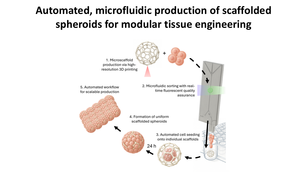
Current tissue engineering approaches typically utilize either scaffold-based or scaffold-free strategies. Recently, a third approach has emerged, offering a synergistic method that leverages the benefits of both approaches while avoiding their usual drawbacks. In this strategy, spheroids are formed within highly porous microscaffolds, which subsequently serve as building blocks for larger tissue constructs produced through self-assembly. This study demonstrates the automated fabrication of large numbers of these building blocks, tested in vessels of up to 1,536-well plates. To achieve this, we first developed a microfluidic device capable of sorting highly porous microscaffolds, each 300 µm in diameter, at high flow speeds of up to 300 mm/s. A fluorescent detection system was integrated to identify only intact microscaffolds and deposit them individually into separate wells of cell culture plates. Subsequently, the system automatically dispensed culture medium containing suspended human adipose-derived stem cells into each well. Spheroids were formed within 24 hours of culture with a formation efficiency of 95%. This study presents the development of an automated microfluidic device capable of depositing single microscaffolds and the cells required to form scaffolded spheroids. By enabling large-scale, high-throughput production, this approach advances the third strategy of tissue engineering and supports applications in both screening and clinical translation.
- Berthiaume F, Maguire TJ, Yarmush ML. Tissue Engineering and Regenerative Medicine: History, Progress, and Challenges. Annu Rev Chem Biomol Eng. 2011;2(1):403-430. doi: 10.1146/annurev-chembioeng-061010-114257
- Langer R, Vacanti JP. Tissue Engineering. Science. 1993;260(5110):920-926. doi: 10.1126/science.8493529
- Fontoura JC, Viezzer C, dos Santos FG, et al. Comparison of 2D and 3D cell culture models for cell growth, gene expression and drug resistance. Mater Sci Eng C. 2020;107:110264. doi: 10.1016/j.msec.2019.110264
- Khademhosseini A, Langer R. A decade of progress in tissue engineering. Nat Protoc. 2016;11(10):1775-1781. doi: 10.1038/nprot.2016.123
- Ovsianikov A, Khademhosseini A, Mironov V. The Synergy of Scaffold-Based and Scaffold-Free Tissue Engineering Strategies. Trends Biotechnol. 2018;36(4):348-357. doi: 10.1016/j.tibtech.2018.01.005
- Daly AC, Riley L, Segura T, Burdick JA. Hydrogel microparticles for biomedical applications. Nat Rev Mater. 2019;5(1):20-43. doi: 10.1038/s41578-019-0148-6
- Hutmacher DW. Scaffold design and fabrication technologies for engineering tissues — state of the art and future perspectives. J Biomater Sci Polym Ed. 2001;12(1):107-124. doi: 10.1163/156856201744489
- Ng WL, Vyas C, Huang B, Yeong WY, Bartolo P. Advanced bioprinting strategies for fabrication of biomimetic tissues and organs. Int J Extrem Manuf. 2025;7(6):062006. doi: 10.1088/2631-7990/adeee0
- Yang G, Mahadik B, Choi JY, Fisher JP. Vascularization in tissue engineering: fundamentals and state-of-art. Prog Biomed Eng. 2020;2(1):012002. doi: 10.1088/2516-1091/ab5637
- Zhang S, Wang Y, Onck P, den Toonder J. A concise review of microfluidic particle manipulation methods. Microfluid Nanofluid. 2020;24(4). doi: 10.1007/s10404-020-2328-5
- Sajeesh P, Sen AK. Particle separation and sorting in microfluidic devices: a review. Microfluid Nanofluid. 2013;17(1):1-52. doi: 10.1007/s10404-013-1291-9
- Zhang P, Chang KC, Abate AR. Precision ejection of microfluidic droplets into air with a superhydrophobic outlet. Lab Chip. 2021;21(8):1484-1491. doi: 10.1039/d0lc01327g
- Kamperman T, Trikalitis VD, Karperien M, Visser CW, Leijten J. Ultrahigh-Throughput Production of Monodisperse and Multifunctional Janus Microparticles Using in-Air Microfluidics. ACS Appl Mater Interfaces. 2018;10(28):23433-23438. doi: 10.1021/acsami.8b05227
- Visser CW, Kamperman T, Karbaat LP, Lohse D, Karperien M. In-air microfluidics enables rapid fabrication of emulsions, suspensions, and 3D modular (bio)materials. Sci Adv. 2018;4(1). doi: 10.1126/sciadv.aao1175
- Torino S, Dhurandhar M, Stroobants A, Claessens R, Efremov RG. Time-resolved cryo-EM using a combination of droplet microfluidics with on-demand jetting. Nat Methods. 2023;20(9):1400-1408. doi: 10.1038/s41592-023-01967-z
- Ferreira DA, Rothbauer M, Conde JP, Ertl P, Oliveira C, Granja PL. A Fast Alternative to Soft Lithography for the Fabrication of Organ‐on‐a‐Chip Elastomeric‐Based Devices and Microactuators. Adv Sci. 2021;8(8). doi: 10.1002/advs.202003273
- Guillaume O, Kopinski-Grünwald O, Weisgrab G, et al. Hybrid spheroid microscaffolds as modular tissue units to build macro-tissue assemblies for tissue engineering. Acta Biomaterialia. 2023;165:72-85. doi: 10.1016/j.actbio.2022.03.010
- Ovsianikov A, Viertl J, Chichkov B, et al. Ultra-Low Shrinkage Hybrid Photosensitive Material for Two-Photon Polymerization Microfabrication. ACS Nano. 2008;2(11):2257-2262. doi: 10.1021/nn800451w
- Tinevez JY, Perry N, Schindelin J, et al. TrackMate: An open and extensible platform for single-particle tracking. Methods. 2017;115:80-90. doi: 10.1016/j.ymeth.2016.09.016
- Schindelin J, Arganda-Carreras I, Frise E, et al. Fiji: an open-source platform for biological-image analysis. Nat Methods. 2012;9(7):676-682. doi: 10.1038/nmeth.2019
- Schroeder WF, Asmussen SL, Cook WD, Vallo CI. Efficiency of 4,4′‐bis(N,N‐diethylamino) benzophenone for the polymerization of dimethacrylate resins in thick sections. Polym Int. 2011;60(9):1362-1369. doi: 10.1002/pi.3089
- National Instruments. NI-DAQmx Python documentation. Accessed October 10, 2021. https://nidaqmx-python.readthedocs.io/en/latest/
- Huang D, Wang H, Wang X, et al. Investigation of Optimal Coupling Velocities of the Sample and Sheath Flows for Hydrodynamic Focusing. J Mar Sci Eng. 2020;8(8):601. doi: 10.3390/jmse8080601
- Pethig R. Review Article—Dielectrophoresis: Status of the theory, technology, and applications. Biomicrofluidics. 2010;4(2). doi: 10.1063/1.3456626
- Grenvall C, Antfolk C, Bisgaard CZ, Laurell T. Two-dimensional acoustic particle focusing enables sheathless chip Coulter counter with planar electrode configuration. Lab Chip. 2014;14(24):4629-4637. doi: 10.1039/c4lc00982g
- Yumoto M, Hemmi N, Sato N, et al. Evaluation of the effects of cell-dispensing using an inkjet-based bioprinter on cell integrity by RNA-seq analysis. Sci Rep. 2020;10(1). doi: 10.1038/s41598-020-64193-z
- Binek A, Rojo D, Godzien J, et al. Flow Cytometry Has a Significant Impact on the Cellular Metabolome. J Proteome Res. doi: 10.1021/acs.jproteome.8b00472
- Binder S, Chalupa-Gantner F, Yoo HW, Zandrini T, Ovsianikov A. Two-photon polymerization system based on a resonant scanner for high-throughput production of tissue engineering microscaffolds. Addit Manuf. 2025;97:104601. doi: 10.1016/j.addma.2024.104601