

Electrohydrodynamically printed microfibrous scaffolds with different pore sizes modulate macrophage polarization and foreign body reaction to enhance bone regeneration

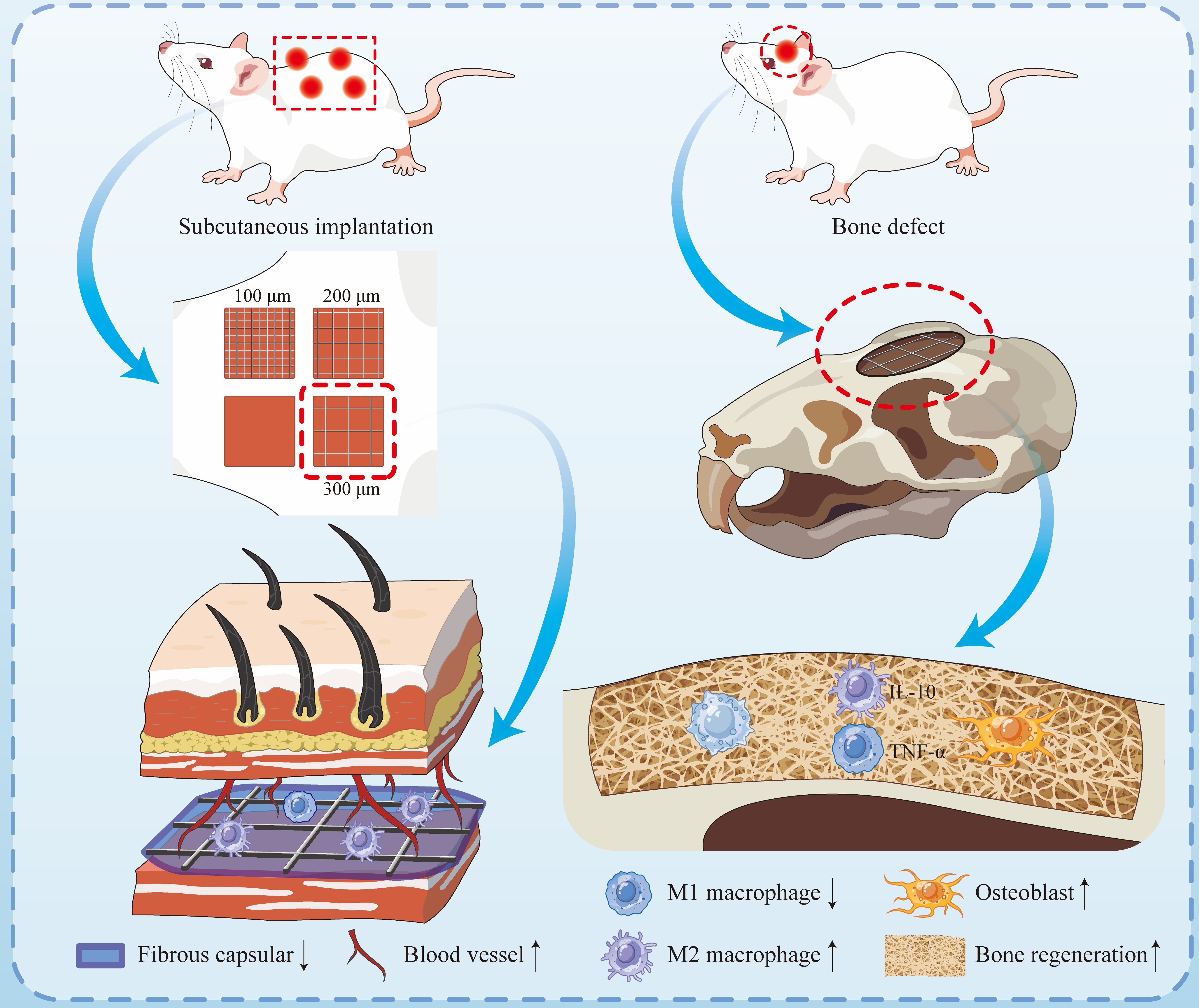
Foreign body reaction (FBR) is a major obstacle to effective osseointegration in bone defect repair. The pore size of scaffolds is a key determinant of FBR; however, its impact on FBR remains controversial, with limited in vivo evidence available. In this study, electrohydrodynamically printed polycaprolactone scaffolds with pore sizes of 100 μm, 200 μm, and 300 μm were fabricated to investigate their effects on macrophage polarization, FBR, and bone regeneration. In vitro experiments showed that the 300 μm group promoted M2 polarization of macrophages, reduced tumor necrosis factor-alpha expression (0.71-fold and 0.81-fold relative to the 100 μm and 200 μm groups, respectively), and increased transforming growth factor-beta 1 expression (1.39-fold and 1.19-fold, respectively), thereby enhancing osteogenic gene expression in MC3T3-E1 cells (Runx2, Col1, and Ocn). Finite element analysis and transcriptomics sequencing revealed that pore size-dependent changes in scaffold stiffness modulate Piezo1 activation, influencing macrophage polarization. In vivo experiments showed that the 300 μm group exhibited the thinnest fibrous capsule (0.78-fold and 0.79-fold relative to the 100 μm and 200 μm groups, respectively), demonstrated enhanced angiogenesis, and achieved better bone regeneration, with increased bone volume/total volume and bone mineral density. These findings indicate that 300 μm pore-sized scaffolds promote bone regeneration by modulating macrophage polarization and attenuating FBR, providing a basis for optimized scaffold design and clinical translation in bone defect repair.
- Liu L, Chen H, Zhao X, et al. Advances in the application and research of biomaterials in promoting bone repair and regeneration through immune modulation. Mater Today Bio. 2024;30:101410. doi: 10.1016/j.mtbio.2024.101410
- Szczodra A, Houaoui A, Agniel R, et al. Boron substitution in silicate bioactive glass scaffolds to enhance bone differentiation and regeneration. Acta Biomater. 2024;186:489-506. doi: 10.1016/j.actbio.2024.07.053
- He X, Liu Y, Dai Z et al. Yoda1 pretreated BMSC derived exosomes accelerate osteogenesis by activating phospho- ErK signaling via Yoda1-mediated signal transmission. J Nanobiotechnol. 2024;22(1):407. doi: 10.1186/s12951-024-02669-0
- Dang Y, Zhang Y, Luo G, et al. The decisive early phase of biomaterial-induced bone regeneration. Appl Mater Today. 2024;38:102236. doi: 10.1016/j.apmt.2024.102236
- Jin S, Wen J, Zhang Y, et al. M2 macrophage-derived exosome-functionalized topological scaffolds regulate the foreign body response and the coupling of angio/osteoclasto/ osteogenesis. Acta Biomater. 2024;177:91-106. doi: 10.1016/j.actbio.2024.01.043
- Qiu D, Cao C, Prasopthum A, et al. Elucidating osseointegration in vivo in 3D printed scaffolds eliciting different foreign body responses. Mater Today Bio. 2023;22:100771. doi: 10.1016/j.mtbio.2023.100771
- Mesa-Restrepo A, Byers E, Brown JL, Ramirez J, Allain JP, Posada VM. Osteointegration of Ti bone implants: a study on how surface parameters control the foreign body response. ACS Biomater Sci Eng. 2024;10(8):4662-4681. doi: 10.1021/acsbiomaterials.4c00114
- Dondossola E, Holzapfel BM, Alexander S, Filippini S, Hutmacher DW, Friedl P. Examination of the foreign body response to biomaterials by nonlinear intravital microscopy. Nat Biomed Eng. 2016;1:0007. doi: 10.1038/s41551-016-0007
- Anderson JM, Rodriguez A, Chang DT. Foreign body reaction to biomaterials. Semin Immunol. 2008;20(2):86-100. doi: 10.1016/j.smim.2007.11.004
- Fu M, Yang C, Sun G. Recent advances in immunomodulatory hydrogels biomaterials for bone tissue regeneration. Mol Immunol. 2023;163:48-62. doi: 10.1016/j.molimm.2023.09.010
- Li W, Dai F, Zhang S, et al. Pore size of 3D-printed polycaprolactone/polyethylene glycol/hydroxyapatite scaffolds affects bone regeneration by modulating macrophage polarization and the foreign body response. ACS Appl Mater Interfaces. 2022;14(18):20693-20707. doi: 10.1021/acsami.2c02001.
- Franz S, Rammelt S, Scharnweber D, Simon JC. Immune responses to implants - a review of the implications for the design of immunomodulatory biomaterials. Biomaterials. 2011;32(28):6692-6709. doi: 10.1016/j.biomaterials.2011.05.078.
- Kastellorizios M, Papadimitrakopoulos F, Burgess DJ. Multiple tissue response modifiers to promote angiogenesis and prevent the foreign body reaction around subcutaneous implants. J Control Release. 2015;214:103-111. doi: 10.1016/j.jconrel.2015.07.021.
- Spiller KL, Nassiri S, Witherel CE, et al. Sequential delivery of immunomodulatory cytokines to facilitate the M1-to-M2 transition of macrophages and enhance vascularization of bone scaffolds. Biomaterials. 2015;37:194-207. doi: 10.1016/j.biomaterials.2014.10.017.
- Li R, Zhang K, Dong C, Wang K, Gu X, Qin Y. Osteoinductivity enhancement by tailoring the surface chemical bond status: a strategy to mobilize host bone growth factors for in situ bone regeneration. Mater Today Bio. 2024;29:101256. doi: 10.1016/j.mtbio.2024.101256.
- Vegas AJ, Veiseh O, Doloff JC, et al. Combinatorial hydrogel library enables identification of materials that mitigate the foreign body response in primates. Nat Biotechnol. 2016;34(6):345-352. doi: 10.1038/nbt0616-666e.
- Xiong S, Zhang Y, Zeng J, et al. DLP fabrication of HA scaffold with customized porous structures to regulate immune microenvironment and macrophage polarization for enhancing bone regeneration. Mater Today Bio. 2024;24:100929. doi: 10.1016/j.mtbio.2023.100929.
- Veiseh O, Doloff JC, Ma M, et al. Size- and shape-dependent foreign body immune response to materials implanted in rodents and non-human primates. Nat Mater. 2015;14(6):643–651. doi: 10.1038/nmat4290.
- Sridharan R, Cavanagh B, Cameron AR, Kelly DJ, O’Brien FJ. Material stiffness influences the polarization state, function and migration mode of macrophages. Acta Biomater. 2019;89:47-59. doi: 10.1016/j.actbio.2019.02.048.
- Xia D, Wang Y, Wu R, et al. The effect of pore size on cell behavior in mesoporous bioglass scaffolds for bone regeneration. Appl Mater Today. 2022;29:101607. doi: 10.1016/j.apmt.2022.101607
- Jiang S, Lyu C, Zhao P, et al. Cryoprotectant enables structural control of porous scaffolds for exploration of cellular mechano-responsiveness in 3D. Nat Commun. 2019;10(1):3491. doi: 10.1038/s41467-019-11397-1.
- Hady TF, Hwang B, Pusic AD, et al. Uniform 40‐μm‐pore diameter precision templated scaffolds promote a pro‐healing host response by extracellular vesicle immune communication. J Tissue Eng Regen Med. 2020; 15(1):24-36. doi: 10.1002/term.3160.
- Meng Z, Yang S, Yin F, et al. 3D-printed biodegradable polycaprolactone rib implants with tissue-specific mechanical properties promote chest wall recovery by stimulating tissue fibrosis. Virtual Phys Prototyp. 2024;19(1):2346816. doi: 10.1080/17452759.2024.2346816.
- He J, Xia P, Li D. Development of melt electrohydrodynamic 3D printing for complex microscale poly (ε-caprolactone) scaffolds. Biofabrication. 2016;8(3):035008. doi: 10.1088/1758-5090/8/3/035008.
- Han K, He J, Fu L, Mao M, Kang Y, Li D. Engineering highly-aligned three-dimensional (3D) cardiac constructs for enhanced myocardial infarction repair. Biofabrication. 2022;15(1):015003. doi: 10.1088/1758-5090/ac94f9.
- Shi Y, Wang L, Sun L, et al. Melt electrospinning writing PCL scaffolds after alkaline modification with outstanding cytocompatibility and osteoinduction. Int J Bioprint. 2023;9(6):1071. doi: 10.36922/ijb.1071.
- Yao C, Qiu Z, Li X, Zhu H, Li D, He J. Electrohydrodynamic printing of microfibrous architectures with cell‐scale spacing for improved cellular migration and neurite outgrowth. Small. 2023;19(19):2207331. doi: 10.1002/smll.202207331.
- Tylek T, Blum C, Hrynevich A, et al. Precisely defined fiber scaffolds with 40 μm porosity induce elongation driven M2- like polarization of human macrophages. Biofabrication. 2020;12(2):025007. doi: 10.1088/1758-5090/ab5f4e.
- Bružauskaitė I, Bironaitė D, Bagdonas E, Bernotienė E. Scaffolds and cells for tissue regeneration: different scaffold pore sizes—different cell effects. Cytotechnology. 2016;68(3):355-369. doi: 10.1007/s10616-015-9895-4.
- Zhang Y, Li R, Wu W, et al. Adhesion and proliferation of osteoblast-like cells on porous polyetherimide scaffolds. Biomed Res Int. 2018;2018:1491028. doi: 10.1155/2018/1491028.
- Lai Y, Cao H, Wang X, et al. Porous composite scaffold incorporating osteogenic phytomolecule icariin for promoting skeletal regeneration in challenging osteonecrotic bone in rabbits. Biomaterials. 2017;153:1-13. doi: 10.1016/j.biomaterials.2017.10.025.
- Hu S, Meng Z, Zhou J, et al. Enhanced attachment and collagen type I deposition of MC3T3-E1 cells via electrohydrodynamic printed sub-microscale fibrous architectures. Int J Bioprint. 2022;8(2):514. doi: 10.18063/ijb.v8i2.514.
- Brennan CM, Eichholz KF, Hoey DA. The effect of pore size within fibrous scaffolds fabricated using melt electrowriting on human bone marrow stem cell osteogenesis. Biomed Mater. 2019;14(6):065016. doi: 10.1088/1748-605x/ab49f2.
- Shi Y, Tao W, Yang W, et al. Calcium phosphate coating enhances osteointegration of melt electrowritten scaffold by regulating macrophage polarization. J Nanobiotechnol. 2024;22(1):47. doi: 10.1186/s12951-024-02310-0.
- Palmieri EM, McGinity C, Wink DA, McVicar DW. Nitric oxide in macrophage immunometabolism: hiding in plain sight. Metabolites. 2020;10(11):429. doi: 10.3390/metabo10110429.
- Zhao DW, Liu C, Zuo KQ, et al. Strontium-zinc phosphate chemical conversion coating improves the osseointegration of titanium implants by regulating macrophage polarization. Chem Eng J. 2020;408:127362. doi: 10.1016/j.cej.2020.127362.
- Lv L, Xie Y, Li K, et al. Unveiling the mechanism of surface hydrophilicity‐modulated macrophage polarization. Adv Healthc Mater. 2018;7(19):1800675. doi: 10.1002/adhm.201800675.
- Abedin E, Lari R, Mahdavi Shahri N, Fereidoni M. Development of a demineralized and decellularized human epiphyseal bone scaffold for tissue engineering: a histological study. Tissue Cell. 2018;55:46-52. doi: 10.1016/j.tice.2018.09.003.
- Wei X, Zhou W, Tang Z, et al. Magnesium surface-activated 3D printed porous PEEK scaffolds for in vivo osseointegration by promoting angiogenesis and osteogenesis. Bioact Mater. 2023;20:16-28. doi: 10.1016/j.bioactmat.2022.05.011.
- Xu J, Guan W, Kong Y, et al. Regulation of macrophage behavior by chitosan scaffolds with different elastic modulus. Coatings. 2022;12(11):1742. doi: 10.3390/coatings12111742.
- Du Y, Xu B, Li Q, Peng C, Yang K. The role of mechanically sensitive ion channel Piezo1 in bone remodeling. Front Bioeng Biotechnol. 2024;12:1342149. doi: 10.3389/fbioe.2024.1342149.
- Li W, Xu F, Dai F, et al. Hydrophilic surface-modified 3D printed flexible scaffolds with high ceramic particle concentrations for immunopolarization-regulation and bone regeneration. Biomater Sci. 2023;11:3976-3997. doi: 10.1039/d3bm00362k.
- Mahon OR, Browe DC, Gonzalez-Fernandez T, et al. Nano-particle mediated M2 macrophage polarization enhances bone formation and MSC osteogenesis in an IL-10 dependent manner. Biomaterials. 2020;239:119833. doi: 10.1016/j.biomaterials.2020.119833.
- Li Y, He J, Zhou J, et al. A conductive photothermal non-swelling nanocomposite hydrogel patch accelerating bone defect repair. Biomater Sci. 2022;10:1326-1341. doi: 10.1039/d1bm01937f.
- Garg K, Pullen NA, Oskeritzian CA, Ryan JJ, Bowlin GL. Macrophage functional polarization (M1/M2) in response to varying fiber and pore dimensions of electrospun scaffolds. Biomaterials. 2013;34(18):4439-4451. doi: 10.1016/j.biomaterials.2013.02.065.
- Yin Y, He XT, Wang J, et al. Pore size-mediated macrophage M1-to-M2 transition influences new vessel formation within the compartment of a scaffold. Appl Mater Today. 2019;18:100466. doi: 10.1016/j.apmt.2019.100466.
- Sridharan R, Cameron AR, Kelly DJ, Kearney CJ, O’Brien FJ. Biomaterial based modulation of macrophage polarization: a review and suggested design principles. Mater Today. 2015;18(6):313-325. doi: 10.1016/j.mattod.2015.01.019.
- Zhang D, Chen Q, Shi C, et al. Dealing with the foreign‐body response to implanted biomaterials: strategies and applications of new materials. Adv Funct Mater. 2021;31(6):2007226.doi: 10.1002/adfm.202007226.
- Liu Y, Suarez-Arnedo A, Riley L, Miley T, Xia J, Segura T. Spatial confinement modulates macrophage response in microporous annealed particle (MAP) scaffolds. Adv Healthcare Mater. 2023;12(26):2300823. doi: 10.1002/adhm.202300823.
- Sheikh Z, Brooks P, Barzilay O, Fine N, Glogauer M. Macrophages, foreign body giant cells and their response to implantable biomaterials. Materials. 2015;8(9):5671-5701. doi: 10.3390/ma8095269
- Ivanova E, Fayzullin A, Minaev N, et al. Surface topography of PLA implants defines the outcome of foreign body reaction: an in vivo study. Polymers. 2023;15(20):4119. doi: 10.3390/polym15204119.
- Zhao SJ, Kong FQ, Jie J, et al. Macrophage MSR1 promotes BMSC osteogenic differentiation and M2-like polarization by activating PI3K/AKT/GSK3β/β-catenin pathway. Theranostics. 2020;10(1):17-35. doi: 10.7150/thno.36930.
- Fan S, Zhang C, Sun X, et al. Metformin enhances osteogenic differentiation of BMSC by modulating macrophage M2 polarization. Sci Rep. 2024;14(1):20267. doi: 10.1038/s41598-024-71318-1.
- Feito MJ, Casarrubios L, Oñaderra M, et al. Response of RAW 264.7 and J774A.1 macrophages to particles and nanoparticles of a mesoporous bioactive glass: a comparative study. Colloids Surf B Biointerfaces. 2021;208:112110. doi: 10.1016/j.colsurfb.2021.112110
- Trentini M, D’Amora U, Ronca A, et al. Bone regeneration revolution: pulsed electromagnetic field modulates macrophage-derived exosomes to attenuate osteoclastogenesis. Int J Nanomed. 2024;19:8695-8707. doi: 10.2147/ijn.s470901.