

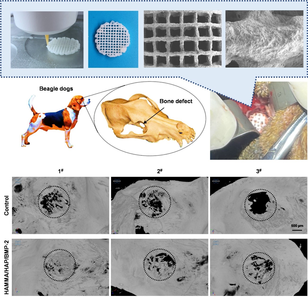
Hydroxyapatite/BMP-2–mineralized decellularized amniotic membrane scaffolds for orbital defect repair

Orbital wall fractures often result in midfacial deformities characterized by herniation of orbital adipose and soft tissues into the maxillary sinus, potentially causing endophthalmitis or subbulbar inflammation. However, current orbital reconstruction materials face critical limitations, including inadequate osteogenic capacity and poor osseointegration, predisposing implants to displacement, immune rejection, and infection. To overcome these challenges, we fabricated a three-dimensional (3D)-printed scaffold based on hydroxyapatite/bone morphogenetic protein-2–mineralized decellularized amniotic membrane for orbital defect repair. By precisely modulating the material composition and leveraging advanced 3D printing techniques, we achieved simultaneous control over the scaffold’s physicochemical properties and biological activity. The resulting constructs feature optimized macro- and micro-architectures. This study establishes a novel strategy for orbital reconstruction, addressing both bone volume restoration and functional regeneration, offering a transformative approach for personalized craniofacial repair.
- Sachs ME. Orbital floor fractures: the maxillary approach. Adv Ophthalmic Plast Reconstr Surg. 1987;6(1):387-391.
- Ni X, Feng J, Liang M, et al. Enhancing bone repair with β-TCP-based composite scaffolds: a review of design strategies and biological mechanisms. Orthop Res Rev. 2025;17:313-340. doi: 10.2147/ORR.S525959
- Al-Khdhairi OBH, Abdulrazaq SS. Is orbital floor reconstruction with titanium mesh safe?. J Craniofac Surg. 2017;28(7):e692-e694. doi: 10.1097/SCS.0000000000003896
- Shah HA, Shipchandler T, Vernon D, et al. Extra-ocular movement restriction and diplopia following orbital fracture repair. Am J Otolaryngol. 2018;39(1):34-36. doi: 10.1016/j.amjoto.2017.08.008
- Kwon H, Kim HJ, Seo BF, Jeong YJ, Jung SN, Shim HS. The role of resorbable plate and artificial bone substitute in reconstruction of large orbital floor defect. Biomed Res Int. 2016;2016:1358312. doi: 10.1155/2016/1358312
- Song X, Li L, Sun Y, Fan X, Li Z. Long-term infectious complications of using porous polyethylene mesh for orbital fracture reconstruction. Medicine (Baltimore). 2016;95(25):e3819. doi: 10.1097/MD.0000000000003819
- Zheng YX, Zhao HY, Jing XB, et al. Reconstruction of orbital floor defect with polylacticglycolide acid/recombinant human bone morphogenetic protein 2 compound implanted material in sheep. [in Chinese] Zhonghua Yan Ke Za Zhi. 2006;42(6):535-539.
- Samadikuchaksaraei A, Mehdipour A, Habibi Roudkenar M, et al. A dermal equivalent engineered with TGF‐ β3 expressing bone marrow stromal cells and amniotic membrane: cosmetic healing of full‐thickness skin wounds in rats. Artif Organs. 2016;40(12):266-279. doi: 10.1111/aor.12807
- Zheng Y, Ji S, Wu H, et al. Topical administration of cryopreserved living micronized amnion accelerates wound healing in diabetic mice by modulating local microenvironment. Biomaterials. 2017;113:56-67. doi: 10.1016/j.biomaterials.2016.10.031
- Etchebarne M, Fricain JC, Kerdjoudj H, et al. Use of amniotic membrane and its derived products for bone regeneration: a systematic review. Front Bioeng Biotechnol. 2021;9: 661332. doi: 10.3389/fbioe.2021.661332
- Leal-Marin S, Kern T, Hofmann N, et al. Human amniotic membrane: a review on tissue engineering, application, and storage. J Biomed Mater Res B Appl Biomater. 2021;109(8):1198-1215. doi: 10.1002/jbm.b.34782
- Dadkhah Tehrani F, Firouzeh A, Shabani I, Shabani A. A review on modifications of amniotic membrane for biomedical applications. Front Bioeng Biotechnol. 2021;8:606982. doi: 10.3389/fbioe.2020.606982
- Suter AJ, Molteno AC, Bevin TH, et al. Long term follows up of bone derived hydroxyapatite orbital implants. Br J Ophthalmol. 2002;86(11):1287-1292. doi: 10.1136/bjo.86.11.1287
- Yan S, Feng L, Zhu Q, et al. Controlled release of BMP- 2 from a Heparin-conjugated strontium-substituted nanohydroxyapatite/silk fibroin scaffold for bone regeneration. ACS Biomater Sci Eng. 2018;4(9):3291-3303. doi: 10.1021/acsbiomaterials.8b00549
- Dewey MJ, Johnson EM, Slater ST, Milner DJ, Wheeler MB, Harley BAC. Mineralized collagen scaffolds fabricated with amniotic membrane matrix increase osteogenesis under inflammatory conditions. Regen Biomater. 2020;7(3):247-258. doi: 10.1093/rb/rbaa005
- Jia Z, Ma H, Liu J, et al. Preparation and characterization of polylactic acid/nano hydroxyapatite/nano hydroxyapatite/ human acellular amniotic membrane (PLA/nHAp/HAAM) hybrid scaffold for bone tissue defect repair. Materials (Basel). 2023;16(5):1937. doi: 10.3390/ma16051937.
- Cole P, Boyd V, Banerji S, Hollier LH. Comprehensive management of orbital fractures. Plast Reconstr Surg. 2007;120(7 Suppl 2):57S-63S. doi: 10.1097/01.prs.0000260752.20481.b4
- Wallace J, Wang M, Thompson P, et al. Validating continuous digital light processing (cDLP) additive manufacturing accuracy and tissue engineering utility of a dye-initiator package. Biofabrication. 2014;6(1):015003. doi: 10.1088/1758-5082/6/1/015003
- Zhong M, Sun J, Wei D, et al. Establishing a cell-affinitive interface and spreading space in a 3D hydrogel by introduction of microcarriers and an enzyme. J Mater Chem B. 2014;2(38):6601-6610. doi: 10.1039/c4tb00887a
- Yang R, Chen B, Zhang X, Bao Z, Yan Q, Luan S. Degradable nanohydroxyapatite-reinforced superglue for rapid bone fixation and promoted osteogenesis. ACS Nano. 2024;18(11):8517-8530. doi: 10.1021/acsnano.4c01214
- Jamnezhad S, Asefnejad A, Motififard M, et al. Development and investigation of novel alginate-hyaluronic acid bone fillers using freeze drying technique for orthopedic field. Nanomed Res J. 2020;5(4):306-315. doi: 10.22034/nmrj.2020.04.001
- Lakkireddy C, Vishwakarma SK, Bardia A, et al. Biofabrication of allogenic bone grafts using cellularized amniotic scaffolds for application in efficient bone healing. Tissue Cell. 2021;73:101631. doi: 10.1016/j.tice.2021.101631
- Dawiec G, Niemczyk W, Wiench R, Niemczyk S, Skaba D. Introduction to amniotic membranes in maxillofacial surgery-a scoping review. Medicina (Kaunas). 2024;60(4):663. doi: 10.3390/medicina60040663
- Han S, Paeng KW, Park S, Jung UW, Cha JK, Hong J. Programmed BMP-2 release from biphasic calcium phosphates for optimal bone regeneration. Biomaterials. 2021;272:120785. doi: 10.1016/j.biomaterials.2021.120785
- Bölgen N, Plieva F, Galaev I, Mattiasson B, Pişkin E. Cryogelation for preparation of novel biodegradable tissue-engineering scaffolds. J Biomater Sci Polym Ed. 2007;18(9):1165-1179. doi: 10.1163/156856207781554046
- Xu M, Qin M, Zhang X, et al. Porous PVA/SA/HA hydrogels fabricated by dual-crosslinking method for bone tissue engineering. J Biomater Sci Polym Ed. 2020;31(6):816-831. doi: 10.1080/09205063.2020.1720155
- Dorman LJ, Tucci M, Benghuzzi H. In vitro effects of bmp- 2, bmp-7, and bmp-13 on proliferation and differentation of mouse mesenchymal stem cells. Biomed Sci Instrum. 2012;48:81-87.
- Kua HY, Liu H, Leong WF, et al. c-Abl promotes osteoblast expansion by differentially regulating canonical and non-canonical BMP pathways and p16INK4a expression. Nat Cell Biol. 2012;14(7):727-737. doi: 10.1038/ncb2528
- Grassi F, Tyagi AM, Calvert JW, et al. Hydrogen sulfide is a novel regulator of bone formation implicated in the bone loss induced by estrogen deficiency. J Bone Miner Res. 2016;31(5):949-963. doi: 10.1002/jbmr.2757
- Matsumoto Y, Otsuka F, Takano-Narazaki M, et al. Estrogen facilitates osteoblast differentiation by upregulating bone morphogenetic protein-4 signaling. Steroids. 2013;78(5):513-520. doi: 10.1016/j.steroids.2013.02.011
- Shen Y, Liu F, Duan J, et al. Biomaterial cues regulated differentiation of neural stem cells into GABAergic neurons through Ca2+/c-Jun/TLX3 signaling promoted by hydroxyapatite nanorods. Nano Lett. 2021;21(17):7371-7378. doi: 10.1021/acs.nanolett.1c02708
- Tang K, Wu J, Xiong Z, Ji Y, Sun T, Guo X. Human acellular amniotic membrane: a potential osteoinductive biomaterial for bone regeneration. J Biomater Appl. 2018;32(6):754-764. doi: 10.1177/0885328217739753
- Akhlaghi F, Hesami N, Rad MR, Nazeman P, Fahimipour F, Khojasteh A. Improved bone regeneration through amniotic membrane loaded with buccal fat pad-derived MSCs as an adjuvant in maxillomandibular reconstruction. J Craniomaxillofac Surg. 2019;47(8):1266-1273. doi: 10.1016/j.jcms.2019.03.030