

Enhancing osteogenesis using 3D-printed porous tantalum scaffolds: A biomechanical, in vivo, and in vitro study

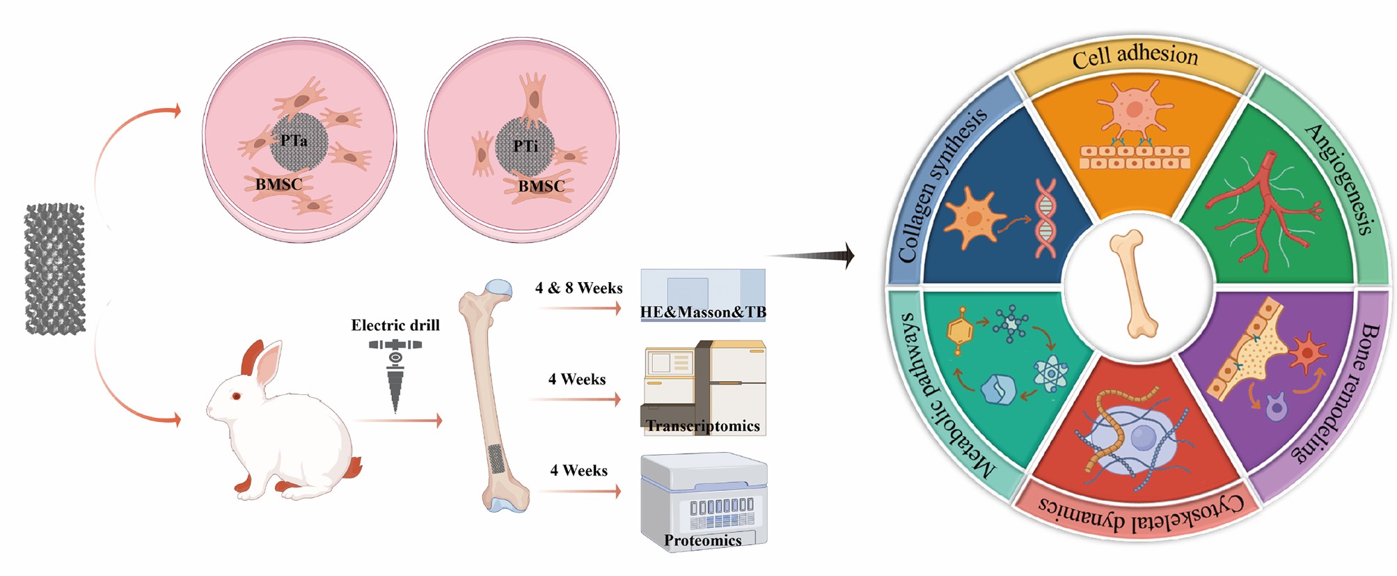
Complex bone defects continue to pose significant challenges in the field of orthopedics, where restoring structural integrity and promoting osteointegration are essential for successful repair outcomes. Three-dimensional (3D) printing offers a robust approach for fabricating patient-specific scaffolds with precise architectural and functional control. In this study, we designed and fabricated porous scaffolds composed of tantalum and titanium alloys, both with identical porosity, utilizing 3D printing technology. We systematically compared their mechanical properties, in vitro osteogenic potential, and in vivo bone integration within a defect model. The porous tantalum (PTa) scaffolds demonstrated exceptional biocompatibility, enhanced cell adhesion, and significantly promoted the osteogenic differentiation of mesenchymal stem cells, as well as extracellular matrix mineralization. In vivo, the PTa scaffolds not only expedited bone repair but also improved osteoconductive ingrowth compared to their titanium counterparts. Multi-omics analyses further elucidated potential biological mechanisms underlying the superior performance of PTa. These findings underscore the potential of 3D-printed PTa as a promising scaffold material for the clinical repair of bone defects.
- Zura R, Xiong Z, Einhorn T, et al. Epidemiology of fracture nonunion in 18 human bones. JAMA Surg. 2016;151(11):e162775. doi: 10.1001/jamasurg.2016.2775
- Miron RJ. Advanced functional materials. Periodontology 2000. 2024;94(1):143-160. doi: 10.1111/prd.12517
- Zadpoor AA. Current trends in metallic orthopedic biomaterials: from additive manufacturing to bio-functionalization, infection prevention, and beyond. IJMS. 2018;19(9):2684. doi: 10.3390/ijms19092684
- Arabnejad S, Johnston B, Tanzer M, Pasini D. Fully porous 3D printed titanium femoral stem to reduce stress- shielding following total hip arthroplasty. J Orthop Res. 2017;35(8):1774-1783. doi: 10.1002/jor.23445
- Wauthle R, Van Der Stok J, Amin Yavari S, et al. Additively manufactured porous tantalum implants. Acta Biomater. 2015;14:217-225. doi: 10.1016/j.actbio.2014.12.003
- Melancon D, Bagheri ZS, Johnston RB, Liu L, Tanzer M, Pasini D. Mechanical characterization of structurally porous biomaterials built via additive manufacturing: experiments, predictive models, and design maps for load-bearing bone replacement implants. Acta Biomater. 2017;63:350-368. doi: 10.1016/j.actbio.2017.09.013
- Levine BR, Sporer S, Poggie RA, Della Valle CJ, Jacobs JJ. Experimental and clinical performance of porous tantalum in orthopedic surgery. Biomaterials. 2006;27(27):4671-4681. doi: 10.1016/j.biomaterials.2006.04.041
- Mao S, Liu Y, Wang F, et al. Design and biomechanical analysis of patientspecific porous tantalum prostheses for knee joint revision surgery. IJB. 2024;9(4):735. doi: 10.18063/ijb.735
- Liu Y, Bao C, Wismeijer D, Wu G. The physicochemical/ biological properties of porous tantalum and the potential surface modification techniques to improve its clinical application in dental implantology. Mater Sci Eng C. 2015;49:323-329. doi: 10.1016/j.msec.2015.01.007
- Sagomonyants KB, Hakim‐Zargar M, Jhaveri A, Aronow MS, Gronowicz G. Porous tantalum stimulates the proliferation and osteogenesis of osteoblasts from elderly female patients. J Orthop Res. 2011;29(4):609-616. doi: 10.1002/jor.21251
- Pertea M, Pertea GM, Antonescu CM, Chang TC, Mendell JT, Salzberg SL. StringTie enables improved reconstruction of a transcriptome from RNA-seq reads. Nat Biotechnol. 2015;33(3):290-295. doi: 10.1038/nbt.3122
- Love MI, Huber W, Anders S. Moderated estimation of fold change and dispersion for RNA-seq data with DESeq2. Genome Biol. 2014;15(12):550. doi: 10.1186/s13059-014-0550-8
- Wiśniewski JR, Zougman A, Nagaraj N, Mann M. Universal sample preparation method for proteome analysis. Nat Methods. 2009;6(5):359-362. doi: 10.1038/nmeth.1322
- Schwanhäusser B, Busse D, Li N, et al. Global quantification of mammalian gene expression control. Nature. 2011;473(7347):337-342. doi: 10.1038/nature10098
- Kanehisa M, Goto S, Sato Y, Furumichi M, Tanabe M. KEGG for integration and interpretation of large-scale molecular data sets. Nucleic Acids Res. 2012;40(D1):D109-D114. doi: 10.1093/nar/gkr988
- Dou X, Wei X, Liu G, et al. Effect of porous tantalum on promoting the osteogenic differentiation of bone marrow mesenchymal stem cells in vitro through the MAPK/ERK signal pathway. J Orthop Transl. 2019;19:81-93. doi: 10.1016/j.jot.2019.03.006
- Qian H, Lei T, Hua L, et al. Fabrication, bacteriostasis and osteointegration properties researches of the additively-manufactured porous tantalum scaffolds loading vancomycin. Bioact Mater. 2023;24:450-462. doi: 10.1016/j.bioactmat.2022.12.013
- Luo C, Wang C, Wu X, et al. Influence of porous tantalum scaffold pore size on osteogenesis and osteointegration: a comprehensive study based on 3D-printing technology. Mater Sci Eng C. 2021;129:112382. doi: 10.1016/j.msec.2021.112382
- Fan L, Chen S, Yang M, Liu Y, Liu J. Metallic materials for bone repair. Adv Healthc Mater. 2024;13(3):2302132. doi: 10.1002/adhm.202302132
- Li JJ, Ebied M, Xu J, Zreiqat H. Current approaches to bone tissue engineering: the interface between biology and engineering. Adv Healthc Mater. 2018;7(6):1701061. doi: 10.1002/adhm.201701061
- Dimitriou R, Jones E, McGonagle D, Giannoudis PV. Bone regeneration: current concepts and future directions. BMC Med. 2011;9(1):66. doi: 10.1186/1741-7015-9-66
- Bekmurzayeva A, Duncanson WJ, Azevedo HS, Kanayeva D. Surface modification of stainless steel for biomedical applications: revisiting a century-old material. Mater Sci Eng C. 2018;93:1073-1089. doi: 10.1016/j.msec.2018.08.049
- Kaur M, Singh K. Review on titanium and titanium based alloys as biomaterials for orthopaedic applications. Mater Sci Eng C. 2019;102:844-862. doi: 10.1016/j.msec.2019.04.064
- Zhang Y, Sun B, Zhao L, Yang G. Design and manufacturing of a novel trabecular tibial implant. Materials. 2023;16(13):4720. doi: 10.3390/ma16134720
- Qian H, Lei T, Lei P, Hu Y. Additively manufactured tantalum implants for repairing bone defects: a systematic review. Tissue Eng B Rev. 2021;27(2):166-180. doi: 10.1089/ten.teb.2020.0134
- Liu B, Ma Z, Li J, et al. Experimental study of a 3D printed permanent implantable porous Ta-coated bone plate for fracture fixation. Bioact Mater. 2022;10:269-280. doi: 10.1016/j.bioactmat.2021.09.009
- Jin J, Wang D, Qian H, et al. Precision pore structure optimization of additive manufacturing porous tantalum scaffolds for bone regeneration: a proof-of-concept study. Biomaterials. 2025;313:122756. doi: 10.1016/j.biomaterials.2024.122756
- Balla VK, Banerjee S, Bose S, Bandyopadhyay A. Direct laser processing of a tantalum coating on titanium for bone replacement structures. Acta Biomater. 2010;6(6):2329-2334. doi: 10.1016/j.actbio.2009.11.021
- Sautet P, Parratte S, Mékidèche T, et al. Antibiotic-loaded tantalum may serve as an antimicrobial delivery agent. Bone Joint J. 2019;101-B(7):848-851. doi: 10.1302/0301-620X.101B7.BJJ-2018-1206.R1
- Wang X, Liu W, Jiang C, et al. Research progress on the osteogenic properties of tantalum in the field of medical implant materials. J Mater Res Technol. 2024;30:1706-1715. doi: 10.1016/j.jmrt.2024.03.200
- Zhou Z, Liu D. Mesenchymal stem cell-seeded porous tantalum-based biomaterial: a promising choice for promoting bone regeneration. Colloids Surf B Biointerfaces. 2022;215:112491. doi: 10.1016/j.colsurfb.2022.112491