

Design and performance of 3D-printed cross-scale metamaterial porous structures for orthopedic implants

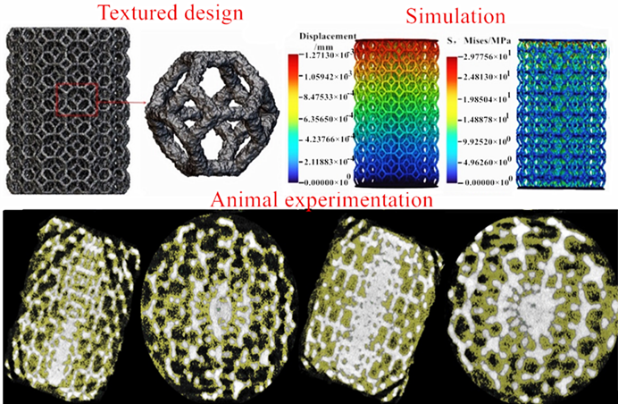
The rising prevalence of orthopedic conditions in aging populations has created a growing demand for advanced implants with enhanced biocompatibility, mechanical performance, and tissue integration. To meet these demands, it is necessary to investigate the metamaterial properties of cross-scale porous structures, including both macroscale architecture and microscale texture. Accordingly, we employed parametric modeling to design porous structures; analyzed blood flow distribution through various multi-level porous designs using mold flow simulation; evaluated their compressive properties through finite element analysis; assessed biocompatibility via animal experiments; and obtained tissue ingrowth data using micro-computed tomography. The results indicated that when fluid flowed through cross-scale porous structures, the overall pressure was low, and the Kelvin cell structure exhibited favorable flow field characteristics under low pressure. When the structures were pressurized, texturization methods involving material removal resulted in larger displacements, while those involving material addition led to smaller displacements. The Kelvin cell structure exhibited extensive tissue ingrowth with a dense tissue pattern internally, and the amount of ingrowth decreased from the inside to the outside. Increasing the roughness of porous structures via material removal increased the surface-to-volume ratio to a certain extent but did not promote tissue ingrowth. In contrast, increasing roughness by material addition favored tissue ingrowth, laying a foundation for the design of cross-scale metamaterial implants.
- Chinese Osteoporosis and Mineral Diseases Society. Guidelines for the diagnosis and treatment of primary osteoporosis (2022). Chin J Gen Pract. 2023;26(14):1671-1691. doi: 10.12114/j.issn.1007-9572.2023.0121.
- Wu Y, Wang Y, Liu M, et al. Design, manufacturing, and application of porous implants based on 3D printing technology. Chin J Med Devices. 2024;48(01):15-19. doi: CNKI: SUN:ZYLZ.0.2024-01-004.
- Salaha ZFM, Ammarullah MI, Abdullah NNAA, et al. Biomechanical effects of the porous structure of gyroid and voronoi hip implants: a finite element analysis using an experimentally validated model. Materials. 2023;16(9):3298. doi: 10.3390/ma16093298
- Jandyal A, Chaturvedi I, Wazir I, Raina A, Haq MIU. 3D printing–a review of processes, materials and applications in industry 4.0. Sustain Oper Comput. 2022;3:33-42. doi: 10.1016/j.susoc.2021.09.004
- Leschok M, Cheibas I, Piccioni V, et al. 3D printing facades: design, fabrication, and assessment methods. Autom Constr. 2023;152:104918. doi: 10.1016/j.autcon.2023.104918
- Li, K, Ji C, Bai S, Jiang B, Pan F. Selective laser melting of magnesium alloys: necessity, formability, performance, optimization and applications. J Mater Sci Technol. 2023;154:65-93. doi: 10.1016/j.jmst.2022.12.053
- Wang H. Comparative Study on the Application of Personalized Titanium Alloy Prostheses and Allogeneic Mandibular Bones for Mandibular Defect Repair in Beagle Dogs. Beijing: Chinese People’s Liberation Army Medical College; 2017.
- Yang J, Li Y, Shi X, et al. Design and analysis of three-dimensional printing of a porous titanium scaffold. BMC Musculoskelet Dis. 2021;22(1):654. doi: 10.1186/s12891-021-04520-1
- Wang Y. Experimental Study on Biomechanical Properties and Biocompatibility of 3D Printed Artificial Trabecular Prosthesis. Zhengzhou University; 2021.
- Wang S, Gao K, Xu Z, et al. 3D printing-assisted traditional steel plate internal fixation for complex tibial plateau fractures. Chin J Tissue Eng Res. 2022;26(18):2823-2827. doi: 10.12307/2022.688.
- Zhang P, Jiang B, Zhang L, et al. Biocompatibility evaluation of 3D printed porous tantalum. Med Equip. 2022;35(09):44-49. doi: CNKI: SUN: YLZB.0. 2022-09-014.
- Liang H, Li R, Liu G, et al. Research and clinical application status of porous structure design for medical orthopedic implants. J Clin Rehabil Tissue Eng Res. 2017;21(15):2410-2417. doi: CNKI: SUN:XDKF.0.2017-15-023.
- Liu C, Wang C, Liu H, et al. Mechanical properties and biocompatibility of 3D printed Ti6Al4V titanium alloy scaffolds. Chin J Nonferrous Metal. 2018; 28(04):758-765. doi: 10.19476/j.ysxb.1004.0609.2018.04.14
- Lei H, Zhou Z, Liu J, et al. Structural optimization of 3D-printed porous titanium implants promotes bone regeneration for enhanced biological fixation. ACS Appl Mater Interfaces. 2025;17(12):18059-18073. doi: 10.1021/acsami.4c22401
- Tamaddon M, Samizadeh S, Wang L, Blunn G, Liu C. Intrinsic osteoinductivity of porous titanium scaffold for bone tissue engineering. Int J Biomater. 2017;2017(1):5093063. doi: 10.1155/2017/5093063
- Zaharin HA, Abdul Rani AM, Azam FI, et al. Effect of unit cell type and pore size on porosity and mechanical behavior of additively manufactured Ti6Al4V scaffolds. Materials. 2018;11(12):2402. doi: 10.3390/ma11122402
- Zadpoor AA. Bone tissue regeneration: the role of scaffold geometry. Biomater Sci. 2015;3(2):231-245. doi: 10.1039/c4bm00291a
- Kumar A, Nune KC, Murr LE, Misra RDK. Biocompatibility and mechanical behaviour of three-dimensional scaffolds for biomedical devices: process–structure–property paradigm. Int Mater Rev. 2016;61(1):20-45. doi: 10.1080/09506608.2015.1128310
- Tu C. Study on the Biocompatibility of 3D Printed Porous Composite Scaffolds. Huazhong University of Science and Technology; 2016.
- Zhao Y, Li H, Guo Y, et al. Preparation and biocompatibility study of mPEG-PDLLA/nHAP/TTCP porous bone scaffolds. J Armed Police Med Coll (Med Ed). 2016;25(12):954-958. doi: 10.16548/j.2095-3720.2016.12.002.
- Jodati H, Yılmaz B, Evis Z. A review of bioceramic porous scaffolds for hard tissue applications: effects of structural features. Ceram Int. 2020;46(10):15725-15739. doi: 10.1016/j.ceramint.2020.03.192
- Zheng H. Research on the Surface Topological Structure Regulation of Degradable Polymer Microspheres and its Effects on Cell Adhesion and Function. Zhejiang University; 2018.
- Lai Y. Basic Research on the Application of Micro-groove Topography and Nano-antibacterial Coating to the Surface of Gingival-crossing Part of Dental Implants. Fujian Medical University; 2014.
- Zhang G, Yang Y, Zhang Z, et al. Optimization design of support structure for laser selective melting forming parts. China Laser. 2016;43(12):59-66. doi: 10.3788/CJL201643.1202002