

3D-bioprinted osteochondral model based on hierarchical polymeric microarchitectures for in vitro osteoarthritis drug screening

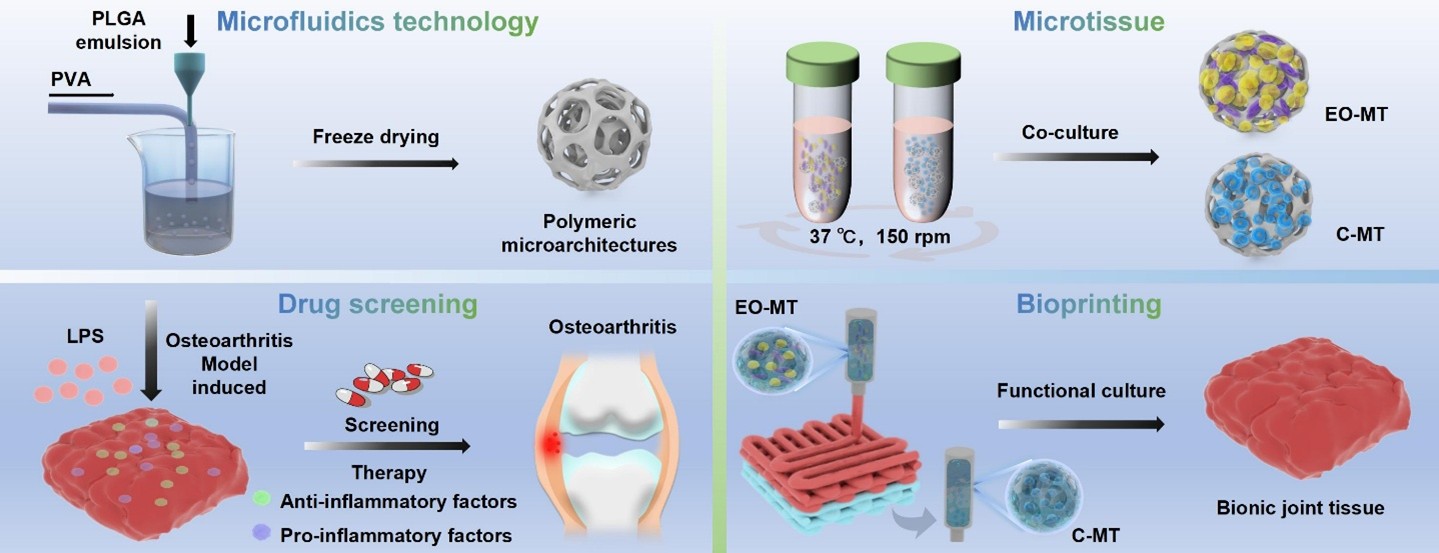
Compared to conventional two-dimensional (2D) or scaffold-free three-dimensional (3D) drug screening models, biomimetic osteochondral constructs offer superior physiological relevance for studying osteoarthritis (OA) and accelerating therapeutic discovery. This study reports the development of a polymeric microarchitecture (PM)- based 3D osteochondral model for drug screening applications. Microfluidics-assisted fabrication enabled the generation of cartilage-like and osteogenic microtissues by encapsulating chondrocytes and endothelial/osteoblast cells within PMs. These multicellular aggregates were embedded in gelatin methacryloyl and assembled via 3D bioprinting into a stratified osteochondral construct. The model exhibited favorable cell viability, high proliferation, and organized microtissue formation, validating its biological functionality. An OA-like microenvironment was induced using lipopolysaccharide, significantly elevating pro-inflammatory cytokines. Treatment with diclofenac, dexamethasone, or curcumin markedly attenuated this response, reducing tumor necrosis factor-alpha, interleukin (IL)-1β, and IL-6 to 42.1, 193.5, and 193.5 pg/mL, respectively, while elevating the anti-inflammatory cytokine IL-10 to 90.2 pg/mL. Overall, this PM-based 3D osteochondral platform reproduces key features of native joint tissue and holds promise for OA research, drug screening, and regenerative medicine.
- Bhardwaj N, Singh YP, Mandal BB. Silk fibroin scaffold-based 3D co-culture model formodulation of chondrogenesis without hypertrophy via reciprocal cross-talk and paracrine signaling. ACS Biomater Sci Eng. 2019;5(10):5240-5254. doi: 10.1021/acsbiomaterials.9b00573
- Wang D, Liu W, Venkatesan JK, Madry H, Cucchiarini M. Therapeutic controlled release strategies for human osteoarthritis. Adv Healthc Mater. 2024;14(2):e2402737. doi: 10.1002/adhm.202402737
- Samvelyan HJ, Hughes D, Stevens C, Staines KA. Models of osteoarthritis: relevance and new insights. Calcif Tissue Int. 2021;109(3):243-256. doi: 10.1007/s00223-020-00670-x
- Yessica Eduviges ZC, Martínez-Nava G, Reyes-Hinojosa D, et al. Impact of cadmium toxicity on cartilage loss in a 3D in vitro model. Environ Toxicol Pharmacol. 2020;74:103307. doi: 10.1016/j.etap.2019.103307
- Jones G, Winzenberg T. Osteoarthritis: a new short-term treatment option? Lancet. 2019;394(10213):1967-1968. doi: 10.1016/s0140-6736(19)32729-1
- Yang D, Xu J, Xu K, Xu P. Skeletal interoception in osteoarthritis. Bone Res. 2024;12(22). doi: 10.1038/s41413-024-00328-6
- Cowan KJ, Kleinschmidt-Dörr K, Gigout A, et al. Translational strategies in drug development for knee osteoarthritis. Drug Discov Today. 2020;25(6): 1054-1064. doi: 10.1016/j.drudis.2020.03.015
- Assi R, Quintiens J, Monteagudo S, Lories RJ. Innovation in targeted intra-articular therapies for osteoarthritis. Drugs. 2023;83(8):649-663. doi: 10.1007/s40265-023-01863-y
- Cao H, Deng S, Chen X, et al. An injectable cartilage-coating composite with long-term protection, effective lubrication and chondrocyte nourishment for osteoarthritis treatment. Acta Biomater. 2024;179:95-105. doi: 10.1016/j.actbio.2024.03.015
- Wu M, Zheng K, Li W, et al. Nature-inspired strategies for the treatment of osteoarthritis. Adv Funct Mater. 2023;34(4):2305603. doi: 10.1002/adfm.202305603
- Yeung P, Cheng KH, Yan CH, Chan BP. Collagen microsphere based 3D culture system for human osteoarthritis chondrocytes (hOACs). Sci Rep. 2019;9(1):12453. doi: 10.1038/s41598-019-47946-3
- Zou Z, Luo X, Chen Z, Zhang YS, Wen C. Emerging microfluidics-enabled platforms for osteoarthritis management: from benchtop to bedside. Theranostics. 2022;12(2):891-909. doi: 10.7150/thno.62685
- Liu H, Wu X, Liu R, Wang W, Zhang D, Jiang Q. Cartilage-on-a-chip with magneto-mechanical transformation for osteoarthritis recruitment. Bioact Mater. 2024; 33:61-68. doi: 10.1016/j.bioactmat.2023.10.030
- Chapman JH, Ghosh D, Attari S, Ude CC, Laurencin CT. Animal models of osteoarthritis: updated models and outcome measures 2016–2023. Regen Eng Transl Med. 2023;10(2):127-146. doi: 10.1007/s40883-023-00309-x
- Singh YP, Moses JC, Bhardwaj N, Mandal BB. Overcoming the dependence on animal models for osteoarthritis therapeutics – the promises and prospects of in vitro models. Adv Healthc Mater. 2021;10(20):2100961. doi: 10.1002/adhm.202100961
- Zhou M, Lozano N, Wychowaniec JK, et al. Graphene oxide: a growth factor delivery carrier to enhance chondrogenic differentiation of human mesenchymal stem cells in 3D hydrogels. Acta Biomater. 2019;96:271-280. doi: 10.1016/j.actbio.2019.07.027
- Ding SL, Zhao XY, Xiong W, et al. Cartilage lacuna‐inspired microcarriers drive hyaline neocartilage regeneration. Adv Mater. 2023;35(30):e2212114. doi: 10.1002/adma.202212114
- Hwang HS, Kim HA. Chondrocyte apoptosis in the pathogenesis of osteoarthritis. In. J Mol Sci. 2015;16(11):26035-26054. doi: 10.3390/ijms161125943
- Ebata T, Terkawi MA, Kitahara K, et al. Noncanonical pyroptosis triggered by macrophage‐derived extracellular vesicles in chondrocytes leading to cartilage catabolism in osteoarthritis. Arthritis Rheumatol. 2023;75(8):1358-1369. doi: 10.1002/art.42505
- Maihemuti A, Zhang H, Lin X, et al. 3D-printed fish gelatin scaffolds for cartilage tissue engineering. Bioact. Mater. 2023;26:77-87. doi: 10.1016/j.bioactmat.2023.02.007
- Korpayev S, Kaygusuz G, Şen M, Orhan K, Oto Ç, Karakeçili A. Chitosan/collagen based biomimetic osteochondral tissue constructs: A growth factor-free approach. Int J Biol Macromol. 2020;156:681-690. doi: 10.1016/j.ijbiomac.2020.04.109
- Singh YP, Moses JC, Bandyopadhyay A, Mandal BB. 3D bioprinted silk‐based in vitro osteochondral model for osteoarthritis therapeutics. Adv Healthc Mater. 2022;11(24):200209. doi: 10.1002/adhm.202200209
- Salehi S, Brambilla S, Rasponi M, Lopa S, Moretti M. Development of a microfluidic vascularized osteochondral model as a drug testing platform for osteoarthritis. Adv Healthc Mater. 2024;13(31):e2402350. doi: 10.1002/adhm.202402350
- Ong LJY, Sun AR, Wang Z, Lee J, Prasadam I, Toh YC. Localized oxygen control in a microfluidic osteochondral interface model recapitulates bone–cartilage crosstalk during osteoarthritis. Adv Funct Mater. 2024;34(28):2315608. doi: 10.1002/adfm.202315608
- Wei Y, Deng Y, Ma S, et al. Local drug delivery systems as therapeutic strategies against periodontitis: a systematic review. J Control Release. 2021;333:269-282. doi: 10.1016/j.jconrel.2021.03.041
- Jo YK, Lee D. Biopolymer microparticles prepared by microfluidics for biomedical applications. Small. 2020;16(9):1903736. doi: 10.1002/smll.201903736
- Jin Z, Zhai Y, Zhou Y, et al. Regulation of mesenchymal stem cell osteogenic potential via microfluidic manipulation of microcarrier surface curvature. Chem Eng J. 2022;448:137739. doi: 10.1016/j.cej.2022.137739
- He Q, Zhang J, Liao Y, et al. Current advances in microsphere based cell culture and tissue engineering. Biotechnol Adv. 2020;39:107459. doi: 10.1016/j.biotechadv.2019.107459
- Hendow EK, Iacoviello F, Casajuana Ester M, Pellet‐Many C, Day RM. Hierarchically structured biodegradable microspheres promote therapeutic angiogenesis. Adv Healthc Mater. 2024;13(31):e2401832. doi: 10.1002/adhm.202401832
- Wang Y, Kankala RK, Zhang J, et al. Modeling endothelialized hepatic tumor microtissues for drug screening. Adv Sci. 2020;7(21):2002002. doi: 10.1002/advs.202002002
- Zhang Y, Ma C, Xie J, Ågren H, Zhang H. Black phosphorus/polymers: status and challenges. Adv Mater. 2021;37(33):2100113. doi: 10.1002/adma.202100113
- Bai L, Han Q, Han Z, et al. Stem cells expansion vector via bioadhesive porous microspheres for accelerating articular cartilage regeneration. Adv Healthc Mater. 2023;13(3):2302327. doi: 10.1002/adhm.202302327
- Dhanabalan KM, Gupta VK, Agarwal R. Rapamycin–PLGA microparticles prevent senescence, sustain cartilage matrix production under stress and exhibit prolonged retention in mouse joints. Biomater Sci. 2020;8(15):4308-4321. doi: 10.1039/D0BM00596G
- Wang Y, Yuan X, Yu K, et al. Fabrication of nanofibrous microcarriers mimicking extracellular matrix for functional microtissue formation and cartilage regeneration. Biomaterials. 2018;171:118-132. doi: 10.1016/j.biomaterials.2018.04.033
- Hu Z, Lin H, Wang Z, et al. 3D printing hierarchical porous nanofibrous scaffold for bone regeneration. Small. 2024; 21(2):2405406. doi: 10.1002/smll.202405406
- Dai W, Li S, Jia H, et al. Indirect 3D printing CDHA scaffolds with hierarchical porous structure to promote osteoinductivity and bone regeneration. J Mater Sci Technol. 2025;207:295-307. doi: 10.1016/j.jmst.2024.04.032
- He J, Sun Y, Gao Q, et al. Gelatin methacryloyl hydrogel, from standardization, performance, to biomedical application. Adv Healthc Mater. 2023;12(23):2300395. doi: 10.1002/adhm.202300395
- Huang Z, Kraus VB. Does lipopolysaccharide-mediated inflammation have a role in OA? Nat Rev Rheumatol. 2016;12(2):123-129. doi: 10.1038/nrrheum.2015.158
- Xue C, Tian J, Cui Z, et al. Reactive oxygen species (ROS)- mediated M1 macrophage-dependent nanomedicine remodels inflammatory microenvironment for osteoarthritis recession. Bioact Mater. 2024;33:545-561. doi: 10.1016/j.bioactmat.2023.10.032
- Yu X, Gholipourmalekabadi M, Wang X, Yuan C, Lin K. Three‐dimensional bioprinting biphasic multicellular living scaffold facilitates osteochondral defect regeneration. Interdiscip Mater. 2024;3(5):738-756. doi: 10.1002/idm2.12181
- Jhun J, Min H-K, Na HS, et al. Combinatmarion treatment with Lactobacillus acidophilus LA-1, vitamin B, and curcumin ameliorates the progression of osteoarthritis by inhibiting the pro-inflammatory mediators. Immunol Lett. 2020;228:112-121. doi: 10.1016/j.imlet.2020.10.008
- Luo W, Bai L, Zhang J, et al. Polysaccharides-based nanocarriers enhance the anti-inflammatory effect of curcumin. Carbohydr Polym. 2023;311:120718. doi: 10.1016/j.carbpol.2023.120718
- Liu X, Chen B, Chen J, et al. A cardiac‐targeted nanozyme interrupts the inflammation‐free radical cycle in myocardial infarction. Adv Mater. 2023;36(2):2308477. doi: 10.1002/adma.202308477
- Yao Q, Yang Y, Hu M, Qiu Y, Shi Y, Kou L. Liposomal dexamethasone for intra-articular therapy: Functional strategies and clinical progress. J Control Release. 2025;385:114040. doi: 10.1016/j.jconrel.2025.114040
- da Costa BR, Pereira TV, Saadat P, et al. Effectiveness and safety of non-steroidal anti-inflammatory drugs and opioid treatment for knee and hip osteoarthritis: network meta-analysis. BMJ. 2021;375:n2321. doi: 10.1136/bmj.n2321
- Chen Y, Chen L-F, Wang Y, et al. Engineered dECM-based microsystem promotes cartilage regeneration in osteoarthritis by synergistically enhancing chondrogenesis of BMSCs and anti-inflammatory effect. Compos B. 2025;290:111974. doi: 10.1016/j.compositesb.2024.111974
- Wu D, Yu Y, Zhao C, et al. NK cell-encapsulated porous microspheres via microfluidic electrospray for tumor immunotherapy. ACS Appl Mater Interfaces. 2019;11(37):33716-33724. doi: 10.1021/acsami.9b12816
- Chen X, Zhang D, Wang X, et al. Preparation of porous GelMA microcarriers by microfluidic technology for stem-cell culture. Chem Eng J. 2023;477:146444. doi: 10.1016/j.cej.2023.146444
- Long J, Yao Z, Zhang W, et al. Regulation of osteoimmune microenvironment and osteogenesis by 3D‐printed PLAG/ black phosphorus scaffolds for bone regeneration. Adv Sci. 2023;10(28):2302539. doi: 10.1002/advs.202302539
- Wei J, Xia X, Xiao S, et al. Sequential dual‐biofactor release from the scaffold of mesoporous HA microspheres and PLGA matrix for boosting endogenous bone regeneration. Adv Healthc Mater. 2023;12(20):2300624. doi: 10.1002/adhm.202300624
- Dong R, Kang M, Qu Y, Hou T, Zhao J, Cheng X. Incorporating hydrogel (with low polymeric content) into 3D‐printed PLGA scaffolds for local and sustained release of BMP2 in repairing large segmental bone defects. Adv Healthc Mater. 2024;14(2):2403613. doi: 10.1002/adhm.202403613
- Kamboj N, Kazantseva J, Rahmani R, Rodríguez MA, Hussainova I. Selective laser sintered bio-inspired silicon-wollastonite scaffolds for bone tissue engineering. Mater Sci Eng C. 2020;116:111223. doi: 10.1016/j.msec.2020.111223
- Zhu S, Bennett S, Kuek V, et al. Endothelial cells produce angiocrine factors to regulate bone and cartilage via versatile mechanisms. Theranostics. 2020;10(13):5957-5965. doi: 10.7150/thno.45422
- Sun T, Feng Z, He W, et al. Novel 3D-printing bilayer GelMA-based hydrogel containing BP, β-TCP and exosomes for cartilage–bone integrated repair. Biofabrication. 2023;16(1):015008. doi: 10.1088/1758-5090/ad04fe
- Liu S, Chen G, Chen Z, Wang F, Lv Y. Research progress on stiffness controllable scaffolds based on gelatin methacryloyl hydrogels for tissue repair and reconstruction. Int J Biol Macromol. 2025;321(Pt 3):146485. doi: 10.1016/j.ijbiomac.2025.146485
- Matai I, Kaur G, Seyedsalehi A, McClinton A, Laurencin CT. Progress in 3D bioprinting technology for tissue/organ regenerative engineering. Biomaterials. 2020;226:119536. doi: 10.1016/j.biomaterials.2019.119536
- Tong W, Chen X, Song X, et al. Resveratrol inhibits LPS-induced inflammation through suppressing the signaling cascades of TLR4-NF-κB/MAPKs/IRF3. Exp Ther Med. 2020;19(3):1824-1834. doi: 10.3892/etm.2019.8396
- Lei J, Fu Y, Zhuang Y, Zhang K. Sema4D aggravated LPS-Induced injury via activation of the MAPK signaling pathway in ATDC5 chondrocytes. Biomed Res Int. 2020;2020:8691534. doi: 10.1155/2020/8691534
- Peng K, Li Y, Lu C, Hu S. ABIN-1 protects chondrocytes from lipopolysaccharide-induced inflammatory injury through the inactivation of NF-κB signalling. Clin Exp Pharmacol Physiol. 2020;47(7):1212-1220. doi: 10.1111/1440-1681.13291
- Yan F, Li H, Zhong Z, et al. Co-delivery of prednisolone and curcumin in human serum albumin nanoparticles for effective treatment of rheumatoid arthritis. Int J Nanomedicine. 2019;14:9113-9125. doi: 10.2147/ijn.S219413