

Development and evaluation of dual-controlled release antibiotic-loaded bone scaffolds

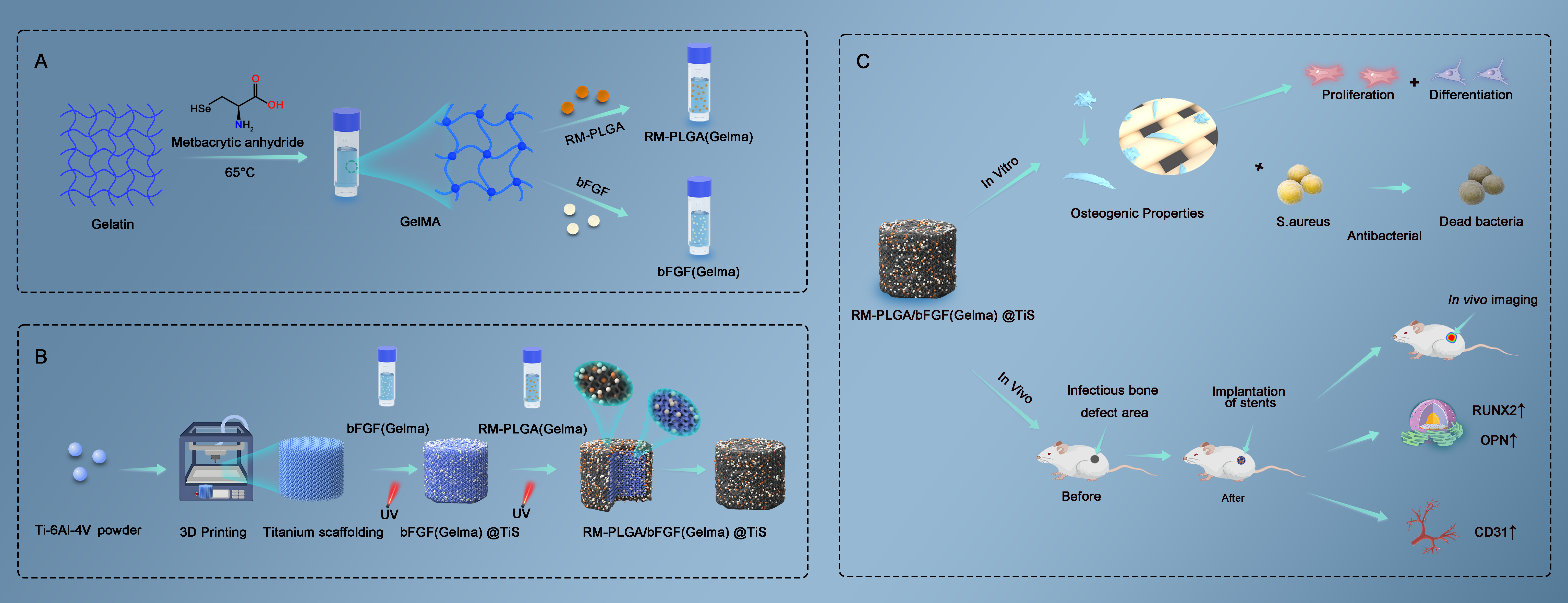
The treatment of infectious bone defects is a major challenge in orthopedics, with infection control and defect repair as the two primary treatment goals. The development of 3D-printed bone scaffolds capable of sustained and stable antibiotic release is an effective strategy for treating such defects. Specifically, the antibiotic loading method and the concentration of released antibiotics significantly affect infection control and bone repair outcomes. In this study, double antibiotic microspheres were prepared via the double emulsion-solvent evaporation method. Moxifloxacin and rifampicin (RM) were encapsulated by poly(lactic-co-glycolic acid) (PLGA), forming RM–PLGA. Subsequently, different concentrations of RM–PLGA and basic fibroblast growth factor (bFGF) were loaded onto a 3D-printed triply periodic minimal surface (TPMS) titanium scaffold (TiS) with a graded porosity design, enabling stable dual-controlled antibiotic release and enhanced release stability. In vitro results revealed that RM–PLGA/bFGFgelatin methacrylate [GelMA])@TiS exhibited strong antimicrobial properties, cytocompatibility, and the capacity for osteoblast differentiation and extracellular mineralization. In vivo, RM–PLGA/bFGF(GelMA)@ TiS was effective in inhibiting infections induced by Staphylococcus aureus while promoting osteogenesis and angiogenesis. These results suggest that RM–PLGA-2/ bFGF(GelMA)@TiS can stably release antibiotics to achieve the therapeutic goals of infection control and induction of both osteogenesis and angiogenesis.
- Zhang S, Lian Z, Chen X, et al. Preparation and properties of chitosan quaternary ammonium salt/α-calcium sulfate hemihydrate/β-tricalcium phosphate composite bone cement. Colloids Surf B Biointerfaces. 2025;253:114738. doi: 10.1016/j.colsurfb.2025.114738
- Zhang P, Liu X, Guo P, et al. Effect of cyclic mechanical loading on immunoinflammatory microenvironment in biofabricating hydroxyapatite scaffold for bone regeneration. Bioact Mater. 2021;6(10):3097-3108. doi: 10.1016/j.bioactmat.2021.02.024
- Li J, Jiang B, Yang L, et al. Dual-functional titanium implants via polydopamine-mediated lithium and copper co-incorporation: synergistic enhancement of osseointegration and antibacterial efficacy. Front Bioeng Biotechnol. 2025;13:1593545. doi: 10.3389/fbioe.2025.1593545
- Farazin A, Mahjoubi S. Dual-functional hydroxyapatite scaffolds for bone regeneration and precision drug delivery. J Mech Behav Biomed Mater. 2024;157:106661. doi: 10.1016/j.jmbbm.2024.106661
- Buchholz T, Siverino C, Moriarty TF, et al. Antibiotic-loaded polymer-calcium phosphate scaffold for treating orthopedic device-related infection in a rabbit segmental bone defect model. J Biomed Mater Res A. 2025; 113(5):e.37917. doi: 10.1002/jbm.a.37917
- Ceccarelli G, Perciballi B, Russo A, et al. Chronic suppressive antibiotic treatment for Staphylococcal bone and joint implant-related infections. Antibiotics. 2023;12(5):937. doi: 10.3390/antibiotics12050937
- Hu K, Olsen BR. Osteoblast-derived VEGF regulates osteoblast differentiation and bone formation during bone repair. J Clin Investig. 2016;126(2):509-526. doi: 10.1172/jci82585
- Eelen G, Treps L, Li X, Carmeliet P. Basic and therapeutic aspects of angiogenesis updated. Circ Res. 2020;127(2):310-329. doi: 10.1161/circresaha.120.316851
- Chen Z, Yang Y, Liu B, Li X, Tian Y. Application of 3D-printed porous prosthesis for the reconstruction of infectious bone defect with concomitant severe soft tissue lesion: a case series of 13 cases. BMC Musculoskelet Disord. 2024;25(1):1090. doi: 10.1186/s12891-024-08248-6
- Wang J, Liu M, Yang C, et al. Photocrosslinked gelatin methacryloyl/hyaluronic acid methacryloyl composite hydrogels loaded with bone morphogenetic protein 2-black phosphorus nanosheets for bone regeneration. J Biomater Sci Polym Ed. 2025:1-23. doi: 10.1080/09205063.2025.2489846
- Pramanik S, Alhomrani M, Alamri AS, et al. Unveiling the versatility of gelatin methacryloyl hydrogels: a comprehensive journey into biomedical applications. Biomed Mater. 2024;19(4):042008. doi: 10.1088/1748-605x/ad4df7
- Chu C, Qiu J, Zhao Q, et al. Injectable dual drug-loaded thermosensitive liposome-hydrogel composite scaffold for vascularised and innervated bone regeneration. Colloids Surf B Biointerfaces. 2024;245:114203. doi: 10.1016/j.colsurfb.2024.114203
- Seifert LB, Aigner A, Abazi S, et al. A 3D printed hydroxyapatite implant for temporal hollowing reconstruction: a patient- specific approach. Craniomaxillofac Trauma Reconstr. 2025;18(2):28. doi: 10.3390/cmtr18020028
- Cidonio G, Glinka M, Kim YH, et al. Nanoclay-based 3D printed scaffolds promote vascular ingrowth ex vivo and generate bone mineral tissue in vitro and in vivo. Biofabrication. 2020;12(3):035010. doi: 10.1088/1758-5090/ab8753
- Li J, Yang Y, Sun Z, et al. Integrated evaluation of biomechanical and biological properties of the biomimetic structural bone scaffold: biomechanics, simulation analysis, and osteogenesis. Mater Today Bio. 2023;24:100934. doi: 10.1016/j.mtbio.2023.100934
- Chi G, Ren H, Lin B, Huang K. Subtrochanteric femoral fracture with postoperative chronic osteomyelitis treated successfully by 1-stage operation: a case report. Ann Transl Med. 2021;9(6):514. doi: 10.21037/atm-21-413
- Graczyk S, Pasławski R, Grzeczka A, et al. Antimicrobial and antiproliferative coatings for stents in veterinary medicine— state of the art and perspectives. Materials. 2023;16(21):6834. doi: 10.3390/ma16216834
- Barua N, Huang L, Li C, et al. Comparative study of two-dimensional (2D) vs. three-dimensional (3D) organotypic kertatinocyte-fibroblast skin models for Staphylococcus aureus (MRSA) infection. Int J Mol Sci. 2021;23(1):299. doi: 10.3390/ijms23010299
- Pinho MG, Foster SJ. Cell growth and division of Staphylococcus aureus. Annu Rev Microbiol. 2024;78(1):293-310. doi: 10.1146/annurev-micro-041222-125931
- Zhang H, Wang Y, Qiang H, et al. Exploring the frontiers: the potential and challenges of bioactive scaffolds in osteosarcoma treatment and bone regeneration. Mater Today Bio. 2024;29:101276. doi: 10.1016/j.mtbio.2024.101276
- Hackemann VCJ, Hagel S, Jandt KD, Rödel J, Löffler B, Tuchscherr L. The controversial effect of antibiotics on methicillin-sensitive S. aureus: a comparative in vitro study. Int J Mol Sci. 2023;24(22):16308. doi: 10.3390/ijms242216308
- Ayre WN, Birchall JC, Evans SL, Denyer SP. A novel liposomal drug delivery system for PMMA bone cements. J Biomed Mater Res B Appl Biomater. 2015;104(8): 1510-1524. doi: 10.1002/jbm.b.33488
- Qiao Z, Yuan Z, Zhang W, Wei D, Hu N. Preparation, in vitro release and antibacterial activity evaluation of rifampicin and moxifloxacin-loaded poly(D,L-lactide-co-glycolide) microspheres. Artif Cells Nanomed Biotechnol. 2019;47(1):790-798. doi: 10.1080/21691401.2019.1581792
- Liang D, Frank S, Schwendeman SP. Aqueous remote loading of model cationic peptides in uncapped poly(lactide-co-glycolide) microspheres for long-term controlled release. Drug Deliv Transl Res. 2023;14(3):696-704. doi: 10.1007/s13346-023-01424-6
- Li S, Shi X, Xu B, et al. In vitro drug release and antibacterial activity evaluation of silk fibroin coated vancomycin hydrochloride loaded poly (lactic-co-glycolic acid) (PLGA) sustained release microspheres. J Biomater Appl. 2022;36(9):1676-1688. doi: 10.1177/08853282211064098
- Vishwa B, Moin A, Gowda DV, et al. Pulmonary targeting of inhalable moxifloxacin microspheres for effective management of tuberculosis. Pharmaceutics. 2021;13(1):79. doi: 10.3390/pharmaceutics13010079
- Lin Y, Liu L, He J, Shen J, Ren Q. Rapid release of high-valent silver ions from water-soluble porphyrin complexes to enhance the direct killing of methicillin-resistant Staphylococcus aureus. Acta Biomater. 2024;192:419-430. doi: 10.1016/j.actbio.2024.12.004
- Chung JH, Baek N, Lim H, et al. Inner surface modification of polyurethane ureteral stents using plasma-enhanced chemical vapor deposition to improve the resistance to encrustation in a pig model. Investig Clin Urol. 2023;64(2):175. doi: 10.4111/icu.20220393
- Vishwakarma A, Sinha N. Determination of the optimum architecture of additively manufactured magnetic bioactive glass scaffolds for bone tissue engineering and drug-delivery applications. ACS Appl Bio Mater. 2024;7(10):6847-6864. doi: 10.1021/acsabm.4c00995
- Panagakis P, Zygogiannis K, Fanourgiakis I, Kalatzis D, Stathopoulos K. The role of the periosteum in bone formation from adolescence to old age. Cureus. 2025;17(1):e76774. doi: 10.7759/cureus.76774
- Fiume E, Tulyaganov DU, Akbarov A, et al. Biological evaluation of a new sodium-potassium silico-phosphate glass for bone regeneration: in vitro and in vivo studies. Materials. 2021;14(16):4546. doi: 10.3390/ma14164546
- Sevinc‐Sasmaz C, Erci F, Torlak E, Yöntem M. Characterization of silver nanoparticles synthesized using Hypericum perforatum L. and their effects on Staphylococcus aureus. Microsc Res Tech. 2025;88(8):2321-2332. doi: 10.1002/jemt.24862
- Wang Y, Hu Y, Pan K, et al. In-vivo imaging revealed antigen-directed gingival B10 infiltration in experimental periodontitis. Biochim Biophys Acta Mol Basis Dis. 2020;1867(1):165991. doi: 10.1016/j.bbadis.2020.165991
- DeMourdant T, Rajkovic CJ, Tracz JA, et al. A novel rodent model of chronic spinal implant-associated infection. Spine J. 2023;23(9):1389-1399.doi: 10.1016/j.spinee.2023.05.014
- Subramanian AK, Sreenivasagan S, Mohanraj KG, Kumar RS. Assessment of toxicity of green synthesized silver nanoparticle-coated titanium mini-implants with uncoated mini-implants: comparison in an animal model study. J Contemp Dent Pract. 2024;24(12):944-950. doi: 10.5005/jp-journals-10024-3577
- Sifi A, Adi-Bessalem S, Laraba-Djebari F. Role of angiotensin II and angiotensin type-1 receptor in scorpion venom-induced cardiac and aortic tissue inflammation. Exp Mol Pathol. 2016;102(1):32-40. doi: 10.1016/j.yexmp.2016.11.006
- Yamamoto J, Deguchi H, Sumiyoshi T, et al. Accumulation and phagocytosis of fluorescently visualized macrophages against Edwardsiella piscicida infection in established mpeg1.1-transgenic Japanese Medaka Oryzias latipes. Mar Biotechnol. 2024;26(4):658-671. doi: 10.1007/s10126-024-10333-9
- Ratnaningtyas NI, Husen F. Therapeutic potential of Coprinus comatus nanogels: antiarthritic and anti-inflammatory effects in rheumatoid arthritis models. Vet World. 2025;18(13):582-597. doi: 10.14202/vetworld.2025.582-597
- Correia MR, Han SW, Escalante T, Moreira V. The role of the cyclooxygenase-2 pathway in tissue ischemia and revascularization following skeletal muscle injury induced by bothropic snake venom. Microvasc Res. 2024; 157:104760. doi: 10.1016/j.mvr.2024.104760
- Beeren IAO, Dijkstra PJ, Lourenço AFH, et al. Installation of click-type functional groups enable the creation of an additive manufactured construct for the osteochondral interface. Biofabrication. 2022;15(1):014106. doi: 10.1088/1758-5090/aca3d4
- Koushik TM, Miller CM, Antunes E. Graded hydroxyapatite triply periodic minimal surface structures for bone tissue engineering applications. Adv Healthc Mater. 2025:e2402953. doi: 10.1002/adhm.202402953
- Chen XJ, Wu X, Lin HH, Liu ZX, Liu S. Effects of methacrylic anhydride gelatin hydrogel loaded with silver and recombinant human basic fibroblast growth factor on deep partial-thickness burn wounds in rabbits. Zhonghua Shao Shang Yu Chuang Mian Xiu Fu Za Zhi. 2022;38(7):640-649. doi: 10.3760/cma.j.cn501120-20210726-00260
- Lin H, Gao Z, Shan T, et al. A review on the promising antibacterial agents in bone cement–from past to current insights. J Orthop Surg Res. 2024;19(1):673. doi: 10.1186/s13018-024-05143-7
- Wang L, Wang P, Liu Y, et al. The effect of different factors on poly(lactic-co-glycolic acid) nanoparticle properties and drug release behaviors when co-loaded with hydrophilic and hydrophobic drugs. Polymers. 2024;16(7):865. doi: 10.3390/polym16070865
- Jackson J. Triple encapsulation and controlled release of vancomycin, rifampicin and silver from poly (methyl methacrylate) or poly (lactic-co-glycolic acid) nanofibers. Bioengineering. 2024;11(6):529. doi: 10.3390/bioengineering11060529
- Li Y, Xu W, Su Q, Wang Q. Microstructure and properties of TI–AL coating on titanium alloy surface assisted by electromagnetic field. ACS Omega. 2024;9(46):46176-46191. doi: 10.1021/acsomega.4c06757