

Three-dimensional-printed collagen scaffold with limbal stem cells derived from adipose-derived mesenchymal stem cells for the treatment of limbal stem cell deficiency

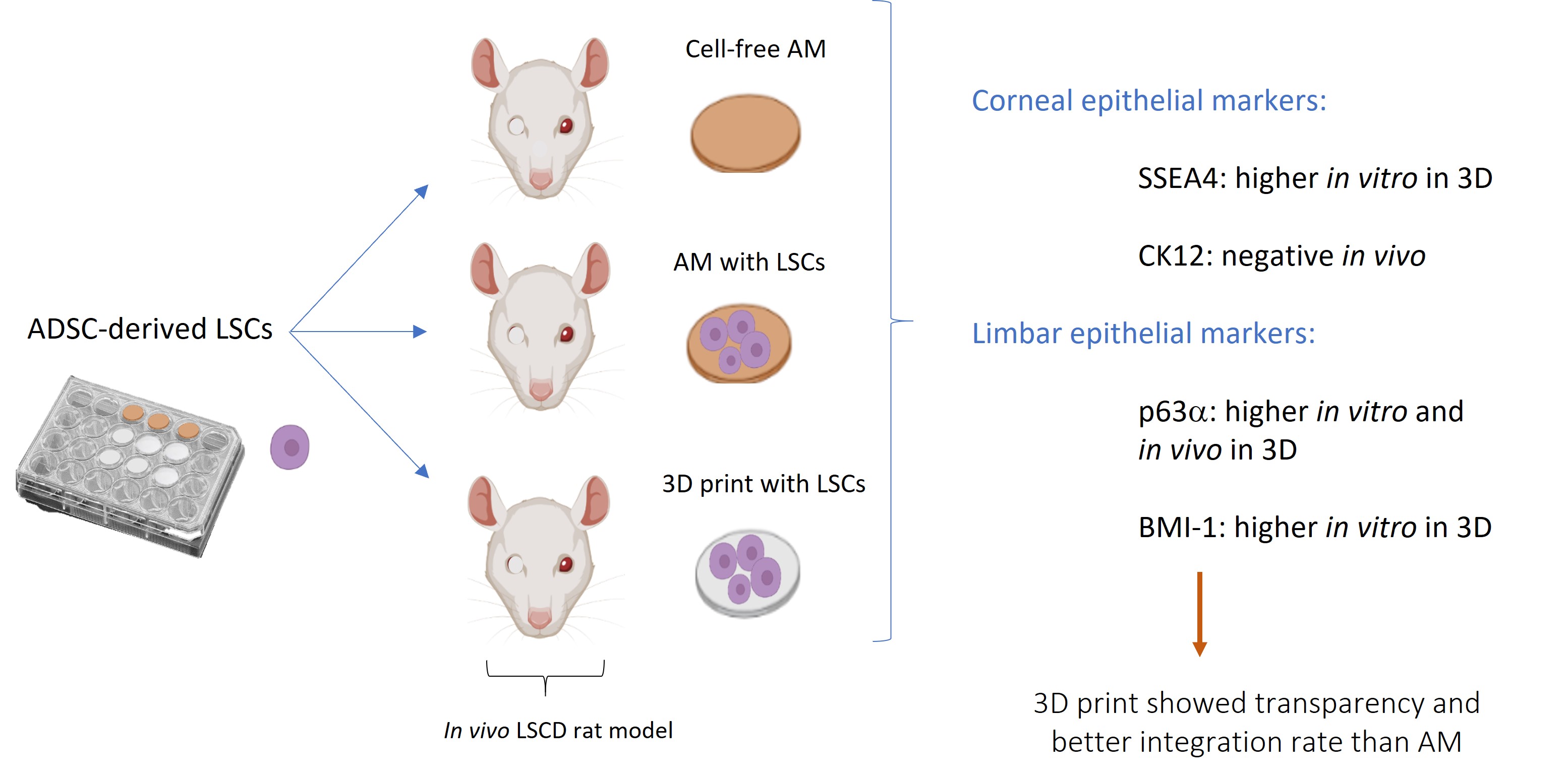
When limbal stem cell deficiency (LSCD) is partial, the standard treatment involves covering the corneal surface with amniotic membrane (AM), which supports the proliferation of the remaining limbal stem cells (LSCs). In cases of complete LSCD, the most common treatment is cultured limbal epithelial transplantation (CLET), although there is a risk of rejection. Studies have shown that mesenchymal stem cell transplantation is equally safe and effective as CLET. Recent research has demonstrated successful differentiation of adipose-derived adult mesenchymal stem cells (ADSCs) into LSCs. Combining AM transplantation with LSCs improves treatment efficacy. However, a limitation of AM use is donor variability and the associated risk of immune rejection. We propose the use of 3D-printed collagen as a scaffold seeded with LSCs derived from ADSCs for the treatment of LSCD in a rat model. The 3D-printed collagen scaffolds exhibited good transparency. In vitro differentiation of ADSCs into LSCs showed morphological changes that were more pronounced and occurred more rapidly on 3D-printed collagen. Among the tested substrates, 3D-printed collagen was the most efficient for differentiation, yielding the highest expression of LSC-specific markers (p63α and BMI-1) and the corneal epithelial marker (SSEA-4). LSCs differentiated in either AM or 3D-printed collagen I scaffolds were transplanted into a rat model of LSCD and compared with the standard, cell-free AM treatment. In all treatment groups, the induced epithelial wound was closed; however, integration of the 3D-printed collagen scaffold was statistically superior to that of AM. However, markers for different corneal structures (PAS, BMI-1, p63α, and cytokeratins 12 and 13) indicated that the generated epithelium was conjunctival rather than corneal, suggesting that the contribution of ADSC-derived LSCs was insufficient for complete corneal re-epithelization.
- De Miguel MP, Casaroli-Marano RP, Nieto-Nicolau N, et al. Frontiers in regenerative medicine for cornea and ocular surface. In: Frontiers in Stem Cell and Regenerative Medicine Research. Vol 1. Bentham Science Publishers; 2015:92-138. doi: 10.2174/9781608059942115010006
- Zavala J, López Jaime GR, Rodríguez Barrientos CA, Valdez- Garcia J. Corneal endothelium: developmental strategies for regeneration. Eye (Basingstoke). 2013;27(5):579-588. doi: 10.1038/eye.2013.15
- Akanda ZZ, Naeem A, Russell E, Belrose J, Si FF, Hodge WG. Graft rejection rate and graft failure rate of penetrating keratoplasty (PKP) vs lamellar procedures: a systematic review. PLoS One. 2015;10(3):e0119934. doi: 10.1371/journal.pone.0119934
- Popova P, Malalana F, Biddolph S, et al. Interspecies comparative morphological evaluation of the corneal epithelial stem cell niche: a pilot observational study. J Vet Sci. 2022;23(4):e62. doi: 10.4142/JVS.22009
- Fernández A, Moreno J, Prósper F, García M, Echeveste J. Regeneration of the ocular surface: stem cells and reconstructive techni-ques. An Sist Sunit Navar. 2008; 31(1): 53-69. doi: 10.4321/s1137-66272008000100005
- Nuzzi A, Pozzo Giuffrida F, Luccarelli S, Nucci P. Corneal epithelial regeneration: old and new perspectives. Int J Mol Sci. 2022;23(21):13114. doi: 10.3390/ijms232113114
- Tanifuji-Terai N, Terai K, Hayashi Y, Chikama TI, Kao WWY. Expression of keratin 12 and maturation of corneal epithelium during development and postnatal growth. Invest Ophthalmol Vis Sci. 2006;47(2):545-551. doi: 10.1167/iovs.05-1182
- Kayama M, Kurokawa MS, Ueno H, Suzuki N. Recent advances in corneal regeneration and possible application of embryonic stem cell-derived corneal epithelial cells. Clin Ophthalmol. 2007;1(4):373-382.
- Truong TT, Huynh K, Nakatsu MN, Deng SX. SSEA4 Is a potential negative marker for the enrichment of human corneal epithelial stem/progenitor cells. Invest Ophthalmol Vis Sci. 2011;52(9):6315-6320. doi: 10.1167/iovs.11-7518
- Barbaro V, Testa A, Di Iorio E, Mavilio F, Pellegrini G, De Luca M. C/EBPδ regulates cell cycle and self-renewal of human limbal stem cells. J Cell Biol. 2007;177(6):1037-1049. doi: 10.1083/jcb.200703003
- Polisetti N, Sharaf L, Schlötzer-Schrehardt U, Schlunck G, Reinhard T. Efficient isolation and functional characterization of niche cells from human corneal limbus. Int J Mol Sci. 2022;23(5):2750. doi: 10.3390/ijms23052750
- Novelli F, Ganini C, Melino G, et al. p63 in corneal and epidermal differentiation. Biochem Biophys Res Commun. 2022;610:15-22. doi: 10.1016/j.bbrc.2022.04.022
- Ramos T, Scott D, Ahmad S. An update on ocular surface epithelial stem cells: cornea and conjunctiva. Stem Cells Int. 2015;2015:601731. doi: 10.1155/2015/601731
- Pellegrini G, Rama P, Matuska S, et al. Biological parameters determining the clinical outcome of autologous cultures of limbal stem cells. Regenerative Med. 2013;8(5):553-567. doi: 10.2217/rme.13.43
- Galindo S, Herreras JM, López-Paniagua M, et al. Therapeutic effect of human adipose tissue-derived mesenchymal stem cells in experimental corneal failure due to limbal stem cell niche damage. Stem Cells. 2017;35(10): 2160-2174. doi: 10.1002/stem.2672
- Nieto-Nicolau N, Martínez-Conesa EM, Fuentes-Julián S, et al. Priming human adipose-derived mesenchymal stem cells for corneal surface regeneration. J Cell Mol Med. 2021;25(11):5124-5137. doi: 10.1111/jcmm.16501
- Calonge M, Pérez I, Galindo S, et al. A proof-of-concept clinical trial using mesenchymal stem cells for the treatment of corneal epithelial stem cell deficiency. Transl Res. 2019;206:18-40. doi: 10.1016/j.trsl.2018.11.003
- Cadenas-Martin M, Arnalich-Montiel F, Miguel MPD. Derivation of limbal stem cells from human adult mesenchymal stem cells for the treatment of limbal stem cell deficiency. Int J Mol Sci. 2023;24(3):2350. doi: 10.3390/ijms24032350
- Edel MJ, Casellas HS, Osete JR, et al. An optimized method to produce human-induced pluripotent stem cell-derived limbal stem cells easily adaptable for clinical use. Stem Cells Dev. 2025;34(3-4):49-60. doi: 10.1089/scd.2024.0172
- Bisevac J, Moe MC, Drolsum L, Kristianslund O, Petrovski G, Noer A. A novel technique of amniotic membrane preparation mimicking limbal epithelial crypts enhances the number of progenitor cells upon expansion. Cells. 2023;12(5):738. doi: 10.3390/cells12050738
- Malhotra C, Jain AK. Human amniotic membrane transplantation: different modalities of its use in ophthalmology. World J Transplant. 2014;4(2):111. doi: 10.5500/wjt.v4.i2.111
- Paolin A, Cogliati E, Trojan D, et al. Amniotic membranes in ophthalmology: long term data on transplantation outcomes. Cell Tissue Bank. 2016;17(1):51-58. doi: 10.1007/s10561-015-9520-y
- Harkin DG, Foyn L, Bray LJ, Sutherland AJ, Li FJ, Cronin BG. Concise reviews: can mesenchymal stromal cells differentiate into corneal cells? A systematic review of published data. Stem Cells. 2015;33(3):785-791. doi: 10.1002/stem.1895
- Nguyen KN, Bobba S, Richardson A, et al. Native and synthetic scaffolds for limbal epithelial stem cell transplantation. Acta Biomater. 2018;65:21-35. doi: 10.1016/j.actbio.2017.10.037
- Tan G, Ioannou N, Mathew E, Tagalakis AD, Lamprou DA, Yu-Wai-Man C. 3D printing in ophthalmology: from medical implants to personalised medicine. Int J Pharm. 2022;625:122094. doi: 10.1016/j.ijpharm.2022.122094
- Boularaoui S, Al Hussein G, Khan KA, Christoforou N, Stefanini C. An overview of extrusion-based bioprinting with a focus on induced shear stress and its effect on cell viability. Bioprinting. 2020;20:e00093. doi: 10.1016/j.bprint.2020.e00093
- Ng WL, Vyas C, Huang B, Yeong WY, Bartolo P. Advanced bioprinting strategies for fabrication of biomimetic tissues and organs. Int J Extreme Manuf. 2025; 7(6):062006. doi: 10.1088/2631-7990/adeee0
- De Miguel MP. European Patent Application No. 22382119.0. Published online 2022.
- He Z, Forest F, Bernard A, et al. Cutting and decellularization of multiple corneal stromal lamellae for the bioengineering of endothelial grafts. Invest Ophthalmol Vis Sci. 2016;57(15):6639-6651. doi: 10.1167/iovs.16-20256
- Zuk PA, Zhu M, Ashjian P, et al. Human adipose tissue is a source of multipotent stem cells. Mol Biol Cell. 2002;13(12):4279-4295. doi: 10.1091/mbc.E02-02-0105
- Mikhailova A, Ilmarinen T, Uusitalo H, Skottman H. Small-molecule induction promotes corneal epithelial cell differentiation from human induced pluripotent stem cells. Stem Cell Reports. 2014;2(2):219-231. doi: 10.1016/j.stemcr.2013.12.014
- Casaroli-Marano RP, Martínez-Conesa EM, Nieto- Nicolau N, Arnalich-Montiel F, Fuentes-Julian S, De Miguel MP. Adipose derived stem cells (ADS) for ocular surface regeneration. Invest Ophthalmol Vis Sci. 2014; 55(13):5183.
- Gerardo H, Lima A, Carvalho J, et al. Soft culture substrates favor stem-like cellular phenotype and facilitate reprogramming of human mesenchymal stem/stromal cells (hMSCs) through mechanotransduction. Sci Rep. 2019;9(1):9086. doi: 10.1038/s41598-019-45352-3
- Maddox JR, Ludlow KD, Li F, Niyibizi C. Breast and abdominal adipose multipotent mesenchymal stromal cells and stage-specific embryonic antigen 4 expression. Cells Tissues Organs. 2012;196(2):107-116. doi: 10.1159/000331332
- Guasti L, Prasongchean W, Kleftouris G, et al. High plasticity of pediatric adipose tissue-derived stem cells: too much for selective skeletogenic differentiation? Stem Cells Transl Med. 2012;1(5):384-395. doi: 10.5966/sctm.2012-0009
- Martínez-Conesa EM, Espel E, Reina M, Casaroli-Marano RP. Characterization of ocular surface epithelial and progenitor cell markers in human adipose stromal cells derived from lipoaspirates. Invest Ophthalmol Vis Sci. 2012;53(1):513-520. doi: 10.1167/iovs.11-7550
- Ahearne M, Masterton S. Donor dependent limbal derived stem cell response to fluidic shear stress. Invest Ophthalmol Vis Sci. 2024;65(7):4462-4462.
- Di Iorio E, Barbaro V, Ruzza A, Ponzin D, Pellegrini G, De Luca M. Isoforms of Np63 and the Migration of Ocular Limbal Cells in Human Corneal Regeneration; 2005. www.pnas.orgcgidoi10.1073pnas.0503437102
- Collin J, Queen R, Zerti D, et al. A single cell atlas of human cornea that defines its development, limbal progenitor cells and their interactions with the immune cells. Ocul Surf. 2021;21:279-298. doi: 10.1016/j.jtos.2021.03.010
- Wang DY, Cheng CC, Kao MH, Hsueh YJ, Ma DHK, Chen JK. Regulation of limbal keratinocyte proliferation and differentiation by TAp63 and ΔNp63 transcription factors. Invest Ophthalmol Vis Sci. 2005;46(9):3102-3108. doi: 10.1167/iovs.05-0051
- Koster MI, Kim S, Mills AA, DeMayo FJ, Roop DR. p63 is the molecular switch for initiation of an epithelial stratification program. Genes Dev. 2004;18(2):126-131. doi: 10.1101/gad.1165104
- Verma S, Lin X, Coulson-Thomas VJ. The potential reversible transition between stem cells and transient-amplifying cells: the limbal epithelial stem cell perspective. Cells. 2024;13(9):748. doi: 10.3390/cells13090748
- Kalha S, Shrestha B, Sanz Navarro M, Jones KB, Klein OD, Michon F. Bmi1+ progenitor cell dynamics in murine cornea during homeostasis and wound healing. Stem Cells. 2018;36(4):562-573. doi: 10.1002/stem.2767
- Liang Q, Le Q, Wang L, et al. Cytokeratin 13 is a new biomarker for the diagnosis of limbal stem cell deficiency. Cornea. 2022;41(7):867-873. doi: 10.1097/ICO.0000000000002903
- Ramirez-Miranda A, Nakatsu MN, Zarei-Ghanavati S, Nguyen CV, Deng SX. Keratin 13 is a more specific marker of conjunctival epithelium than keratin 19. Mol Vis. 2011;17:1652-1661. http://www.molvis.org/molvis/v17/a183
- De Miguel MP, Cadenas-Martin M, Stokking M, Martin- Gonzalez AI. Biomedical application of MSCs in corneal regeneration and repair. Int J Mol Sci. 2025;26(2):695. doi: 10.3390/ijms26020695
- Dietrich-Ntoukas T, Hofmann-Rummelt C, Kruse FE, Schlötzer-Schrehardt U. Comparative analysis of the basement membrane composition of the human limbus epithelium and amniotic membrane epithelium. Cornea. 2012;31(5):564-569. doi: 10.1097/ICO.0b013e3182254b78
- Yazdanpanah G, Haq Z, Kang K, Jabbehdari S, Rosenblatt Ml, Djalilian AR. Strategies for reconstructing the limbal stem cell niche. Ocular Surf. 2019;17(2):230-240. doi: 10.1016/j.jtos.2019.01.002
- Dong C, Lv Y. Application of collagen scaffold in tissue engineering: Recent advances and new perspectives. Polymers (Basel). 2016;8(2):42. doi: 10.3390/polym8020042
- Wang X, Majumdar S, Soiberman U, et al. Multifunctional synthetic Bowman’s membrane-stromal biomimetic for corneal reconstruction. Biomaterials. 2020;241:119880. doi: 10.1016/j.biomaterials.2020.119880
- Simpson FC, McTiernan CD, Islam MM, et al. Collagen analogs with phosphorylcholine are inflammation-suppressing scaffolds for corneal regeneration from alkali burns in mini-pigs. Commun Biol. 2021;4(1):608. doi: 10.1038/s42003-021-02108-y
- Levis HJ, Daniels JT. Recreating the human limbal epithelial stem cell niche with bioengineered limbal crypts. Curr Eye Res. 2016;41(9):1153-1160. doi: 10.3109/02713683.2015.1095932
- Zhang B, Xue Q, Li J, et al. 3D bioprinting for artificial cornea: challenges and perspectives. Med Eng Phys. 2019;71:68-78. doi: 10.1016/j.medengphy.2019.05.002
- Bonnet C, Gonzalez S, Deng SX. Limbal stem cell therapy. Curr Opin Ophthalmol. 2024;35(4):309-314. doi: 10.1097/ICU.0000000000001061