

3D-printed PETG/BC scaffolds for bone tissue repair

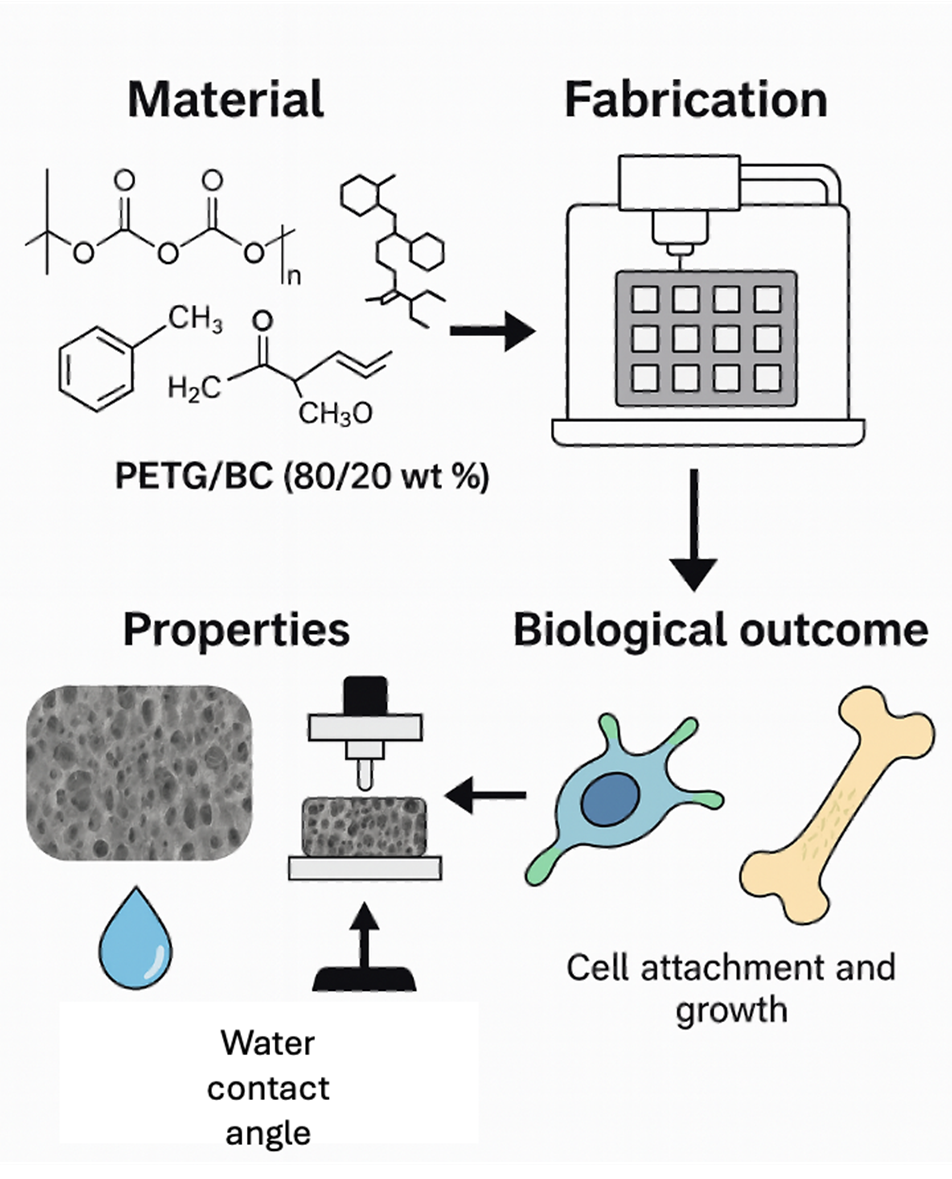
Bone tissue supports the body, enables movement, protects organs, produces blood cells, and stores minerals. In regenerative medicine, bone’s natural healing ability drives the need for engineered solutions to treat fractures, defects, and support implants. This study explores the development of polyethylene terephthalate glycol (PETG) and PETG/bacterial cellulose (BC) composite scaffolds with varying BC contents (10, 15, and 20 wt%) for bone tissue engineering (TE). Scanning electron microscopy and atomic force microscopy revealed porous structures with increasing surface roughness as BC content increased. Water contact angle analysis revealed enhanced hydrophilicity in PETG/BC composites, particularly at higher BC levels. Fourier transform infrared spectroscopy, X-ray diffraction, and differential scanning calorimetry confirmed successful BC integration and interactions with PETG, along with increased crystallinity. Mechanical testing indicated that compressive strength improved with higher BC content, with 20 wt% BC achieving optimal performance. Biological tests using human adipose-derived stem cells displayed enhanced proliferation, differentiation, and mineralization on PETG/BC scaffolds. Among the tested BC scaffolds, the 20 wt% BC scaffold demonstrated the most favorable physical, mechanical, and biological properties. Overall, PETG/BC scaffolds, especially those with 20 wt% BC, display strong potential for future bone TE applications.
- Wang Y, Pereira FR, Peach C, Huang B, Vyas C, Bartolo P. Robotic in situ bioprinting for cartilage tissue engineering. Int J Extreme Manuf. 2023;5(3):032004. doi: 10.1088/2631-7990/acda67
- Dwivedi R, Mehrotra D. 3D bioprinting and craniofacial regeneration. J Oral Biol Craniofac Res. 2020;10(4): 650-659. doi: 10.1016/j.jobcr.2020.08.011
- Baptista R, Guedes M. Morphological and mechanical characterization of 3D printed PLA scaffolds with controlled porosity for trabecular bone tissue replacement. Mater Sci Eng C. 2021;118:111528. doi: 10.1016/j.msec.2020.111528
- Loh QL, Choong C. Three-dimensional scaffolds for tissue engineering applications: role of porosity and pore size. Tissue Eng Part B Rev. 2013;19(6):485-502. doi: 10.1089/ten.teb.2012.0437
- Rahmati M, Silva EA, Reseland JE, Heyward CA, Haugen HJ. Biological responses to physicochemical properties of biomaterial surface. Chem Soc Rev. 2020;49(15): 5178–5224. doi: 10.1039/D0CS00103A
- Wang C, Huang W, Zhou Y, et al. 3D printing of bone tissue engineering scaffolds. Bioact Mater. 2020;5(1): 82-91. doi: 10.1016/j.bioactmat.2020.01.004
- Ribas RG, Schatkoski VM, Montanheiro TLA, et al. Current advances in bone tissue engineering concerning ceramic and bioglass scaffolds: a review. Ceram Int. 2019;45(17):21051-21061. doi: 10.1016/j.ceramint.2019.07.096
- Vyas C, Ates G, Aslan E, Hart J, Huang B, Bartolo B. Three-dimensional printing and electrospinning dual-scale polycaprolactone scaffolds with low-density and oriented fibers to promote cell alignment. 3D Print Addit Manuf. 2020;7(3):105-113. doi: 10.1089/3dp.2019.0091
- Huang B, Aslan E, Jiang Z, et al. Engineered dual-scale poly(ε-caprolactone) scaffolds using 3D printing and rotational electrospinning for bone tissue regeneration. Addit Manuf. 2020;36:101452. doi: 10.1016/j.addma.2020.101452
- Caetano G, Violante R, Sant’Ana AB, et al. Cellularized versus decellularized scaffolds for bone regeneration. Mater Lett. 2016;182:318-322. doi: 10.1016/j.matlet.2016.05.152
- Hassan MH, Omar AM, Daskalakis E, et al. The potential of polyethylene terephthalate glycol as biomaterial for bone tissue engineering. Polymers. 2020;12(12):3045. doi: 10.3390/polym12123045
- Martins RF, Branco R, Martins M, et al. Mechanical properties of additively manufactured polymeric materials— PLA and PETG—for biomechanical applications. Polymers. 2024;16(13):1868. doi: 10.3390/polym16131868
- Moreno Nieto D, Alonso-García M, Pardo-Vicente MA, Rodríguez-Parada L. Product design by additive manufacturing for water environments: study of degradation and absorption behavior of PLA and PETG. Polymers. 2021;13(7):1036. doi: 10.3390/polym13071036
- Habibi Y, Lucia LA, Rojas OJ. Cellulose nanocrystals: chemistry, self-assembly, and applications. Chem Rev. 2010;110(6):3479-3500. doi: 10.1021/cr900339w
- Saxena IM, Brown RM. Cellulose biosynthesis: current views and evolving concepts. Ann Bot. 2005;96(1):9–21. doi: 10.1093/aob/mci155
- Bhatnagar A, Sain M. Processing of cellulose nanofiber-reinforced composites. J Reinf Plast Compos. 2005;24(12):1259-1268. doi: 10.1177/0731684405049864
- Wu Q, Henriksson M, Liu X, Berglund LA. A high strength nanocomposite based on microcrystalline cellulose and polyurethane. Biomacromolecules. 2007;8(12):3687-3692. doi: 10.1021/bm701061t
- Utoiu E, Manoiu VS, Oprita EI, Craciunescu O. Bacterial cellulose: a sustainable source for hydrogels and 3D-printed scaffolds for tissue engineering. Gels. 2024;10(6):387. doi: 10.3390/gels10060387
- Shrivastav P, Pramanik S, Vaidya G, et al. Bacterial cellulose as a potential biopolymer in biomedical applications: a state-of-the-art review. J Mater Chem B. 2022;10(17): 3199-3241. doi: 10.1039/d1tb02709c
- Torgbo S, Sukyai P. Biodegradation and thermal stability of bacterial cellulose as biomaterial: the relevance in biomedical applications. Polym Degrad Stabil. 2020;179:109232. doi: 10.1016/j.polymdegradstab.2020.109232
- Wu Y, Wang Y, Wang F, Huang Y, He J. Preparation of 3D printed polylactic acid/bacterial cellulose composite scaffolds for tissue engineering applications. Polymers. 2022;14(21):4756. doi: 10.3390/polym14214756
- Panaitescu MD, Frone NA, Chiulan I, Gabor RA, Spataru IC, Căşărică A. Biocomposites from polylactic acid and bacterial cellulose nanofibers obtained by mechanical treatment. Bioresources. 2017;12(1):662-672. doi: 10.15376/biores.12.1.662-672
- Yodsanga S, Poeaim S, Chantarangsu S, Swasdison S. Investigation of biodegradation and biocompatibility of chitosan–bacterial cellulose composite scaffold for bone tissue engineering applications. Cells. 2025;14(10):723. doi: 10.3390/cells14100723
- Saska S, Barud SH, Gaspar MMA, Marchetto R, Ribeiro SJL, Messaddeq Y. Bacterial cellulose-hydroxyapatite nanocomposites for bone regeneration. Int J Biomater. 2011;2011:1-8. doi: 10.1155/2011/175362
- Boyetey BM, Torgbo S, Sukyai P. Bio-scaffold for bone tissue engineering with focus on bacterial cellulose, biological materials for hydroxyapatite synthesis and growth factors. Eur Polym J. 2023;194:112168. doi: 10.1016/j.eurpolymj.2023.112168
- Wang X, Zhang Y, Luo J, et al. Printability of hybridized composite from maleic acid-treated bacterial cellulose with gelatin for bone tissue regeneration. Adv Compos Hybrid Mater. 2023;6:134. doi: 10.1007/s42114-023-00711-7
- Lee CM, Gu J, Kafle K, Catchmark J, Kim SH. Cellulose produced by Gluconacetobacter xylinus strains ATCC 53524 and ATCC 23768: pellicle formation, post-synthesis aggregation and fiber density. Carbohydr Polym. 2015;133:270-276. doi: 10.1016/j.carbpol.2015.06.091
- Yan C, Kleiner C, Tabigue A, et al. PETG: applications in modern medicine. Eng Regen. 2024;5(1):45-55. doi: 10.1016/j.engreg.2023.11.001
- Daicho K, Kobayashi K, Fujisawa S, Saito T. Crystallinity-independent yet modification-dependent true density of nanocellulose. Biomacromolecules. 2019;21(2):939-945. doi: 10.1021/acs.biomac.9b01584
- Saxena P, Shukla P, Gaur M. Thermal analysis of polymer blends and double layer by DSC. Polym Polym Compos. 2020;29(9):S11-S18. doi: 10.1177/0967391120984606
- Martínez Cortizas A, López-Costas O. Linking structural and compositional changes in archaeological human bone collagen: an FTIR-ATR approach. Sci. Rep. 2020;10(1): 17888. doi: 10.1038/s41598-020-74993-y
- Figueiredo M, Fernando A, Martins G, Freitas J, Judas F, Figueiredo H. Effect of the calcination temperature on the composition and microstructure of hydroxyapatite derived from human and animal bone. Ceram Int. 2010;36(8):2383–2393. doi: 10.1016/j.ceramint.2010.07.016
- Zhu X, Chen T, Feng B, et al. Biomimetic bacterial cellulose-enhanced double-network hydrogel with excellent mechanical properties applied for the osteochondral defect repair. ACS Biomater Sci Eng. 2018;4(10):3534-3544. doi: 10.1021/acsbiomaterials.8b00682
- Loskot J, Jezbera D, Loskot R, et al. Influence of print speed on the microstructure, morphology, and mechanical properties of 3D-printed PETG products. Polym Test. 2023;123:108055. doi: 10.1016/j.polymertesting.2023.108055
- Aguayo MG, Fernández Pérez A, Reyes G, et al. Isolation and characterization of cellulose nanocrystals from rejected fibers originated in the Kraft pulping process. Polymers. 2018;10(10):1145. doi: 10.3390/polym10101145
- Techawinyutham L, Tengsuthiwat J, Srisuk R, Techawinyutham W, Rangappa SM, Siengchin S. Recycled LDPE/PETG blends and HDPE/PETG blends: mechanical, thermal, and rheological properties. J Mater Res Technol. 2021;15:2445-2458. doi: 10.1016/j.jmrt.2021.09.052
- Bhandari S, Lopez-Anido RA, Gardner DJ. Enhancing the interlayer tensile strength of 3D printed short carbon fiber reinforced PETG and PLA composites via annealing. Addit Manuf. 2019;30:100922. doi: 10.1016/j.addma.2019.100922
- Rand B, Robinson R. Surface characteristics of carbon fibres from PAN. Carbon. 1977;15(4):257-263. doi: 10.1016/0008-6223(77)90011-2
- Calore AR, Srinivas V, Groenendijk L, et al. Manufacturing of scaffolds with interconnected internal open porosity and surface roughness. Acta Biomater. 2023;156:158-176. doi: 10.1016/j.actbio.2022.07.017
- Ahn J-H, Kim J, Han G, et al. 3D-printed biodegradable composite scaffolds with significantly enhanced mechanical properties via the combination of binder jetting and capillary rise infiltration process. Addit Manuf. 2021;41:101988. doi: 10.1016/j.addma.2021.101988
- McAdam B, Fournet MB, McDonald P, Mojicevic M. Production of polyhydroxybutyrate (PHB) and factors impacting its chemical and mechanical characteristics. Polymers. 2020;12(12):2908. doi: 10.3390/polym12122908
- Oh JE, Park N-M. Hydrophilic, transparent, and stretchable film using unmodified cellulose fibers. Mater Lett. 2022;309:131385. doi: 10.1016/j.matlet.2021.131385
- Huang K, Zhu T, Nie J, et al. Microporous spongy scaffolds based on biodegradable elastic polyurethanes for the migration and growth of host cells. ACS Appl Polym Mater. 2022;4(5):3942-3951. doi: 10.1021/acsapm.2c00398
- Zou M, Zhao X, Zhang X, Zhao Y, Zhang C, Shi K. Bio-inspired multiple composite film with anisotropic surface wettability and adhesion for tissue repair. Chem Eng J. 2020;398:125563. doi: 10.1016/j.cej.2020.125563
- Conrad TL, Jaekel DJ, Kurtz SM, Roeder RK. Effects of the mold temperature on the mechanical properties and crystallinity of hydroxyapatite whisker-reinforced polyetheretherketone scaffolds. J Biomed Mater Res B. 2013;101(4):576-583. doi: 10.1002/jbm.b.32859
- Bhattarai N, Li Z, Edmondson D, Zhang M. Alginate-based nanofibrous scaffolds: structural, mechanical, and biological properties. Adv Mater. 2006;18(11):1463-1467. doi: 10.1002/adma.200502537
- Hassan MH, Omar AM, Daskalakis E, Liu F, Bartolo P. Preliminary studies on the suitability of PETG for 4D printing applications. MATEC Web Conf. 2020;318:01010. doi: 10.1051/matecconf/202031801010
- Lanyi FJ, Wenzke N, Kaschta J, Schubert DW. On the determination of the enthalpy of fusion of α‐crystalline isotactic polypropylene using differential scanning calorimetry, X‐ray diffraction, and Fourier‐transform infrared spectroscopy: an old story revisited. Adv Eng Mater. 2019;22(9):1900796. doi: 10.1002/adem.201900796
- Paszkiewicz S, Szymczyk A, Pawlikowska D, et al. Synthesis and characterization of poly(ethylene terephthalate-co-1,4-cyclohexanedimethylene terephthalate)-block-poly(tetramethylene oxide) copolymers. RSC Adv. 2017;7(66):41745-41754. doi: 10.1039/c7ra07172h
- Atykyan N, Revin V, Shutova V. Raman and FT-IR spectroscopy investigation of the cellulose structural differences from bacteria gluconacetobacter sucrofermentans during different regimes of cultivation on a molasses media. AMB Express. 2020;10(1):84. doi: 10.1186/s13568-020-01020-8
- Bagwan JK, Ahuja BB, Mulay AV, Jawale KJ. Geometrical analysis of extrusion-based (additively manufactured) 3D designed scaffold for bone tissue engineering: a finite element approach, Mater Today Proc. 2022;50(5):1465-1471. doi: 10.1016/j.matpr.2021.09.049
- Yao Y, Chen S. The effects of fiber’s surface roughness on the mechanical properties of fiber-reinforced polymer composites. J. Compos. Mater. 2012;47(23):2909-2923. doi: 10.1177/0021998312459871
- Shuai C, Wang C, Qi F, et al. Enhanced crystallinity and antibacterial of PHBV scaffolds incorporated with zinc oxide. J Nanomater. 2020:1-12. doi: 10.1155/2020/6014816
- Wang G, Qi F, Yang W, et al. Crystallinity and reinforcement in poly-L-lactic acid scaffold induced by carbon nanotubes. Adv Polym Technol. 2019:1-10. doi: 10.1155/2019/8625325
- Huang B, Wang Y, Vyas C, Bartolo B. Crystal growth of 3D poly(ε-caprolactone) based bone scaffolds and its effects on the physical properties and cellular interactions. Adv Sci. 2020;10(1):2203183. doi: 10.1002/advs.202203183
- Ustunel S, Prévôt ME, Webb G, et al. Mechanically tunable elastomer and cellulose nanocrystal composites as scaffolds for in vitro cell studies. Mater Adv. 2021;2(1):464-476. doi: 10.1039/d0ma00676a
- Lužanin O, Gudurić V, Bernhardt A, et al. Impact of in-process crystallinity of biodegradable scaffolds fabricated by material extrusion on the micro- and nanosurface topography, viability, proliferation, and differentiation of human mesenchymal stromal cells. Polymers. 2023;15(6):1468. doi: 10.3390/polym15061468
- Wen JH, Vincent LG, Fuhrmann A, et al. Interplay of matrix stiffness and protein tethering in stem cell differentiation. Nat Mater. 2014;13(10):979-987. doi: 10.1038/nmat4051
- Morgan EF, Unnikrisnan GU, Hussein AI. Bone mechanical properties in healthy and diseased states. Annu Rev Biomed Eng. 2018;20:119-143. doi: 10.1146/annurev-bioeng-062117-121139
- Öhman‐Mägi C, Holub O, Wu D, Hall RM, Persson C. Density and mechanical properties of vertebral trabecular bone—a review. JOR Spine. 2021;4(4):e1176. doi: 10.1002/jsp2.1176