

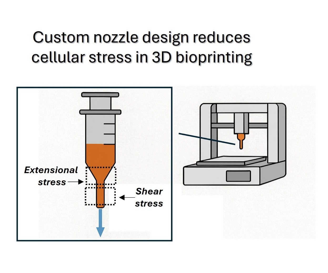
Optimizing nozzle design in extrusion-based 3D bioprinting to minimize mechanical stress and enhance cell viability

Extrusion-based three-dimensional bioprinting is a widely used technique for fabricating cell-laden constructs in tissue engineering and regenerative medicine. However, the mechanical stresses experienced by cells during the printing process can negatively affect their viability. This study examines the influence of nozzle geometry—specifically contraction angle and outlet diameter—on stress distribution and its effects on cell survival. Through a combination of experimental analysis and theoretical modeling, the impacts of nozzle design on the balance between shear and extensional stresses during bioprinting are explored. The findings highlight the importance of optimizing nozzle parameters to minimize mechanical damage and enhance post-printing cell viability. The proposed model provides a framework for guiding nozzle design, offering insights into the development of customized bioprinting strategies that enhance construct fidelity and biological functionality. These results contribute to advancing bioprinting techniques for applications in tissue engineering and regenerative medicine.
- Jim´enez M, Romero L, Dom´ınguez IA, Espinosa MdM, Dom´ınguez M. Additive manufacturing technologies: an overview about 3D printing methods and future prospects. Complexity. 2019;2019:9656938. doi: 10.1155/2019/9656938
- Gu BK, Choi DJ, Park SJ, Kim MS, Kang CM, Kim CH. 3-Dimensional bioprinting for tissue engineering applications. Biomater Res. 2016;20(1):1–8. doi: 10.1186/s40824-016-0058-2
- Jose RR, Rodriguez MJ, Dixon TA, Omenetto F, Kaplan DL. Evolution of bioinks and additive manufacturing technologies for 3D bio-printing. ACS Biomater Sci Eng. 2016;2(10):1662–1678. doi: 10.1021/acsbiomaterials.6b00088
- Bertassoni LE, Bertassoni LE. Bioprinting of complex multicellular organs with advanced functionality— recent progress and challenges ahead. Adv Mater. 2022; 34(3):2101321. doi: 10.1002/ADMA.202101321
- Parihar A, Pandita V, Kumar A, et al. 3D printing: advancement in biogenerative engineering to combat shortage of organs and bioapplicable materials. Regen Eng Transl Med. 2021;8:173–199. doi: 10.1007/S40883-021-00219-W
- Lam EHY, Yu F, Zhu S, Wang Z. 3D bioprinting for next-generation personalized medicine. Int J Mol Sci. 2023;24(7):6357. doi: 10.3390/IJMS24076357
- Mazzocchi A, Soker S, Skardal A. 3D bioprinting for high-throughput screening: drug screening, disease modeling, and precision medicine applications. Appl Phys Rev. 2019;6:1–12. doi: 10.1063/1.5056188
- Chen A, Wang W, Mao Z, et al. Multimaterial 3D and 4D bioprinting of heterogenous constructs for tissue engineering. Adv Mater. 2024;36(34):2307686. doi: 10.1002/adma.202307686
- You S, Xiang Y, Hwang HH, et al. High cell density and high-resolution 3D bioprinting for fabricating vascularized tissues. Sci Adv. 2023;9(8):eade7923. doi: 10.1126/sciadv.ade7923
- Scalzone A, Tonda-Turo C, Ferreira AM, Gentile P. 3D-printed soft hydrogels for cell encapsulation. In: Azevedo HS, Mano JF, Borges J, eds. Soft Matter for Biomedical Applications. Royal Society of Chemistry. London, UK; 2021:594–625. doi: 10.1039/9781839161124-00594. Soft Matter Series.
- Levato R, Jungst T, Scheuring RG, Blunk T, Groll J, Malda J. From shape to function: the next step in bioprinting. Adv Mater. 2020;32(12):1906423. doi: 10.1002/adma.201906423
- Ying G, Jiang N, Yu C, Zhang YS. Three-dimensional bioprinting of gelatin methacryloyl (GelMA). Bio Des Manuf. 2018;1:215–224. doi: 10.1007/S42242-018-0028-8
- Pedde RD, Mirani B, Navaei A, et al. Emerging biofabrication strategies for engineering complex tissue constructs. Adv Mater. 2017;29(19):1606061. doi: 10.1002/adma.201606061
- Tian X, Li M, Chen X. Bio-rapid-prototyping of tissue engineering scaffolds and the process-induced cell damage. J Biomim Biomater Tissue Eng. 2013;17:1–23. doi: 10.4028/www.scientific.net/JBBTE.17.1
- Lucas L, Aravind A, Emma P, Christophe M, Edwin-Joffrey C. Rheology, simulation and data analysis toward bioprinting cell viability awareness. Bioprinting. 2021;21:e00119. doi: 10.1016/J.BPRINT.2020.E00119
- Ning L, Betancourt N, Schreyer DJ, Chen X. Characterization of cell damage and proliferative ability during and after bioprinting. ACS Biomater Sci Eng. 2018;4(11):3906–3918. doi: 10.1021/acsbiomaterials.8b00714
- Bae YB, Jang HK, Shin TH, et al. Microfluidic assessment of mechanical cell damage by extensional stress. Lab Chip. 2016;16(1):96–103. doi: 10.1039/C5LC01006C
- Ning L, Chen X. A brief review of extrusion-based tissue scaffold bio-printing. Biotechnol J. 2017;12(8):1600671. doi: 10.1002/BIOT.201600671
- Grace HP. Dispersion phenomena in high viscosity immiscible fluid systems and application of static mixers as dispersion devices in such systems. Chem Eng Commun. 1982;14(3-6):225–277. doi: 10.1080/00986448208911047
- Aguado BA, Mulyasasmita W, Su J, Lampe KJ, Heilshorn SC. Improving viability of stem cells during syringe needle flow through the design of hydrogel cell carriers Tissue Eng Part A. 2012;18(7-8):806–815. doi: 10.1089/TEN.TEA.2011.0391
- Down LA, Papavassiliou DV, O’Rear EA. Significance of extensional stresses to red blood cell lysis in a shearing flow. Ann Biomed Eng. 2011;39:1632–1642. doi: 10.1007/S10439-011-0262-0
- Boularaoui S, Al Hussein G, Khan KA, Christoforou N, Stefanini C. An overview of extrusion-based bioprinting with a focus on induced shear stress and its effect on cell viability. Bioprinting. 2020;20:e00093. doi: 10.1016/J.BPRINT.2020.E00093
- Cogswell FN. Measuring the extensional rheology of polymer melts. Trans Soc Rheol. 1972;16(3):383–403. doi: 10.1122/1.549257
- Cogswell FN. Converging flow of polymer melts in extrusion dies. Polym Eng Sci. 1972;12(1):64–73. doi: 10.1002/PEN.760120111
- Alam K, Iqbal M, Hasan A, Al-Maskari N. Rheological characterization of biological hydrogels in aqueous state. J Appl Biotechnol Rep. 2020;7(3):171–175. doi: 10.30491/JABR.2020.109994
- Villone MM, Maffettone PL. Dynamics, rheology, and applications of elastic deformable particle suspensions: a review. Rheol Acta. 2019;58:109–130. doi: 10.1007/S00397-019-01134-2
- Tan SCW, Pan WX, Ma G, Cai N, Leong KW, Liao K. Viscoelastic behaviour of human mesenchymal stem cells. BMC Cell Biol. 2008;9(9):40. doi: 10.1186/1471-2121-9-40
- Nikolaev NI, Mu¨ller T, Williams DJ, Liu Y. Changes in the stiffness of human mesenchymal stem cells with the progress of cell death as measured by atomic force microscopy. J Biomech. 2014;47(3):625–630. doi: 10.1016/j.jbiomech.2013.12.004