

Three-dimensional bioprinted silk fibroin-hydroxypropyl cellulose scaffold loaded with tendon stem/progenitor cells for the prevention of heterotopic ossification following Achilles tendon injury

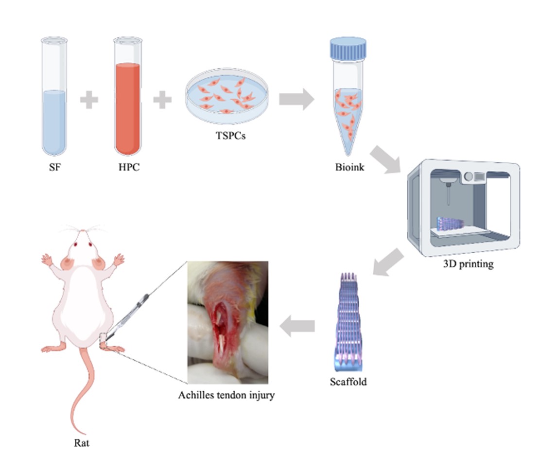
Achilles tendon injury is a common musculoskeletal disorder, particularly prevalent among athletes and middle-aged/elderly populations. Heterotopic ossification (HO) following Achilles tendon injury is a frequent complication that significantly compromises patients’ quality of life and athletic performance. Conventional conservative treatments and surgical interventions for HO often yield suboptimal outcomes, failing to restore native tendon functionality. Tissue engineering strategies integrating biomaterials and cells offer promising solutions for tendon regeneration and functional recovery. Three-dimensional bioprinting presents unique advantages in fabricating tissue-engineered scaffolds through precise control of architectural geometry and internal microstructure. In this study, we developed a novel silk fibroin (SF)–hydroxypropyl cellulose (HPC)–tendon stem/progenitor cell (TSPC) bioink with exceptional cytocompatibility and rheological properties. This bioink demonstrated superior printability for fabricating porous Achilles tendon scaffolds with high mechanical strength (elastic modulus: 85 MPa), controlled biodegradability, and optimal porosity (91%). In vitro experiments revealed that SF– HPC–TSPCs scaffolds promoted TSPC survival, migration, proliferation, and tenogenic differentiation within the scaffold microenvironment. In vivo assessments demonstrated that the scaffolds exhibited excellent biocompatibility, elicited no systemic inflammatory or immune responses, and effectively prevented HO in rat models of Achilles tendon injury. This study establishes a groundbreaking approach for addressing post-traumatic HO in tendon regeneration.
- Traweger A, Scott A, Kjaer M, et al. Achilles tendinopathy. Nat Rev Dis Primers. 2025;11(1):20. doi: 10.1038/s41572-025-00602-9
- Fischer S. Acute rupture of the Achilles tendon: diagnostics, treatment and aftercare. Unfallchirurgie (Heidelb). 2024;127(8):597-606. doi: 10.1007/s00113-024-01454-w
- Geuskens W, Caekebeke P, VAN Riet R. Prevalence and clinical implications of heterotopic ossification after distal biceps tendon repair. Acta Orthop Belg. 2023;89(4):695-700. doi: 10.52628/89.4.12447
- Fischer CS, Porsche J, Leyder D, Schüll D, Histing T, Ziegler P. Heterotopic ossification following severe radial head fractures: clinical outcome and associated factors. Jt Dis Relat Surg. 2025;36(1):47-55. doi: 10.52312/jdrs.2025.1992
- Dai G, Li Y, Liu J, et al. Higher BMP Expression in tendon stem/progenitor cells contributes to the increased heterotopic ossification in achilles tendon with aging. Front Cell Dev Biol. 2020;8:570605. doi: 10.3389/fcell.2020.570605
- Yu T, Zhang J, Zhu W, et al. Chondrogenesis mediates progression of ankylosing spondylitis through heterotopic ossification. Bone Res. 2021;9(1):19. doi: 10.1038/s41413-021-00140-6
- Wang Z, Yi X, Liu Y, Liu Q, Li Z, Yu A. Differential expression profiles and functional prediction of circRNA in mice with traumatic heterotopic ossification. Front Immunol. 2022;13:1090529. doi: 10.3389/fimmu.2022.1090529
- Xu L, Xu K, Wu Z, et al. Pioglitazone attenuates advanced glycation end products-induced apoptosis and calcification by modulating autophagy in tendon-derived stem cells. J Cell Mol Med. 2020;24(3):2240-2251. doi: 10.1111/jcmm.14901
- Tachibana N, Chijimatsu R, Okada H, et al. RSPO2 defines a distinct undifferentiated progenitor in the tendon/ ligament and suppresses ectopic ossification. Sci Adv. 2022;8(33):eabn2138. doi: 10.1126/sciadv.abn2138
- Wang T, Chen P, Chen L, et al. Reduction of mechanical loading in tendons induces heterotopic ossification and activation of the β-catenin signaling pathway. J Orthop Transl. 2021;29:42-50. doi: 10.1016/j.jot.2021.03.004
- Pierantoni M, Hammerman M, Silva Barreto I, et al. Spatiotemporal and microstructural characterization of heterotopic ossification in healing rat Achilles tendons. FASEB J. 2023;37(6). doi: 10.1096/fj.202201018RRR
- Yamaguchi H, Li M, Kitami M, Swaminathan S, Mishina Y, Komatsu Y. Enhanced BMP signaling in Cathepsin K-positive tendon progenitors induces heterotopic ossification. Biochem Biophys Res Commun. 2023;688:149147. doi: 10.1016/j.bbrc.2023.149147
- Magnusson SP, Agergaard AS, Couppé C, et al. Heterotopic ossification after an achilles tendon rupture cannot be prevented by early functional rehabilitation: a cohort study. Clin Orthop Relat Res. 2020;478(5):1101-1108. doi: 10.1097/corr.0000000000001085
- Agarwal S, Loder S, Levi B. Heterotopic ossification following upper extremity injury. Hand Clin. 2017;33(2): 363-373. doi: 10.1016/j.hcl.2016.12.013
- Delgado Caceres M, Angerpointner K, Galler M, et al. Tenomodulin knockout mice exhibit worse late healing outcomes with augmented trauma-induced heterotopic ossification of Achilles tendon. Cell Death Dis. 2021;12(11):1049. doi: 10.1038/s41419-021-04298-z
- Walden G, Liao X, Donell S, Raxworthy MJ, Riley GP, Saeed A. A clinical, biological, and biomaterials perspective into tendon injuries and regeneration. Tissue Eng Part B Rev. 2017;23(1):44-58. doi: 10.1089/ten.TEB.2016.0181
- No YJ, Castilho M, Ramaswamy Y, Zreiqat H. Role of biomaterials and controlled architecture on tendon/ligament repair and regeneration. Adv Mater. 2020;32(18):e1904511. doi: 10.1002/adma.201904511
- Sensini A, Cristofolini L. Biofabrication of electrospun scaffolds for the regeneration of tendons and ligaments. Materials (Basel). 2018;11(10):1963. doi: 10.3390/ma11101963
- Yan K, Zhang X, Liu Y, et al. 3D-bioprinted silk fibroin-hydroxypropyl cellulose methacrylate porous scaffold with optimized performance for repairing articular cartilage defects. Mater Des. 2023;225:111531. doi: 10.1016/j.matdes.2022.111531
- Zhang X, Liu Y, Zuo Q, et al. 3D bioprinting of biomimetic bilayered scaffold consisting of decellularized extracellular matrix and silk fibroin for osteochondral repair. Int J Bioprint. 2021;7(4):401. doi: 10.18063/ijb.v7i4.401
- Park W, Gao G, Cho DW. Tissue-specific decellularized extracellular matrix bioinks for musculoskeletal tissue regeneration and modeling using 3d bioprinting technology. Int J Mol Sci. 2021;22(15):7837. doi: 10.3390/ijms22157837
- Jiu J, Liu H, Li D, et al. 3D bioprinting approaches for spinal cord injury repair. Biofabrication. 2024;16(3). doi: 10.1088/1758-5090/ad3a13
- Zhang Y, Sun Y, Luo J, et al. Hydrogel-based materials for mandibular reconstruction. MSAM. 2025;4(2). doi: 10.36922/msam025070006
- Li H, Zeng S, Zhou L. Biocompatible nanogels with tunable size and tailorable properties: a simple synthesis by self-assembly and disulfide crosslinking of amphiphilic hyperbranched peach gum polysaccharide. Int J Biol Macromol. 2025;309(Pt 4):143083. doi: 10.1016/j.ijbiomac.2025.143083
- Zhang DKY, Brockman JM, Adu-Berchie K, et al. Subcutaneous biodegradable scaffolds for restimulating the antitumour activity of pre-administered CAR-T cells. Nat Biomed Eng. 2025;9(2):268-278. doi: 10.1038/s41551-024-01216-4
- Xu Y, Huang J, Mai Y, et al. CBD-conjugated BMP-inhibiting exosomes on collagen scaffold dual-target Achilles tendon repair: synergistic regeneration and heterotopic ossification prevention. Mater Today Bio. 2025;32:101790. doi: 10.1016/j.mtbio.2025.101790
- Avenoso A, Bruschetta G, D’Ascola A, et al. Hyaluronan fragments produced during tissue injury: A signal amplifying the inflammatory response. Arch Biochem Biophys. 2019;663:228-238. doi: 10.1016/j.abb.2019.01.015
- Kim JK, Go EJ, Ko KW, et al. PLGA microspheres containing hydrophobically modified magnesium hydroxide particles for acid neutralization-mediated anti-inflammation. Tissue Eng Regen Med. 2021;18(4):613-622. doi: 10.1007/s13770-021-00338-z
- Wang Y, Feng X, Chen X. Autonomous bioelectronic devices based on silk fibroin. Adv Mater. 2025;37(22):e2500073. doi: 10.1002/adma.202500073
- Shen C, Zhou Z, Li R, et al. Silk fibroin-based hydrogels for cartilage organoids in osteoarthritis treatment. Theranostics. 2025;15(2):560-584. doi: 10.7150/thno.103491
- Wang F, Lei H, Tian C, et al. An efficient biosynthetic system for developing functional silk fibroin-based biomaterials. Adv Mater. 2025;37(7):e2414878. doi: 10.1002/adma.202414878
- Cai G, Zhao W, Zhu T, Oliveira AL, Yao X, Zhang Y. Effects of protein conformational transition accompanied with crosslinking density cues in silk fibroin hydrogels on the proliferation and chondrogenesis of encapsulated stem cells. Regen Biomater. 2025;12:rbaf019. doi: 10.1093/rb/rbaf019
- Ben X, Lu X, Zhao G, Wei Z, Yang J, Kan Y. Internal secondary structural conformational states of silk fibroin studied by raman spectroscopy with band deconvolution analysis. Biomacromolecules. 2025;26(3):1992-2002. doi: 10.1021/acs.biomac.4c01827
- Ji X, Li Y, Wang J, et al. Silk protein gene engineering and its applications: recent advances in biomedicine driven by molecular biotechnology. Drug Des Devel Ther. 2025;19:599-626. doi: 10.2147/dddt.S504783
- Yin J, Fang Y, Xu L, Ahmed A. High-throughput fabrication of silk fibroin/hydroxypropyl methylcellulose (SF/HPMC) nanofibrous scaffolds for skin tissue engineering. Int J Biol Macromol. 2021;183:1210-1221. doi: 10.1016/j.ijbiomac.2021.05.026
- Su D, Yao M, Liu J, Zhong Y, Chen X, Shao Z. Enhancing mechanical properties of silk fibroin hydrogel through restricting the growth of β-sheet domains. ACS Appl Mater Interfaces. 2017;9(20):17489-17498. doi: 10.1021/acsami.7b04623
- Bi Y, Ehirchiou D, Kilts TM, et al. Identification of tendon stem/progenitor cells and the role of the extracellular matrix in their niche. Nat Med. 2007;13(10):1219-1227. doi: 10.1038/nm1630
- He W, Jiang C, Zhou P, Hu X, Gu X, Zhang S. Role of tendon-derived stem cells in tendon and ligament repair: focus on tissue engineer. Front Bioeng Biotechnol. 2024;12: 1357696. doi: 10.3389/fbioe.2024.1357696
- Chen J, Jiang C, Yin L, et al. A review of the role of tendon stem cells in tendon-bone regeneration. Med Sci Monit. 2023;29:e940805. doi: 10.12659/msm.940805
- Lu J, Chen H, Lyu K, et al. The functions and mechanisms of tendon stem/progenitor cells in tendon healing. Stem Cells Int. 2023;2023:1258024. doi: 10.1155/2023/1258024
- Jahani A, Nourbakhsh MS, Ebrahimzadeh MH, Mohammadi M, Yari D, Moradi A. Biomolecules-loading of 3D-printed alginate-based scaffolds for cartilage tissue engineering applications: a review on current status and future prospective. Arch Bone Jt Surg. 2024;12(2):92-101. doi: 10.22038/abjs.2023.73275.3396
- Li M, Shi T, Yao D, Yue X, Wang H, Liu K. High-cytocompatible semi-IPN bio-ink with wide molecular weight distribution for extrusion 3D bioprinting. Sci Rep. 2022;12(1):6349. doi: 10.1038/s41598-022-10338-1
- Delkash Y, Gouin M, Rimbeault T, et al. Bioprinting and in vitro characterization of an eggwhite-based cell-laden patch for endothelialized tissue engineering applications. J Funct Biomater. 2021;12(3):45. doi: 10.3390/jfb12030045
- Kolluru PV, Lipner J, Liu W, et al. Strong and tough mineralized PLGA nanofibers for tendon-to-bone scaffolds. Acta Biomater. 2013;9(12):9442-9450. doi: 10.1016/j.actbio.2013.07.042
- Jacob S, Reshmy R, Antony S, et al. Nanocellulose in tissue engineering and bioremediation: mechanism of action. Bioengineered. 2022;13(5):12823-12833. doi: 10.1080/21655979.2022.2074739
- Veronesi F, Giavaresi G, Bellini D, Casagranda V, Pressato D, Fini M. Evaluation of a new collagen-based medical device (ElastiCo®) for the treatment of acute Achilles tendon injury and prevention of peritendinous adhesions: An in vitro biocompatibility and in vivo investigation. J Tissue Eng Regen Med. 2020;14(8):1113-1125. doi: 10.1002/term.3085
- Webb WR, Dale TP, Lomas AJ, et al. The application of poly(3-hydroxybutyrate-co-3-hydroxyhexanoate) scaffolds for tendon repair in the rat model. Biomaterials. 2013;34(28):6683-6694. doi: 10.1016/j.biomaterials.2013.05.041
- Kim SH, Park JH, Kwon JS, et al. NIR fluorescence for monitoring in vivo scaffold degradation along with stem cell tracking in bone tissue engineering. Biomaterials. 2020;258:120267. doi: 10.1016/j.biomaterials.2020.120267
- Salehi S, Koeck K, Scheibel T. Spider silk for tissue engineering applications. Molecules. 2020;25(3):737. doi: 10.3390/molecules25030737
- Xie Y, Zhang F, Akkus O, King MW. A collagen/PLA hybrid scaffold supports tendon-derived cell growth for tendon repair and regeneration. J Biomed Mater Res B Appl Biomater. 2022;110(12):2624-2635. doi: 10.1002/jbm.b.35116
- Shirosaki Y, Tsukatani Y, Okamoto K, Hayakawa S, Osaka A. Preparation and drug release profile of chitosan-siloxane hybrid capsules coated with hydroxyapatite. Pharmaceutics. 2022;14(5):1111. doi: 10.3390/pharmaceutics14051111
- Fei Y, Ma Y, Zhang H, Li H, Feng G, Fang J. Nanotechnology for research and treatment of the intestine. J Nanobiotechnol. 2022;20(1):430. doi: 10.1186/s12951-022-01517-3
- Sadeghzadeh H, Mehdipour A, Dianat-Moghadam H, et al. PCL/Col I-based magnetic nanocomposite scaffold provides an osteoinductive environment for ADSCs in osteogenic cues-free media conditions. Stem Cell Res Ther. 2022;13(1):143. doi: 10.1186/s13287-022-02816-0
- Zheng A, Wang X, Xin X, et al. Promoting lacunar bone regeneration with an injectable hydrogel adaptive to the microenvironment. Bioact Mater. 2023;21:403-421. doi: 10.1016/j.bioactmat.2022.08.031
- Eggermont LJ, Rogers ZJ, Colombani T, Memic A, Bencherif SA. Injectable cryogels for biomedical applications. Trends Biotechnol. 2020;38(4):418-431. doi: 10.1016/j.tibtech.2019.09.008
- Yao X, Yang Y, Zhou Z. Non-mulberry silk fiber-based composite scaffolds containing millichannels for auricular cartilage regeneration. ACS Omega. 2022;7(17):15064-15073. doi: 10.1021/acsomega.2c00846
- Wang Y, Jin S, Luo D, et al. Functional regeneration and repair of tendons using biomimetic scaffolds loaded with recombinant periostin. Nat Commun. 2021;12(1):1293. doi: 10.1038/s41467-021-21545-1
- Markel DC, Dietz P, Provenzano G, Bou-Akl T, Ren WP. Attachment and growth of fibroblasts and tenocytes within a porous titanium scaffold: a bioreactor approach. Arthroplast Today. 2022;14:231-236.e1. doi: 10.1016/j.artd.2021.12.003
- Lin L, Shen Q, Xue T, Yu C. Heterotopic ossification induced by Achilles tenotomy via endochondral bone formation: expression of bone and cartilage related genes. Bone. 2010;46(2):425-431. doi: 10.1016/j.bone.2009.08.057
- Cai Z, Wu B, Ye G, et al. Enhanced osteogenic differentiation of human bone marrow mesenchymal stem cells in ossification of the posterior longitudinal ligament through activation of the BMP2-Smad1/5/8 pathway. Stem Cells Dev. 2020;29(24):1567-1576. doi: 10.1089/scd.2020.0117
- Zhang H, Liu MF, Liu RC, Shen WL, Yin Z, Chen X. Physical microenvironment-based inducible scaffold for stem cell differentiation and tendon regeneration. Tissue Eng Part B Rev. 2018;24(6):443-453. doi: 10.1089/ten.TEB.2018.0018
- Brownley RC, Agarwal S, Loder S, et al. Characterization of heterotopic ossification using radiographic imaging: evidence for a paradigm shift. PLoS One. 2015;10(11):e0141432. doi: 10.1371/journal.pone.0141432
- Foley KL, Hebela N, Keenan MA, Pignolo RJ. Histopathology of periarticular non-hereditary heterotopic ossification. Bone. 2018;109:65-70. doi: 10.1016/j.bone.2017.12.006
- Ng J, Wei Y, Zhou B, et al. Ectopic implantation of juvenile osteochondral tissues recapitulates endochondral ossification. J Tissue Eng Regen Med. 2018;12(2):468-478. doi: 10.1002/term.2500
- Fujihara R, Chiba Y, Nakagawa T, et al. Histomorphometry of ectopic mineralization using undecalcified frozen bone sections. Microsc Res Tech. 2018;81(11):1318-1324. doi: 10.1002/jemt.23140
- Yuan J, Sun W, Zhang Z, et al. 5-Fluorouracil/curcumin loaded silk fibroin hydrogel for the adjuvant therapy in colorectal cancer. Biomater Adv. 2025;168:214108. doi: 10.1016/j.bioadv.2024.214108
- Yeo D, Hwang SJ, Song YS, Lee HJ. Humulene inhibits acute gastric mucosal injury by enhancing mucosal integrity. Antioxidants (Basel). 2021;10(5):761. doi: 10.3390/antiox10050761
- Jian J, Sun L, Cheng X, Hu X, Liang J, Chen Y. Calycosin-7- O-β-d-glucopyranoside stimulates osteoblast differentiation through regulating the BMP/WNT signaling pathways. Acta Pharm Sin B. 2015;5(5):454-460. doi: 10.1016/j.apsb.2015.06.005
- Winterbourn CC, Kettle AJ, Hampton MB. Reactive oxygen species and neutrophil function. Annu Rev Biochem. 2016;85:765-792. doi: 10.1146/annurev-biochem-060815-014442
- Hong SY, Jiang HC, Xu WC, Zeng HS, Wang SG, Qin BL. Bioinformatics analysis reveals the potential role of matrix metalloproteinases in immunity and urolithiasis. Front Immunol. 2023;14:1158379. doi: 10.3389/fimmu.2023.1158379
- Lu P, Chen Z, Wu M, et al. Type I collagen extracellular matrix facilitates nerve regeneration via the construction of a favourable microenvironment. Burns Trauma. 2024;12:tkae049. doi: 10.1093/burnst/tkae049
- Gong SQ, Tang L, Liu Z, et al. NDGA enhances the physicochemical and anti-biodegradation performance of dentin collagen. Oral Dis. 2023;29(8):3525-3539. doi: 10.1111/odi.14453
- Georgopoulou A, Papadogiannis F, Batsali A, et al. Chitosan/ gelatin scaffolds support bone regeneration. J Mater Sci Mater Med. 2018;29(5):59. doi: 10.1007/s10856-018-6064-2
- Chachlioutaki K, Karavasili C, Adamoudi E, et al. Silk sericin/PLGA electrospun scaffolds with anti-inflammatory drug-eluting properties for periodontal tissue engineering. Biomater Adv. 2022;133:112723. doi: 10.1016/j.msec.2022.112723