

Three-dimensional-printed hydrogel scaffolds with neuregulin-1 sustained-release microspheres for enhanced dedifferentiation and myelin regeneration in peripheral nerve injury

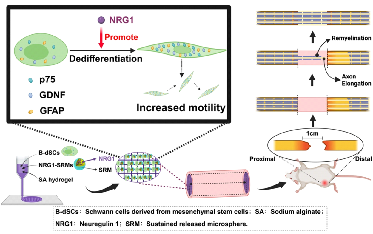
Remyelination is critical for functional recovery following peripheral nerve injury. Although autologous Schwann cell transplantation promotes effective myelin repair, its clinical translation remains limited due to donor scarcity and associated morbidity. Bone marrow-derived Schwann-like cells (B-dSCs) offer a promising alternative; however, their limited dedifferentiation capacity significantly constrains therapeutic outcomes. Neuregulin-1 (NRG1), a key axonal signal, effectively induces Schwann cell dedifferentiation but requires precise, sustained delivery to exert optimal effects. Here, we developed a three-dimensional (3D)-printed hydrogel scaffold incorporating NRG1-loaded sustained-release microspheres to achieve localized, prolonged NRG1 delivery. In vitro studies demonstrated that NRG1 significantly enhanced the dedifferentiation and remyelination capacity of B-dSCs in a dorsal root ganglion co-culture system. Mechanistically, NRG1 promoted dedifferentiation by activating the c-Jun N-terminal kinase (JNK) signaling pathway—a pivotal regulator of Schwann cell plasticity. Pharmacological inhibition of JNK markedly suppressed NRG1-induced dedifferentiation and downregulated myelin-associated gene expression, confirming pathway specificity. Furthermore, the 3D-printed scaffold effectively maintained uniform NRG1 distribution, facilitating enhanced axonal regeneration and improved myelin integrity. Collectively, these findings highlight the essential role of JNK signaling in NRG1-driven Schwann cell dedifferentiation and underscore the therapeutic promise of combining sustained-release systems with engineered cell therapies to advance peripheral nerve repair.
- Zhang Y, Yi D, Hong Q, et al. Platelet-rich plasma-derived exosomes boost mesenchymal stem cells to promote peripheral nerve regeneration. J Control Release. 2024;367 (2):265-282. doi: 10.1016/j.jconrel.2024.01.043.
- Dai Y, Lu T, Li L, et al. Electrospun composite PLLA-PPSB nanofiber nerve conduits for peripheral nerve defects repair and regeneration. Adv Healthc Mater. 2024;13(10):e2303539. doi: 10.1002/adhm.202303539.
- Rodríguez FJ, Verdú E, Ceballos D, Navarro X. Nerve guides seeded with autologous schwann cells improve nerve regeneration. Exp Neurol. 2000;161(2):571-584. doi: 10.1006/exnr.1999.7315.
- Bachelin C, Lachapelle F, Girard C, et al. Efficient myelin repair in the macaque spinal cord by autologous grafts of Schwann cells. Brain. 2005;128(3):540-549. doi: 10.1093/brain/awh406.
- Sun X, Zhu Y, Yin HY, et al. Differentiation of adipose-derived stem cells into Schwann cell-like cells through intermittent induction: potential advantage of cellular transient memory function. Stem Cell Res Ther. 2018;9:133. doi: 10.1186/s13287-018-0884-3.
- Wei C, Guo Y, Ci Z, Li M, Zhang Y, Zhou Y. Advances of Schwann cells in peripheral nerve regeneration: from mechanism to cell therapy. Biomed Pharmacother. 2024;175:116645. doi: 10.1016/j.biopha.2024.116645.
- Hou B, Ye Z, Ji W, et al. Comparison of the effects of BMSC-derived Schwann cells and autologous Schwann cells on remyelination using a rat sciatic nerve defect model. Int J Biol Sci. 2018;14(14):1910-1922. doi: 10.7150/ijbs.26765.
- Li R, Li D, Wu C, et al. Nerve growth factor activates autophagy in Schwann cells to enhance myelin debris clearance and to expedite nerve regeneration. Theranostics. 2020;10(4):1649-1677. doi: 10.7150/thno.40919.
- Kataria H, Alizadeh A, Karimi-Abdolrezaee S. Neuregulin-1/ ErbB network: an emerging modulator of nervous system injury and repair. Prog Neurobiol. 2019;180:101643. doi: 10.1016/j.pneurobio.2019.101643.
- Soto J, Monje PV. Axon contact-driven Schwann cell dedifferentiation. Glia. 2017;65(6):864-882. doi: 10.1002/glia.23131.
- Li X, Zhang T, Li C, et al. Electrical stimulation accelerates Wallerian degeneration and promotes nerve regeneration after sciatic nerve injury. Glia. 2023;71(4):758-774. doi: 10.1002/glia.24309.
- Stoll G, Müller HW. Nerve injury, axonal degeneration and neural regeneration: basic insights. Brain Pathol. 1999;9(2):313-325. doi: 10.1111/j.1750-3639.1999.tb00229.x.
- Ding Z, Dai C, Shan W, et al. TNF-α up-regulates Nanog by activating NF-κB pathway to induce primary rat spinal cord astrocytes dedifferentiation. Life Sci. 2021;287:120126. doi: 10.1016/j.lfs.2021.120126.
- Boerboom A, Reusch C, Pieltain A, Chariot A, Franzen R. KIAA1199: a novel regulator of MEK/ERK-induced Schwann cell dedifferentiation. Glia. 2017;65(10): 1682-1696. doi: 10.1002/glia.23188.
- Honorati MC, Cattini L, Facchini A. IL-17, IL-1beta and TNF-alpha stimulate VEGF production by dedifferentiated chondrocytes. Osteoarthritis Cartilage. 2004;12(8):683-691. doi: 10.1016/j.joca.2004.05.009.
- Dinarello CA. Proinflammatory cytokines. Chest. 2000;118(2):503-508. doi: 10.1378/chest.118.2.503.
- Jayaraman T, Paget A, Shin YS, et al. TNF-alpha-mediated inflammation in cerebral aneurysms: a potential link to growth and rupture. Vasc Health Risk Manag. 2008;4:805-817. doi: 10.2147/vhrm.s2700.
- Zhou Y, Xu Z, Liu Z. Role of IL-33-ST2 pathway in regulating inflammation: current evidence and future perspectives. J Transl Med. 2023;21:902. doi: 10.1186/s12967-023-04782-4.
- Min B, Kim D, Feige MJ. IL-30(†) (IL-27A): a familiar stranger in immunity, inflammation, and cancer. Exp Mol Med. 2021;53:823-834. doi: 10.1038/s12276-021-00630-x.
- Wang P, Qian H, Xiao M, Lv J. Role of signal transduction pathways in IL-1β-induced apoptosis: pathological and therapeutic aspects. Immun Inflamm Dis. 2023;11(5):e762. doi: 10.1002/iid3.762.
- Kang R, Li R, Dai P, Li Z, Li Y, Li C. Deoxynivalenol induced apoptosis and inflammation of IPEC-J2 cells by promoting ROS production. Environ Pollut. 2019;251:689-698. doi: 10.1016/j.envpol.2019.05.026.
- Newbern J, Birchmeier C. Nrg1/ErbB signaling networks in Schwann cell development and myelination. Semin Cell Dev Biol. 2010;21(8-9):922-928. doi: 10.1016/j.semcdb.2010.08.008.
- Birchmeier C. ErbB receptors and the development of the nervous system. Exp Cell Res. 2009;315(4):611-618. doi: 10.1016/j.yexcr.2008.10.035.
- Boerboom A, Dion V, Chariot A, Franzen R. Molecular mechanisms involved in Schwann cell plasticity. Front Mol Neurosci. 2017;10:38. doi: 10.3389/fnmol.2017.00038.
- Fornasari BE, El Soury M, Nato G, et al. Fibroblasts colonizing nerve conduits express high levels of soluble neuregulin1, a factor promoting schwann cell dedifferentiation. Cells. 2020;9(6):1366. doi: 10.3390/cells9061366.
- Monje PV, Soto J, Bacallao K, Wood PM. Schwann cell dedifferentiation is independent of mitogenic signaling and uncoupled to proliferation: role of cAMP and JNK in the maintenance of the differentiated state. J Biol Chem. 2010;285(42):31024-31036. doi: 10.1074/jbc.M110.116970.
- Kong L, Hassinan CW, Gerstner F, et al. Boosting neuregulin 1 type-III expression hastens SMA motor axon maturation. Acta Neuropathol Commun. 2023;11:53. doi: 10.1186/s40478-023-01551-8.
- Michailov GV, Sereda MW, Brinkmann BG, et al. Axonal neuregulin-1 regulates myelin sheath thickness. Science. 2004;304(5671):700-703. doi: 10.1126/science.1095862.
- Kim H, Park H, Lee SJ. Effective method for drug injection into subcutaneous tissue. Sci Rep. 2017;7:9613. doi: 10.1038/s41598-017-10110-w.
- Yin PT, Han E, Lee KB. Engineering stem cells for biomedical applications. Adv Healthc Mater. 2016;5(1):10-55. doi: 10.1002/adhm.201400842.
- Liu L, Wannemuehler MJ, Narasimhan B. Biomaterial nanocarrier-driven mechanisms to modulate anti-tumor immunity. Curr Opin Biomed Eng. 2021;20:100322. doi: 10.1016/j.cobme.2021.100322.
- Gutierrez AM, Frazar EM, Klaus MVX, Paul P, Hilt JZ. Hydrogels and hydrogel nanocomposites: enhancing healthcare through human and environmental treatment. Adv Healthc Mater. 2022;11(2):e2101820. doi: 10.1002/adhm.202101820.
- Qian Y, Lu S, Meng J, Chen W, Li J. Thermo-responsive hydrogels coupled with photothermal agents for biomedical applications. Macromol Biosci. 2023;23(3):e2300214. doi: 10.1002/mabi.202300214.
- Gu D, O’Connor AJ, Qiao GHG, Ladewig K. Hydrogels with smart systems for delivery of hydrophobic drugs. Expert Opin Drug Deliv. 2017;14(8):879-895. doi: 10.1080/17425247.2017.1245290.
- Asadi N, Alizadeh E, Salehi R, Khalandi B, Davaran S, Akbarzadeh A. Nanocomposite hydrogels for cartilage tissue engineering: a review. Artif Cells Nanomed Biotechnol. 2018;46(6):465-471. doi: 10.1080/21691401.2017.1345924.
- Buonanno A, Fischbach GD. Neuregulin and ErbB receptor signaling pathways in the nervous system. Curr Opin Neurobiol. 2001;11(2):287-296. doi: 10.1016/s0959-4388(00)00210-5.
- Jorissen RN, Walker F, Pouliot N, Garrett TPJ, Ward CW, Burgess AW. Epidermal growth factor receptor: mechanisms of activation and signalling. Exp Cell Res. 2003; 284(1):31-53. doi: 10.1016/s0014-4827(02)00098-8.
- Wen J, Hou B, Lin W, et al. 3D-printed hydrogel scaffold-loaded granulocyte colony-stimulating factor sustained-release microspheres and their effect on endometrial regeneration. Biomater Sci. 2022;10(12):3346-3358. doi: 10.1039/d2bm00109h.
- Griffith LG, Naughton G. Tissue engineering—current challenges and expanding opportunities. Science. 2002;295(5557):1009-1014. doi: 10.1126/science.1069210.
- Fraher JP. Myelin-axon relationships in the rat phrenic nerve: longitudinal variation and lateral asymmetry. J Comp Neurol. 1992;323(3):551-557. doi: 10.1002/cne.903230407.
- Su Q, Nasser MI, He J, et al. Engineered schwann cell-based therapies for injury peripheral nerve reconstruction. Front Cell Neurosci. 2022;16:865266. doi: 10.3389/fncel.2022.865266.
- Hui T, Wang C, Yu L, Zhou C, Qiu M. Phosphorene hydrogel conduits as “neurotrophin reservoirs” for promoting regeneration of peripheral nerves. J Mater Chem B. 2023;11(18):3808-3815. doi: 10.1039/d3tb00340j.
- Yasui G, Yamamoto Y, Shichinohe R, et al. Neuregulin-1 released by biodegradable gelatin hydrogels can accelerate facial nerve regeneration and functional recovery of traumatic facial nerve palsy. J Plast Reconstr Aesthet Surg. 2016;69(2):328-334. doi: 10.1016/j.bjps.2015.10.037.
- Jung N, Park S, Choi Y, et al. Tonsil-derived mesenchymal stem cells differentiate into a Schwann cell phenotype and promote peripheral nerve regeneration. Int J Mol Sci. 2016;17(11):1867. doi: 10.3390/ijms17111867.
- Shin YK, Jang SY, Park JY, et al. The neuregulin-Rac-MKK7 pathway regulates antagonistic c-jun/Krox20 expression in Schwann cell dedifferentiation. Glia. 2013;61(5):892-904. doi: 10.1002/glia.22482.
- Kaempchen K, Mielke K, Utermark T, Langmesser S, Hanemann CO. Upregulation of the Rac1/JNK signaling pathway in primary human schwannoma cells. Hum Mol Genet. 2003;12(10):1211-1221. doi: 10.1093/hmg/ddg146.