

Fabrication of an in vitro three-dimensional tumor model using liver-derived decellularized extracellular matrix/gelatin methacrylate bioink for investigating cancer characteristics and drug resistance

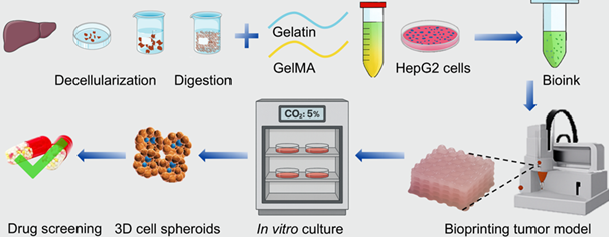
Three-dimensional (3D) printing has emerged as a promising technique for creating in vitro tumor models that replicate the tumor microenvironment, with the potential to reduce or replace the use of experimental animals. The incorporation of 3D decellularized extracellular matrix (dECM) hydrogels significantly enhances cellular responsiveness and functionality in drug screening. However, the limited printability of dECM restricts its application in ex vivo 3D disease models. To address this limitation, researchers have developed a blended bioink composed of dECM, gelatin methacrylate (GelMA), and gelatin, specifically tailored for direct ink writing-based 3D bioprinting. This formulation exhibits favorable shear-thinning behavior, enhanced viscosity, and thermal-sensitive properties, making it suitable for 3D bioprinting. The combination of dECM with GelMA and gelatin not only improves the printability of the bioink but also enhances the resolution of the printed scaffolds. Furthermore, dECM demonstrated positive effects on human hepatocellular carcinoma (HepG2) cells, promoting proliferation, migration, and cell spheroid formation. A 3D liver cancer model was successfully created in vitro by printing HepG2 cells encapsulated in the bioink containing dECM. This model exhibited characteristics akin to in vivo solid tumors, including notable cell proliferation, protein secretion, and substantial cell spheroid formation (up to 78.83 ± 9.41 μm on day 8). Additionally, it showed drug resistance, with 46.23% and 31.34% cell viability observed at 100 μg/mL concentrations of doxorubicin and paclitaxel, respectively. These findings underscore the potential of bioprinted 3D tumor models composed of GelMA, gelatin, and dECM as valuable platforms for the evaluation of anticancer drugs.
- De Santibañes M, Pekolj J, Sanchez Claria R, De Santibañes E, Mazza OM. Technical implications for surgical resection in locally advanced pancreatic cancer. Cancers. 2023;15(5):1509. doi: 10.3390/cancers15051509
- Zaher A, Foromera J, Capanu M, Chou J, Faleck DM. Sa1848 extent of surgical resection for ibd-associated neoplasia and colorectal cancer outcomes. Gastroenterology. 2024;166(5):S-549-S-550. doi: 10.1016/S0016-5085(24)01733-5
- Yu X, Wang X, Sun L, Yamazaki A, Li X. Tumor microenvironment regulation - enhanced radio - immunotherapy. Biomater Adv. 2022;138:212867. doi: 10.1016/j.bioadv.2022.212867
- Pan S, Sun Z, Zhao B, et al. Therapeutic application of manganese-based nanosystems in cancer radiotherapy. Biomaterials. 2023;302:122321. doi: 10.1016/j.biomaterials.2023.122321
- Moy B, Rumble RB, Carey LA, for the Chemotherapy and Targeted Therapy for Endocrine-Pretreated or Hormone Receptor–Negative Metastatic Breast Cancer Expert Panel. Chemotherapy and targeted therapy for endocrine-pretreated or hormone receptor–negative metastatic breast cancer: ASCO guideline rapid recommendation update. JCO. 2023;41(6):1318-1320. doi: 10.1200/JCO.22.02807
- Garg V, Kumar L. Metronomic chemotherapy in ovarian cancer. Cancer Lett. 2023;579:216469. doi: 10.1016/j.canlet.2023.216469
- Michalczyk K, Pawlik J, Czekawy I, Kozłowski M, Cymbaluk- Płoska A. Complementary methods in cancer treatment— cure or curse? IJERPH. 2021;18(1):356. doi: 10.3390/ijerph18010356
- Li Y, Xu X. Nanomedicine solutions to intricate physiological-pathological barriers and molecular mechanisms of tumor multidrug resistance. J Control Release. 2020;323:483-501. doi: 10.1016/j.jconrel.2020.05.007
- Mao S, He J, Zhao Y, et al. Bioprinting of patient-derived in vitro intrahepatic cholangiocarcinoma tumor model: establishment, evaluation and anti-cancer drug testing. Biofabrication. 2020;12(4):045014. doi: 10.1088/1758-5090/aba0c3
- Xu X, Liu Y, Liu Y, et al. Functional hydrogels for hepatocellular carcinoma: Therapy, imaging, and in vitro model. J Nanobiotechnol. 2024;22(1):381. doi: 10.1186/s12951-024-02547-9
- Li W, Hu X, Yang S, et al. A novel tissue-engineered 3D tumor model for anti-cancer drug discovery. Biofabrication. 2018;11(1):015004. doi: 10.1088/1758-5090/aae270
- Vitale S, Calapà F, Colonna F, et al. Advancements in 3D in vitro models for colorectal cancer. Adv Sci. 2024;11(32):2405084. doi: 10.1002/advs.202405084
- Lv J, Du X, Wang M, Su J, Wei Y, Xu C. Construction of tumor organoids and their application to cancer research and therapy. Theranostics. 2024;14(3):1101-1125. doi: 10.7150/thno.91362
- Van Tienderen GS, Conboy J, Muntz I, et al. Tumor decellularization reveals proteomic and mechanical characteristics of the extracellular matrix of primary liver cancer. Biomater Adv. 2023;146:213289. doi: 10.1016/j.bioadv.2023.213289
- Krujatz F, Dani S, Windisch J, et al. Think outside the box: 3D bioprinting concepts for biotechnological applications – recent developments and future perspectives. Biotechnol Adv. 2022;58:107930. doi: 10.1016/j.biotechadv.2022.107930
- Levato R, Dudaryeva O, Garciamendez-Mijares CE, et al. Light-based vat-polymerization bioprinting. Nat Rev Methods Primers. 2023;3(1):47. doi: 10.1038/s43586-023-00231-0
- Kumar S, Tharayil A, Thomas S. 3D bioprinting of nature-inspired hydrogel inks based on synthetic polymers. ACS Appl Polym Mater. 2021;3(8):3685-3701. doi: 10.1021/acsapm.1c00567
- Yang K, Wang L, Vijayavenkataraman S, Yuan Y, Tan ECK, Kang L. Recent applications of three-dimensional bioprinting in drug discovery and development. Adv Drug Deliv Rev. 2024;214:115456. doi: 10.1016/j.addr.2024.115456
- Bian S, Hu X, Zhu H, et al. 3D bioprinting of artificial skin substitute with improved mechanical property and regulated cell behavior through integrating patterned nanofibrous films. ACS Nano. 2024;18(28):18503-18521. doi: 10.1021/acsnano.4c04088
- Kronemberger GS, Spagnuolo FD, Karam AS, Chattahy K, Storey KJ, Kelly DJ. Rapidly degrading hydrogels to support biofabrication and 3D bioprinting using cartilage microtissues. ACS Biomater Sci Eng. 2024;10(10):6441-6450. doi: 10.1021/acsbiomaterials.4c00819
- Pérez Del Río E, Rey-Vinolas S, Santos F, et al. 3D printing as a strategy to scale-up biohybrid hydrogels for T cell manufacture. ACS Appl Mater Interfaces. 2024;16(38):50139-50146. doi: 10.1021/acsami.4c06183
- Pramanick A, Hayes T, Sergis V, McEvoy E, Pandit A, Daly AC. 4D bioprinting shape‐morphing tissues in granular support hydrogels: Sculpting structure and guiding maturation. Adv Funct Mater. 2025;35(5):2414559. doi: 10.1002/adfm.202414559
- Patel ZH, Charania AA, Punjani Z, et al. Evaluating anticancer agents on 3D bioprinted organoid tumors (BOT) to reduce cost and accelerate therapeutic discovery. JCO. 2022;40(16_suppl):e13500. doi: 10.1200/JCO.2022.40.16_suppl.e13500
- Kankala RK, Zhang YS, Kang L, Ambrosio L. Editorial: Polymeric microarchitectures for tissue regeneration and drug screening. Front Bioeng Biotechnol. 2023;11:1144991. doi: 10.3389/fbioe.2023.1144991
- Wang Y, Jeon H. 3D cell cultures toward quantitative high-throughput drug screening. Trends Pharmacol Sci. 2022;43(7):569-581. doi: 10.1016/j.tips.2022.03.014
- González-Callejo P, García-Astrain C, Herrero-Ruiz A, et al. 3D bioprinted tumor-stroma models of triple-negative breast cancer stem cells for preclinical targeted therapy evaluation. ACS Appl Mater Interfaces. 2024;16(21): 27151-27163. doi: 10.1021/acsami.4c04135
- Ferreira LP, Gaspar VM, Mano JF. Decellularized extracellular matrix for bioengineering physiomimetic 3D in vitro tumor models. Trends Biotechnol. 2020;38(12): 1397-1414. doi: 10.1016/j.tibtech.2020.04.006
- García-Astrain C, Henriksen-Lacey M, Lenzi E, et al. A scaffold-assisted 3D cancer cell model for surface-enhanced Raman scattering-based real-time sensing and imaging. ACS Nano. 2024;18(17):11257-11269. doi: 10.1021/acsnano.4c00543
- Ebrahimighaei R, Tarassova N, Bond SC, et al. Extracellular matrix stiffness controls cardiac fibroblast proliferation via the nuclear factor-Y (NF-Y) transcription factor. Biochim Biophys Acta Mol Cell Res. 2024;1871(2):119640. doi: 10.1016/j.bbamcr.2023.119640
- Fu Y, Zhou Y, Wang K, Li Z, Kong W. Extracellular matrix interactome in modulating vascular homeostasis and remodeling. Circ Res. 2024;134(7):931-949. doi: 10.1161/CIRCRESAHA.123.324055
- Guo WY, Wang WH, Xu PY, Kankala RK. Decellularised extracellular matrix-based injectable hydrogels for tissue engineering applications. Biomater Transl. 2024;5(2):114-128. doi: 10.12336/biomatertransl.2024.02.003
- Naba A. Mechanisms of assembly and remodelling of the extracellular matrix. Nat Rev Mol Cell Biol. 2024;25(11): 865-885. doi: 10.1038/s41580-024-00767-3
- Zhou Z, Zhang Y, Zeng Y, et al. Effects of nanomaterials on synthesis and degradation of the extracellular matrix. ACS Nano. 2024;18(11):7688-7710. doi: 10.1021/acsnano.3c09954
- Bhar B, Ranta P, Samudrala PK, Mandal BB. Omentum extracellular matrix-silk fibroin hydroscaffold promotes wound healing through vascularization and tissue remodeling in the diabetic rat model. ACS Biomater Sci Eng. 2024;10(2):1090-1105. doi: 10.1021/acsbiomaterials.3c01877
- Tam NW, Becker A, Mangiarotti A, Cipitria A, Dimova R. Extracellular vesicle mobility in collagen I hydrogels is influenced by matrix-binding integrins. ACS Nano. 2024;18(43):29585-29601. doi: 10.1021/acsnano.4c07186
- Puistola P, Kethiri A, Nurminen A, et al. Cornea-specific human adipose stem cell-derived extracellular matrix for corneal stroma tissue engineering. ACS Appl Mater Interfaces. 2024;16(13):15761-15772. doi: 10.1021/acsami.3c17803
- Na J, Yang Z, Shi Q, et al. Extracellular matrix stiffness as an energy metabolism regulator drives osteogenic differentiation in mesenchymal stem cells. Bioact Mater. 2024;35:549-563. doi: 10.1016/j.bioactmat.2024.02.003
- Tao M, Ao T, Mao X, et al. Sterilization and disinfection methods for decellularized matrix materials: review, consideration and proposal. Bioact Mater. 2021;6(9):2927-2945. doi: 10.1016/j.bioactmat.2021.02.010
- Snyder Y, Jana S. Strategies for development of decellularized heart valve scaffolds for tissue engineering. Biomaterials. 2022;288:121675. doi: 10.1016/j.biomaterials.2022.121675
- Lian L, Xie M, Luo Z, et al. Rapid volumetric bioprinting of decellularized extracellular matrix bioinks. Adv Mater. 2024;36(34):2304846. doi: 10.1002/adma.202304846
- Niu R, Xin Q, Xu E, Yao S, Chen M, Liu D. Nanostarch-stimulated cell adhesion in 3D bioprinted hydrogel scaffolds for cell cultured meat. ACS Appl Mater Interfaces. 2024;16(18):23015-23026. doi: 10.1021/acsami.4c03585
- Bae M, Kim JJ, Kim J, Cho DW. Decellularized extracellular matrix for three- dimensional bioprinted in vitro disease modeling. Int J Bioprint. 2024;10(2):1970. doi: 10.36922/ijb.1970
- Shin M, Galarraga JH, Kwon MY, Lee H, Burdick JA. Gallol-derived ECM-mimetic adhesive bioinks exhibiting temporal shear-thinning and stabilization behavior. Acta Biomater. 2019;95:165-175. doi: 10.1016/j.actbio.2018.10.028
- Ouyang L, Yao R, Zhao Y, Sun W. Effect of bioink properties on printability and cell viability for 3D bioplotting of embryonic stem cells. Biofabrication. 2016;8(3):035020. doi: 10.1088/1758-5090/8/3/035020
- Brumberg VA, Bikmulina PY, Pozdnyakov AA, et al. Scaling liver bioprinting: a guide for usage of the hepatic extracellular matrix as a bioink. Int J Bioprint. 2025;11(1):57-83. doi: 10.36922/ijb.4343
- Ouyang L, Yao R, Zhao Y, Sun W. Effect of bioink properties on printability and cell viability for 3D bioplotting of embryonic stem cells. Biofabrication. 2016;8(3):035020. doi: 10.1088/1758-5090/8/3/035020
- Choudhury D, Tun HW, Wang T, Naing MW. Organ-derived decellularized extracellular matrix: a game changer for bioink manufacturing? Trends Biotechnol. 2018;36(8):787-805. doi: 10.1016/j.tibtech.2018.03.003
- Xu P, Kankala RK, Wang S, Chen A. Decellularized extracellular matrix-based composite scaffolds for tissue engineering and regenerative medicine. Regen Biomater. 2023;11:rbad107. doi: 10.1093/rb/rbad107
- Khati V, Ramachandraiah H, Pati F, Svahn HA, Gaudenzi G, Russom A. 3D bioprinting of multi-material decellularized liver matrix hydrogel at physiological temperatures. Biosensors. 2022;12(7):521. doi: 10.3390/bios12070521
- Myojin Y, Hikita H, Sugiyama M, et al. Hepatic stellate cells in hepatocellular carcinoma promote tumor growth via growth differentiation factor 15 production. Gastroenterology. 2021;160(5):1741-1754.e16. doi: 10.1053/j.gastro.2020.12.015
- Wang Y, Kankala RK, Zhang J, et al. Modeling endothelialized hepatic tumor microtissues for drug screening. Adv Sci. 2020;7(21):2002002. doi: 10.1002/advs.202002002
- Baharvand H, Hashemi SM, Kazemi Ashtiani S, Farrokhi A. Differentiation of human embryonic stem cells into hepatocytes in 2D and 3D culture systems in vitro. Int J Dev Biol. 2006;50(7):645-652. doi: 10.1387/ijdb.052072hb
- Krueger WH, Tanasijevic B, Barber V, et al. Cholesterol-secreting and statin-responsive hepatocytes from human ES and iPS cells to model hepatic involvement in cardiovascular health. PLoS ONE. 2013;8(7):e67296. doi: 10.1371/journal.pone.0067296
- Gevaert E, Billiet T, Declercq H, Dubruel P, Cornelissen R. Galactose‐functionalized gelatin hydrogels improve the functionality of encapsulated Hepg2 cells. Macromol Biosci. 2014;14(3):419-427. doi: 10.1002/mabi.2013003