

Immunomodulation and bone repair of 3D-printed SrBG/PCL scaffolds with PDA coating

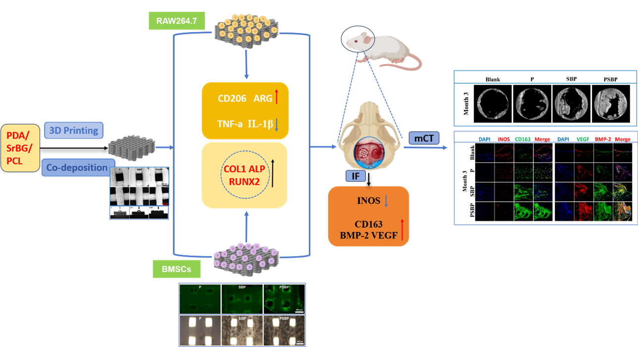
3D-printed polycaprolactone (PCL) scaffolds are widely used for bone tissue engineering but suffer from deficiencies, such as difficulty in cell adhesion, lack of osteogenic activity, and poor immunomodulatory capacity. Enhancing the biological responsiveness of PCL scaffolds remains a key focus in bone tissue engineering. In this study, the following three types of scaffolds were prepared: (i) PCL, (ii) strontium (Sr)-doped bioactive glass (SrBG)/PCL, and (iii) polydopamine (PDA)/SrBG/PCL. The scaffolds were assayed in vitro for their effect on the expression of osteoinductive differentiation markers (ALP, RUNX2, and COL1), and their influence on macrophage (MP) (CD206, ARG, TNF-α, IL1β, IL-10, and IL-12) behavior was evaluated. Their effect on bone defect repair was assessed in vivo using micro-computed tomography (micro- CT), hematoxylin and eosin (HE) staining, Masson staining, and immunofluorescence staining (iNOS, CD163, BMP-2, and VEGF). The results demonstrated that PDA/SrBG/ PCL scaffolds significantly promoted the proliferation and osteogenic differentiation of bone marrow mesenchymal stem cells (BMSCs), inhibited the differentiation of MPs to the M1 phenotype, and promoted the differentiation of MPs to the M2 phenotype, resulting in better pro-osteogenic, immunomodulatory, and angiogenic effects in vivo. This observation may be associated with the release of Sr²+ from SrBG, and surface modification with PDA further enhanced the immunomodulation and bone repair ability of the scaffold. The study demonstrated that the PDA/SrBG/PCL scaffolds exhibit excellent bone repair capabilities and hold strong potential for applications in bone tissue engineering.
- Chi J, Wang M, Chen J, et al. Topographic orientation of scaffolds for tissue regeneration: recent advances in biomaterial design and applications. Biomimetics (Basel). 2022;7(3):131. doi: 10.3390/biomimetics7030131
- Yang X, Wang Y, Zhou Y, et al. The application of polycaprolactone in three-dimensional printing scaffolds for bone tissue engineering. Polymers (Basel). 2021;13(16):2754. doi: 10.3390/polym13162754
- Mancuso E, Shah L, Jindal S, et al. Additively manufactured BaTiO(3) composite scaffolds: a novel strategy for load bearing bone tissue engineering applications. Mater Sci Eng C Mater Biol Appl. 2021;126:112192. doi: 10.1016/j.msec.2021.112192
- Gómez-Lizárraga KK, Flores-Morales C, Del Prado-Audelo ML, et al. Polycaprolactone- and polycaprolactone/ceramic-based 3D-bioplotted porous scaffolds for bone regeneration: a comparative study. Mater Sci Eng C Mater Biol Appl. 2017;79:326-335. doi: 10.1016/j.msec.2017.05.003
- Zhao QM, Sun YY, Wu CS, et al. Enhanced osteogenic activity and antibacterial ability of manganese-titanium dioxide microporous coating on titanium surfaces. Nanotoxicology. 2020;14(3):289-309. doi: 10.1080/17435390.2019.1690065
- Zurita-Méndez NN, Carbajal-De la Torre G, Flores-Merino MV, Espinosa-Medina MA. Development of bioactive glass-collagen-hyaluronic acid-polycaprolactone scaffolds for tissue engineering applications. Front Bioeng Biotechnol. 2022;10:825903. doi: 10.3389/fbioe.2022.825903
- Tian B, Wang N, Jiang Q, et al. The immunogenic reaction and bone defect repair function of ε-poly-L-lysine (EPL)- coated nanoscale PCL/HA scaffold in rabbit calvarial bone defect. J Mater Sci Mater Med. 2021;32(6):63. doi: 10.1007/s10856-021-06533-7
- Blum C, Taskin MB, Shan J, et al. Appreciating the first line of the human innate immune defense: a strategy to model and alleviate the neutrophil elastase-mediated attack toward bioactivated biomaterials. Small. 2021;17(13):e2007551. doi: 10.1002/smll.202007551
- Moore EM, Maestas DR, Jr., Cherry CC, et al. Biomaterials direct functional B cell response in a material-specific manner. Sci Adv. 2021;7(49):eabj5830. doi: 10.1126/sciadv.abj5830
- Knecht RS, Bucher CH, Van Linthout S, et al. Mechanobiological principles influence the immune response in regeneration: implications for bone healing. Front Bioeng Biotechnol. 2021;9:614508. doi: 10.3389/fbioe.2021.614508
- Zhang B, Su Y, Zhou J, et al. Toward a better regeneration through implant-mediated immunomodulation: harnessing the immune responses. Adv Sci (Weinh). 2021;8(16): e2100446. doi: 10.1002/advs.202100446
- Ben Letaifa R, Klaylat T, Tarchala M, et al. Osteoimmunology: an overview of the interplay of the immune system and the bone tissue in fracture healing. Surgeries. 2024;5(2): 402-414. doi: 10.3390/surgeries5020033
- Jiang F, Zhao H, Zhang P, et al. Challenges in tendon-bone healing: emphasizing inflammatory modulation mechanisms and treatment. Front Endocrinol (Lausanne). 2024;15:1485876. doi: 10.3389/fendo.2024.1485876
- Sadowska JM, Ginebra MP. Inflammation and biomaterials: role of the immune response in bone regeneration by inorganic scaffolds. J Mater Chem B. 2020;8(41):9404-9427. doi: 10.1039/d0tb01379j
- Wei S, Wang Y, Sun Y, et al. Biodegradable silk fibroin scaffold doped with mineralized collagen induces bone regeneration in rat cranial defects. Int J Biol Macromol. 2023;235:123861. doi: 10.1016/j.ijbiomac.2023.123861
- Kawcher Alam M, Sahadat Hossain M, Anisur Rahman Dayan M, et al. Fabrication and characterization of a bioscaffold using hydroxyapatite and unsaturated polyester resin. ACS Omega. 2024;9(13):15210-15221. doi: 10.1021/acsomega.3c09599
- Ye B, Wu B, Su Y, et al. Recent advances in the application of natural and synthetic polymer-based scaffolds in musculoskeletal regeneration. Polymers (Basel). 2022; 14(21):4566. doi: 10.3390/polym14214566
- Dong C, Tan G, Zhang G, et al. The function of immunomodulation and biomaterials for scaffold in the process of bone defect repair: a review. Front Bioeng Biotechnol. 2023;11:1133995. doi: 10.3389/fbioe.2023.1133995
- Handley EL, Callanan A. Effects of electrospun fibers containing ascorbic acid on oxidative stress reduction for cardiac tissue engineering. J Appl Polym Sci. 2023;140(32): e54242. doi: 10.1002/app.54242
- Homaeigohar S, Boccaccini AR. Nature-derived and synthetic additives to poly(ε-caprolactone) nanofibrous systems for biomedicine; an updated overview. Front Chem. 2021;9:809676. doi: 10.3389/fchem.2021.809676
- Gniesmer S, Brehm R, Hoffmann A, et al. In vivo analysis of vascularization and biocompatibility of electrospun polycaprolactone fibre mats in the rat femur chamber. J Tissue Eng Regen Med. 2019;13(7):1190-1202. doi: 10.1002/term.2868
- Han Y, Jia B, Lian M, et al. High-precision, gelatin-based, hybrid, bilayer scaffolds using melt electro-writing to repair cartilage injury. Bioact Mater. 2021;6(7):2173-2186. doi: 10.1016/j.bioactmat.2020.12.018
- Zhao S, Xie K, Guo Y, et al. Fabrication and biological activity of 3D-printed polycaprolactone/magnesium porous scaffolds for critical size bone defect repair. ACS Biomater Sci Eng. 2020;6(9):5120-5131. doi: 10.1021/acsbiomaterials.9b01911
- Tang C, Shen Y, Xing Y, et al. 3D-printed stents loaded with Panax notoginseng saponin for promoting re-endothelialization and reducing local inflammation in the carotid artery of rabbits. ACS Biomater Sci Eng. 2024;10(10):6483-6497. doi: 10.1021/acsbiomaterials.4c00925
- Liu J, Du G, Yu H, et al. Synthesis of hierarchically porous bioactive glass and its mineralization activity. Molecules. 2023;28(5):2224. doi: 10.3390/molecules28052224
- Liang J, Lu X, Zheng X, et al. Modification of titanium orthopedic implants with bioactive glass: a systematic review of in vivo and in vitro studies. Front Bioeng Biotechnol. 2023; 11:1269223. doi: 10.3389/fbioe.2023.1269223
- Zhao H, Wang X, Jin A, et al. Reducing relapse and accelerating osteogenesis in rapid maxillary expansion using an injectable mesoporous bioactive glass/fibrin glue composite hydrogel. Bioact Mater. 2022;18:507-525. doi: 10.1016/j.bioactmat.2022.03.001
- Oliver JN, Su Y, Lu X, et al. Bioactive glass coatings on metallic implants for biomedical applications. Bioact Mater. 2019;4:261-270. doi: 10.1016/j.bioactmat.2019.09.002
- Chakraborty A, Bodhak S, Tah I, et al. Tailored bioactive glass coating: navigating devitrification toward a superior implant performance. ACS Biomater Sci Eng. 2024;10(8): 5300-5312. doi: 10.1021/acsbiomaterials.4c01032
- Wa Q, Luo Y, Tang Y, et al. Mesoporous bioactive glass-enhanced MSC-derived exosomes promote bone regeneration and immunomodulation in vitro and in vivo. J Orthop Translat. 2024;49:264-282. doi: 10.1016/j.jot.2024.09.009
- Lin K, Wang X, Zhang N, Shen Y. Strontium (Sr) strengthens the silicon (Si) upon osteoblast proliferation, osteogenic differentiation and angiogenic factor expression. J Mater Chem B. 2016;4(21):3632-3638. doi: 10.1039/c6tb00735j
- Liu X, Huang H, Zhang J, et al. Recent advance of strontium functionalized in biomaterials for bone regeneration. Bioengineering (Basel). 2023;10(4):414. doi: 10.3390/bioengineering10040414
- Zhao F, Lei B, Li X, et al. Promoting in vivo early angiogenesis with sub-micrometer strontium-contained bioactive microspheres through modulating macrophage phenotypes. Biomaterials. 2018;178:36-47. doi: 10.1016/j.biomaterials.2018.06.004
- Ding J, Zhao J, Wang L, et al. Regulated contribution of local and systemic immunity to new bone regeneration by modulating B/Sr concentration of bioactive borosilicate glass. Mater Today Bio. 2023;19:100585. doi: 10.1016/j.mtbio.2023.100585
- Li M, Wang M, Wen Y, et al. Signaling pathways in macrophages: molecular mechanisms and therapeutic targets. MedComm. (2020). 2023;4(5):e349. doi: 10.1002/mco2.349
- Terzopoulou Z, Baciu D, Gounari E, et al. Composite membranes of poly(ε-caprolactone) with bisphosphonate-loaded bioactive glasses for potential bone tissue engineering applications. Molecules. 2019;24(17):3067. doi: 10.3390/molecules24173067
- Sheng X, Li C, Wang Z, et al. Advanced applications of strontium-containing biomaterials in bone tissue engineering. Mater Today Bio. 2023;20:100636. doi: 10.1016/j.mtbio.2023.100636
- Poh PS, Hutmacher DW, Stevens MM, Woodruff MA. Fabrication and in vitro characterization of bioactive glass composite scaffolds for bone regeneration. Biofabrication. 2013;5(4):045005. doi: 10.1088/1758-5082/5/4/045005
- Ma T, Wang CX, Ge XY, Zhang Y. Applications of polydopamine in implant surface modification. Macromol Biosci. 2023;23(10):e2300067. doi: 10.1002/mabi.202300067
- Qin D, Zhao Y, Cheng R, et al. Mussel-inspired immunomodulatory and osteoinductive dual-functional hydroxyapatite nanoplatform for promoting bone regeneration. J Nanobiotechnology. 2024;22(1):320. doi: 10.1186/s12951-024-02593-3
- Jin L, Yuan F, Chen C, et al. Degradation products of polydopamine restrained inflammatory response of LPS-stimulated macrophages through mediation TLR-4-MYD88 dependent signaling pathways by antioxidant. Inflammation. 2019;42(2):658-671. doi: 10.1007/s10753-018-0923-3
- Park J, Lee SJ, Jung TG, et al. Surface modification of a three-dimensional polycaprolactone scaffold by polydopamine, biomineralization, and BMP-2 immobilization for potential bone tissue applications. Colloids Surf B Biointerfaces. 2021;199:111528. doi: 10.1016/j.colsurfb.2020.111528
- Ye G, Bao F, Zhang X, et al. Nanomaterial-based scaffolds for bone tissue engineering and regeneration. Nanomedicine (Lond). 2020;15(20):1995-2017. doi: 10.2217/nnm-2020-0112
- Bharadwaz A, Jayasuriya AC. Recent trends in the application of widely used natural and synthetic polymer nanocomposites in bone tissue regeneration. Mater Sci Eng C Mater Biol Appl. 2020;110:110698. doi: 10.1016/j.msec.2020.110698
- Fazeli N, Arefian E, Irani S, et al. 3D-printed PCL scaffolds coated with nanobioceramics enhance osteogenic differentiation of stem cells. ACS Omega. 2021;6(51): 35284-35296. doi: 10.1021/acsomega.1c04015
- Abd El-Hamid HK, El-Kheshen AA, Abdou AM, Elwan RL. Incorporation of strontium borosilicate bioactive glass in calcium aluminate biocement: physicomechanical, bioactivity and antimicrobial properties. J Mech Behav Biomed Mater. 2023;144:105976. doi: 10.1016/j.jmbbm.2023.105976
- Zhao Q, Gao S. Poly (butylene succinate)/silicon nitride nanocomposite with optimized physicochemical properties, biocompatibility, degradability, and osteogenesis for cranial bone repair. J Funct Biomater. 2022;13(4):231. doi: 10.3390/jfb13040231
- Wu Y, Liu J, Kang L, et al. An overview of 3D printed metal implants in orthopedic applications: present and future perspectives. Heliyon. 2023;9(7):e17718. doi: 10.1016/j.heliyon.2023.e17718
- Zhang L, Yang G, Johnson BN, Jia X. Three-dimensional (3D) printed scaffold and material selection for bone repair. Acta Biomater. 2019;84:16-33. doi: 10.1016/j.actbio.2018.11.039
- Toosi S, Javid-Naderi MJ, Tamayol A, et al. Additively manufactured porous scaffolds by design for treatment of bone defects. Front Bioeng Biotechnol. 2023;11:1252636. doi: 10.3389/fbioe.2023.1252636
- Bouakaz I, Drouet C, Grossin D, et al. Hydroxyapatite 3D-printed scaffolds with gyroid-triply periodic minimal surface porous structure: fabrication and an in vivo pilot study in sheep. Acta Biomater. 2023;170:580-595. doi: 10.1016/j.actbio.2023.08.041
- Du J, Zhou Y, Bao X, et al. Surface polydopamine modification of bone defect repair materials: characteristics and applications. Front Bioeng Biotechnol. 2022;10:974533. doi: 10.3389/fbioe.2022.974533
- Ghalia MA, Alhanish A. Mechanical and biodegradability of porous PCL/PEG copolymer-reinforced cellulose nanofibers for soft tissue engineering applications. Med Eng Phys. 2023;120:104055. doi: 10.1016/j.medengphy.2023.104055
- Xiao L, Li Y, Geng R, et al. Polymer composite microspheres loading (177)Lu radionuclide for interventional radioembolization therapy and real-time SPECT imaging of hepatic cancer. Biomater Res. 2023;27(1):110. doi: 10.1186/s40824-023-00455-x
- Mahnavi A, Shahriari-Khalaji M, Hosseinpour B, et al. Evaluation of cell adhesion and osteoconductivity in bone substitutes modified by polydopamine. Front Bioeng Biotechnol. 2022;10:1057699. doi: 10.3389/fbioe.2022.1057699
- Wang H, Yuan C, Lin K, et al. Modifying a 3D-printed Ti6Al4V implant with polydopamine coating to improve BMSCs growth, osteogenic differentiation, and in situ osseointegration in vivo. Front Bioeng Biotechnol. 2021;9:761911. doi: 10.3389/fbioe.2021.761911
- Zhao ZH, Ma XL, Ma JX, et al. Sustained release of naringin from silk-fibroin-nanohydroxyapatite scaffold for the enhancement of bone regeneration. Mater Today Bio. 2022;13:100206. doi: 10.1016/j.mtbio.2022.100206
- Devi VK A, Ray S, Arora U, et al. Dual drug delivery platforms for bone tissue engineering. Front Bioeng Biotechnol. 2022;10:969843. doi: 10.3389/fbioe.2022.969843
- S S, R G AP, Bajaj G, et al. A review on the recent applications of synthetic biopolymers in 3D printing for biomedical applications. J Mater Sci Mater Med. 2023;34(12):62. doi: 10.1007/s10856-023-06765-9
- Wang S, Gu R, Wang F, et al. 3D-Printed PCL/Zn scaffolds for bone regeneration with a dose-dependent effect on osteogenesis and osteoclastogenesis. Mater Today Bio. 2022;13:100202. doi: 10.1016/j.mtbio.2021.100202
- Murugan S, Parcha SR. Fabrication techniques involved in developing the composite scaffolds PCL/HA nanoparticles for bone tissue engineering applications. J Mater Sci Mater Med. 2021;32(8):93. doi: 10.1007/s10856-021-06564-0
- Labbaf S, Tsigkou O, Müller KH, et al. Spherical bioactive glass particles and their interaction with human mesenchymal stem cells in vitro. Biomaterials. 2011;32(4): 1010-1018. doi: 10.1016/j.biomaterials.2010.08.082
- Ma YX, Jiao K, Wan QQ, et al. Silicified collagen scaffold induces semaphorin 3A secretion by sensory nerves to improve in-situ bone regeneration. Bioact Mater. 2022;9: 475-490. doi: 10.1016/j.bioactmat.2021.07.016
- Qiu P, Li M, Chen K, et al. Periosteal matrix-derived hydrogel promotes bone repair through an early immune regulation coupled with enhanced angio- and osteogenesis. Biomaterials. 2020;227:119552. doi: 10.1016/j.biomaterials.2019.119552
- Zhang Y, Yu W, Ba Z, et al. 3D-printed scaffolds of mesoporous bioglass/gliadin/polycaprolactone ternary composite for enhancement of compressive strength, degradability, cell responses and new bone tissue ingrowth. Int J Nanomed. 2018;13:5433-5447. doi: 10.2147/ijn.S164869
- Qiu H, Xiong H, Zheng J, et al. Sr-incorporated bioactive glass remodels the immunological microenvironment by enhancing the mitochondrial function of macrophage via the PI3K/AKT/mTOR signaling pathway. ACS Biomater Sci Eng. 2024;10(6):3923-3934. doi: 10.1021/acsbiomaterials.4c00228
- Ajita J, Saravanan S, Selvamurugan N. Effect of size of bioactive glass nanoparticles on mesenchymal stem cell proliferation for dental and orthopedic applications. Mater Sci Eng C Mater Biol Appl. 2015;53:142-149. doi: 10.1016/j.msec.2015.04.041
- Feito MJ, Casarrubios L, Oñaderra M, et al. Response of RAW 264.7 and J774A.1 macrophages to particles and nanoparticles of a mesoporous bioactive glass: a comparative study. Colloids Surf B Biointerfaces. 2021;208:112110. doi: 10.1016/j.colsurfb.2021.112110
- Xu H, Zhu Y, Hsiao AW, et al. Bioactive glass-elicited stem cell-derived extracellular vesicles regulate M2 macrophage polarization and angiogenesis to improve tendon regeneration and functional recovery. Biomaterials. 2023;294:121998. doi: 10.1016/j.biomaterials.2023.121998
- Römer P, Desaga B, Proff P, et al. Strontium promotes cell proliferation and suppresses IL-6 expression in human PDL cells. Ann Anat. 2012;194(2):208-211. doi: 10.1016/j.aanat.2011.09.008
- Buache E, Velard F, Bauden E, et al. Effect of strontium-substituted biphasic calcium phosphate on inflammatory mediators production by human monocytes. Acta Biomater. 2012;8(8):3113-3119. doi: 10.1016/j.actbio.2012.04.045
- Palczewska-Komsa MP, Gapiński B, Nowicka A. The influence of new bioactive materials on pulp-dentin complex regeneration in the assessment of cone bone computed tomography (CBCT) and computed micro-tomography (Micro-CT) from a present and future perspective-a systematic review. J Clin Med. 2022;11(11):3091. doi: 10.3390/jcm11113091
- Mohan S, Karunanithi P, Raman Murali M, et al. Potential use of 3D CORAGRAF-loaded PDGF-BB in PLGA microsphere seeded mesenchymal stromal cells in enhancing the repair of calvaria critical-size bone defect in rat model. Mar Drugs. 2022;20(9):561. doi: 10.3390/md20090561
- Lee JB, Seo MS. Mandibular incisors with two canals are associated with the presence of the distolingual root in mandibular first molars: a cone-beam computed tomographic study. BMC Oral Health. 2022;22(1):145. doi: 10.1186/s12903-022-02184-4
- Song W, Zhao L, Gao Y, et al. Dual growth factor-modified microspheres nesting human-derived umbilical cord mesenchymal stem cells for bone regeneration. J Biol Eng. 2023;17(1):43. doi: 10.1186/s13036-023-00360-w
- Wang C, Wu J, Liu L, et al. Improving osteoinduction and osteogenesis of Ti6Al4V alloy porous scaffold by regulating the pore structure. Front Chem. 2023;11:1190630. doi: 10.3389/fchem.2023.1190630
- Okizaki S, Ito Y, Hosono K, et al. Suppressed recruitment of alternatively activated macrophages reduces TGF-β1 and impairs wound healing in streptozotocin-induced diabetic mice. Biomed Pharmacother. 2015;70:317-325. doi: 10.1016/j.biopha.2014.10.020
- Zhu C, Kros JM, van der Weiden M, et al. Expression site of P2RY12 in residential microglial cells in astrocytomas correlates with M1 and M2 marker expression and tumor grade. Acta Neuropathol Commun. 2017;5(1):4. doi: 10.1186/s40478-016-0405-5
- Lisi L, Ciotti GM, Braun D, et al. Expression of iNOS, CD163 and ARG-1 taken as M1 and M2 markers of microglial polarization in human glioblastoma and the surrounding normal parenchyma. Neurosci Lett. 2017;645:106-112. doi: 10.1016/j.neulet.2017.02.076
- Wu S, Dong R, Xie Y, et al. CO-loaded hemoglobin/ EGCG nanoparticles functional coatings for inflammation modulation of vascular implants. Regen Biomater. 2025;12: rbae148. doi: 10.1093/rb/rbae148
- Luo R, Su J, Zhang W, et al. Targeted delivery of NO donor and ROS scavenger for synergistic treatment of rheumatoid arthritis. Biomed Pharmacother. 2024;174:116540. doi: 10.1016/j.biopha.2024.116540
- Chen X, Wang M, Chen F, et al. Correlations between macrophage polarization and osteoinduction of porous calcium phosphate ceramics. Acta Biomater. 2020;103:318-332. doi: 10.1016/j.actbio.2019.12.019
- Alhamdi JR, Peng T, Al-Naggar IM, et al. Controlled M1- to-M2 transition of aged macrophages by calcium phosphate coatings. Biomaterials. 2019;196:90-99. doi: 10.1016/j.biomaterials.2018.07.012
- Ye J, Xie C, Wang C, et al. Promoting musculoskeletal system soft tissue regeneration by biomaterial-mediated modulation of macrophage polarization. Bioact Mater. 2021;6(11): 4096-4109. doi: 10.1016/j.bioactmat.2021.04.017
- Gélébart P, Cuenot S, Sinquin C, et al. Microgels based on Infernan, a glycosaminoglycan-mimetic bacterial exopolysaccharide, as BMP-2 delivery systems. Carbohydr Polym. 2022;284:119191. doi: 10.1016/j.carbpol.2022.119191
- Kathami N, Moreno-Vicente C, Martín P, et al. rhBMP-2 induces terminal differentiation of human bone marrow mesenchymal stromal cells only by synergizing with other signals. Stem Cell Res Ther. 2024;15(1):124. doi: 10.1186/s13287-024-03735-y
- Li T, Peng M, Yang Z, et al. 3D-printed IFN-γ-loading calcium silicate-β-tricalcium phosphate scaffold sequentially activates M1 and M2 polarization of macrophages to promote vascularization of tissue engineering bone. Acta Biomater. 2018;71:96-107. doi: 10.1016/j.actbio.2018.03.012
- Liu Y, Li Z, Li W, et al. Discovery of β-sitosterol’s effects on molecular changes in rat diabetic wounds and its impact on angiogenesis and macrophages. Int Immunopharmacol. 2024;126:111283.doi: 10.1016/j.intimp.2023.111283
- Mao L, Xia L, Chang J, et al. The synergistic effects of Sr and Si bioactive ions on osteogenesis, osteoclastogenesis and angiogenesis for osteoporotic bone regeneration. Acta Biomater. 2017;61:217-232. doi: 10.1016/j.actbio.2017.08.015
- Zhu X, Wu S, Yang K, et al. Polydopamine-modified konjac glucomannan scaffold with sustained release of vascular endothelial growth factor to promote angiogenesis. Int J Biol Macromol. 2024;271(Pt 1):132333. doi: 10.1016/j.ijbiomac.2024.132333
- Cheng S, Wang D, Ke J, et al. Improved in vitro angiogenic behavior of human umbilical vein endothelial cells with oxidized polydopamine coating. Colloids Surf B Biointerfaces. 2020;194:111176. doi: 10.1016/j.colsurfb.2020.111176