

High-resolution 3D printing of collagen I-based scaffolds via Schiff-base interaction for enhanced osteogenic differentiation

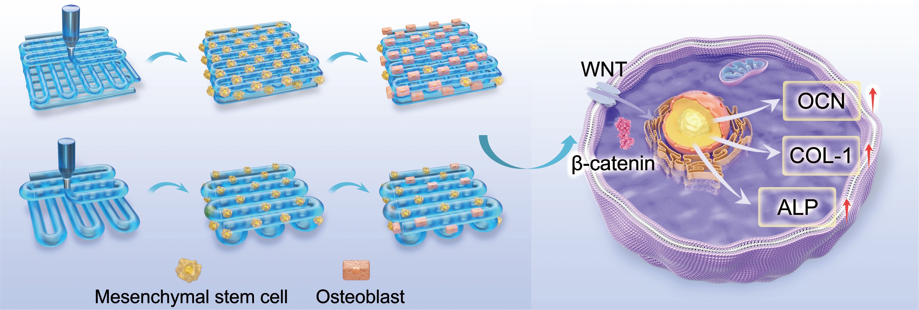
Collagen I is a key extracellular matrix (ECM) component in bone tissue and one of the most important biomaterials for bone tissue engineering applications. However, printing high-resolution mesh scaffold from collagen I remains challenging due to its relatively weak ink shape fidelity. While previous efforts have attempted to improve printability by increasing ink viscosity, such approaches often compromise ink flowability and yield only modest improvements in printing resolution. To solve this issue, we blended oxidized cellulose with collagen I to form a Schiff-base interaction. The resulting hydrogel exhibited lower viscosity but a more apparent linear rheological characteristic, as demonstrated by our large amplitude oscillation sweep results. This enhanced rheological profile enabled the fabrication of scaffolds with a printing resolution approaching 150 μm—one of the highest reported for collagen I-based scaffolds. Scaffolds with this scale of rod diameter and pore size greatly enhanced the proliferation and osteogenic differentiation of mesenchymal stem cells. Correspondingly, the expression of key osteogenic markers, including N-cadherin, HIF-1α, and β-catenin, was upregulated. These findings broaden our understanding of scaffold design and processing optimization of collagen I-based scaffolds and may advance their application in bone tissue engineering.
- Dille MJ, Haug IJ, Draget KI. Chapter 34—gelatin and collagen. In: Phillips GO, Williams PA, eds. Handbook of Hydrocolloids. 3rd ed. Duxford, United Kingdom: Woodhead Publishing; 2021:1073-1097.
- Marques CF, Diogo GS, Pina S, Oliveira JM, Silva TH, Reis RL. Collagen-based bioinks for hard tissue engineering applications: a comprehensive review. J Mater Sci Mater Med. 2019;30(3):32 doi: 10.1007/s10856-019-6234-x.
- Hersel U, Dahmen C, Kessler H. RGD modified polymers: biomaterials for stimulated cell adhesion and beyond. Biomaterials. 2003;24(24):4385-4415. doi: 10.1016/S0142-9612(03)00343-0
- Ruoslahti E, Pierschbacher MD. New perspectives in cell adhesion: RGD and integrins. Science. 1987;238(4826):491-497 doi: 10.1126/science.2821619.
- Kim NR, Lee DH, Chung P-H, Yang H-C. Distinct differentiation properties of human dental pulp cells on collagen, gelatin, and chitosan scaffolds. Oral Surg Oral Med Oral Pathol Oral Radiol Endod. 2009;108(5):e94-e100. doi: 10.1016/j.tripleo.2009.07.031.
- Suo H, Zhang J, Xu M, Wang L. Low-temperature 3D printing of collagen and chitosan composite for tissue engineering. Mater Sci Eng C. 2021;123(12): 111963.doi: 10.1016/j.msec.2021.111963.
- Bacakova L, Novotna K, Hadraba D, Musilkova J, Slepicka P, Beran M. Influence of biomimetically mineralized collagen scaffolds on bone cell proliferation and immune activation. Polymers. 2022;14(3):602. doi: 10.3390/polym14030602
- Dewey MJ, Johnson EM, Slater ST, Milner DJ, Wheeler MB, Harley BA, Mineralized collagen scaffolds fabricated with amniotic membrane matrix increase osteogenesis under inflammatory conditions. Regen Biomater. 2020;7(3):247-258. doi: 10.1093/rb/rbaa005
- Eviana Putri NR, Wang X, Chen Y, Li X, Kawazoe N, Chen G. Preparation of PLGA-collagen hybrid scaffolds with controlled pore structures for cartilage tissue engineering. Prog Nat Sci Mater Int. 2020;30(5):642-650. doi: 10.1016/j.pnsc.2020.07.003.
- Gómez-Guillén MC, Giménez B, López-Caballero ME, Montero MP. Functional and bioactive properties of collagen and gelatin from alternative sources: a review. Food Hydrocoll. 2011;25(8):1813-1827. doi: 10.1016/j.foodhyd.2011.02.007.
- Gurumurthy B, Janorkar AV. Improvements in mechanical properties of collagen-based scaffolds for tissue engineering. Curr Opin Biomed Eng. 2021;17:100253. doi: 10.1016/j.cobme.2020.100253.
- Kokol V, Pottathara YB, Mihelčič M, Perše LS. Rheological properties of gelatine hydrogels affected by flow- and horizontally-induced cooling rates during 3D cryo-printing. Colloids Surf A Physicochem Eng Asp. 2021;616(3):126356. doi: 10.1016/j.colsurfa.2021.126356.
- Gautieri A, Vesentini S, Redaelli A, Buehler MJ. Viscoelastic properties of model segments of collagen molecules. Matrix Biol. 2012;31(2):141-149. doi: 10.1016/j.matbio.2011.11.005.
- Huang H, Li K, Hou J, Shen C. A study of the temperature-dependent stress yielding behavior of a gelatin-based hydrogel ink and its effects on the enhancement of the 3D printing resolution. Polym Test. 2024;137(3):108501. doi: 10.1016/j.polymertesting.2024.108501.
- Sobral JM, Caridade SG, Sousa RA, Mano JF, Reis RL. Three-dimensional plotted scaffolds with controlled pore size gradients: effect of scaffold geometry on mechanical performance and cell seeding efficiency. Acta Biomater. 2011;7(3):1009-1018. doi: 10.1016/j.actbio.2010.11.003.
- Harley BAC, Kim HD, Zaman MH, Yannas IV, Lauffenburger DA, Gibson LJ. Microarchitecture of three-dimensional scaffolds influences cell migration behavior via junction interactions. Biophys J. 2008;95(8):4013-4024. doi: 10.1529/biophysj.107.122598.
- Lee H, Yang GH, Kim M, Lee J, Huh J, Kim G. Fabrication of micro/nanoporous collagen/dECM/silk-fibroin biocomposite scaffolds using a low temperature 3D printing process for bone tissue regeneration. Mater Sci Eng C. 2018;84:140-147. doi: 10.1016/j.msec.2017.11.013
- Yang L, Jin S, Shi L, et al. Cryogenically 3D printed biomimetic scaffolds containing decellularized small intestinal submucosa and Sr2+/Fe3+ co-substituted hydroxyapatite for bone tissue engineering. Chem Eng J. 2022;431(4):133459. doi: 10.1016/j.cej.2021.133459
- Jiang S, Yu Z, Zhang L, et al. Effects of different aperture-sized type I collagen/silk fibroin scaffolds on the proliferation and differentiation of human dental pulp cells. Regen Biomater. 2021;8(4):rbab028. doi: 10.1093/rb/rbab028.
- Kim G, Ahn S, Yoon H, Kim Y, Chun W. A cryogenic direct-plotting system for fabrication of 3D collagen scaffolds for tissue engineering. J Mater Chem. 2009;19(46):8817-8823. doi: 10.1039/B914187A.
- Dutta SD, Hexiu J, Patel DK, Ganguly K, Lim KT. 3D-printed bioactive and biodegradable hydrogel scaffolds of alginate/ gelatin/cellulose nanocrystals for tissue engineering. Int J Biol Macromol. 2021;167:644-658. doi: 10.1016/j.ijbiomac.2020.12.011.
- Liu D, Dong X, Han B, Huang H, Qi M. Cellulose nanocrystal/collagen hydrogels reinforced by anisotropic structure: shear viscoelasticity and related strengthening mechanism. Comp Commun. 2020;21:100374. doi: 10.1016/j.coco.2020.100374.
- Zhang W, Shi K, Yang J, et al. 3D printing of recombinant collagen/chitosan methacrylate/nanoclay hydrogels loaded with Kartogenin nanoparticles for cartilage regeneration. Regen Biomater. 2024;11:rbae097. doi: 10.1093/rb/rbae097.
- Townsend JM, Beck EC, Gehrke SH, Berkland CJ, Detamore MS. Flow behavior prior to crosslinking: the need for precursor rheology for placement of hydrogels in medical applications and for 3D bioprinting. Prog Polym Sci. 2019;91(43):126-140. doi: 10.1016/j.progpolymsci.2019.01.003.
- Huang H, Dean D. 3-D printed porous cellulose acetate tissue scaffolds for additive manufacturing. Addit Manuf. 2020;31:100927. doi: 10.1016/j.addma.2019.100927
- Wang Y, Yang S, Cai H, et al. A dual-crosslinking electroactive hydrogel based on gelatin methacrylate and dibenzaldehyde-terminated telechelic polyethylene glycol for 3D bio-printing. Sci Rep. 2024;14(1):4118. doi: 10.1038/s41598-024-54853-9.
- Heid S, Becker K, Byun J, et al. Bioprinting with bioactive alginate dialdehyde-gelatin (ADA-GEL) composite bioinks: time-dependent in-situ crosslinking via addition of calcium- silicate particles tunes in vitro stability of 3D bioprinted constructs. Bioprinting. 2022;26:e00200. doi: 10.1016/j.bprint.2022.e00200.
- Jiang Y, Zhou J, Yang Z, et al. Dialdehyde cellulose nanocrystal/gelatin hydrogel optimized for 3D printing applications. J Mater Sci. 2018;53(16):11883-11900. doi: 10.1007/s10853-018-2407-0.
- Aghajanzadeh MS, Imani R, Nazarpak MH. In situ forming aldehyde-modified xanthan/gelatin hydrogel for tissue engineering applications: synthesis, characterization, and optimization. J Mater Sci. 2023;58(35):14187-14206. doi: 10.1007/s10853-023-08878-6.
- Cheng Q-P, Hsu S-h. A self-healing hydrogel and injectable cryogel of gelatin methacryloyl-polyurethane double network for 3D printing. Acta Biomater. 2023; 164(3):124-138. doi: 10.1016/j.actbio.2023.04.023.
- Tohamy H-AS, Taha G, Sultan M. Dialdehyde cellulose/ gelatin hydrogel as a packaging material for manganese oxides adsorbents for wastewater remediation: characterization and performance evaluation. Int J Biol. Macromol. 2023;248:125931. doi: 10.1016/j.ijbiomac.2023.125931.
- Lu Y, Zhao M, Peng Y, et al. A physicochemical double-cross-linked gelatin hydrogel with enhanced antibacterial and anti-inflammatory capabilities for improving wound healing. J Nanobiotechnol. 2022;20(1):426. doi: 10.1186/s12951-022-01634-z.
- Kim MH, Lee YW, Jung W-K, Oh J, Nam SY. Enhanced rheological behaviors of alginate hydrogels with carrageenan for extrusion-based bioprinting. J Mech Behav Biomed Mater. 2019;98:187-194. doi: 10.1016/j.jmbbm.2019.06.014.
- Basu P, Saha N, Saha P. Swelling and rheological study of calcium phosphate filled bacterial cellulose‐based hydrogel scaffold. J Appl Polym Sci. 2020;137(14):48522. doi: 10.1002/app.48522
- Li Q, Xu S, Feng Q, et al. 3D printed silk-gelatin hydrogel scaffold with different porous structure and cell seeding strategy for cartilage regeneration. Bioact Mater. 2021;6(10):3396-3410. doi: 10.1016/j.bioactmat.2021.03.013.
- Huang H, Ayariga J, Ning H, Nyairo E, Dean D. Freeze-printing of pectin/alginate scaffolds with high resolution, overhang structures and interconnected porous network. Addit Manuf. 2021;4:102120. doi: 10.1016/j.addma.2021.102120.
- Townsend AK, Wilson HJ. Small- and large-amplitude oscillatory rheometry with bead–spring dumbbells in Stokesian dynamics to mimic viscoelasticity. J Non-Newton Fluid Mech. 2018;261(1):136-152. doi: 10.1016/j.jnnfm.2018.08.010.
- Hyun K, Wilhelm M, Klein CO, et al. A review of nonlinear oscillatory shear tests: analysis and application of large amplitude oscillatory shear (LAOS). Prog Polym Sci. 2011;36(12):1697-1753. doi: 10.1016/j.progpolymsci.2011.02.002.
- Wang Y, Selomulya C. Food rheology applications of large amplitude oscillation shear (LAOS). Trends Food Sci Technol. 2022;127(4):221-244. doi: 10.1016/j.tifs.2022.05.018.
- Liu D, Nikoo M, Boran G, Zhou P, Regenstein JM. Collagen and gelatin. Annu Rev Food Sci Technol. 2015;6:527-557. doi: 10.1146/annurev-food-031414-111800.
- Komsa-Penkova R, Koynova R, Kostov G, Tenchov B. Discrete reduction of type I collagen thermal stability upon oxidation. Biophys Chem. 2000;83(3):185-195. doi: 10.1016/S0301-4622(99)00135-0.
- Offeddu GS, Ashworth JC, Cameron RE, Oyen ML. Structural determinants of hydration, mechanics and fluid flow in freeze-dried collagen scaffolds. Acta Biomater. 2016;41:193-203. doi: 10.1016/j.actbio.2016.05.024.
- Varley MC, Neelakantan S, Clyne TW, Dean J, Brooks RA, Markaki AE. Cell structure, stiffness and permeability of freeze-dried collagen scaffolds in dry and hydrated states. Acta Biomater. 2016;33:166-175. doi: 10.1016/j.actbio.2016.01.041.
- O’Brien FJ, Harley BA, Yannas IV, Gibson L. Influence of freezing rate on pore structure in freeze-dried collagen- GAG scaffolds. Biomaterials. 2004;25(6):1077-1086. doi: 10.1016/S0142-9612(03)00630-6.
- Sionkowska A, Kozłowska J. Properties and modification of porous 3-D collagen/hydroxyapatite composites. Int J Biol Macromol. 2013;52(1):250-259. doi: 10.1016/j.ijbiomac.2012.10.002.
- Solorio L, Zwolinski C, Lund AW, Farrell MJ, Stegemann JP. Gelatin microspheres crosslinked with genipin for local delivery of growth factors. J Tissue Eng Regen Med. 2010;4(7):514-523. doi: 10.1002/term.267.
- Nickerson MT, Patel J, Heyd DV, Rousseau D, Paulson AT. Kinetic and mechanistic considerations in the gelation of genipin-crosslinked gelatin. Int J Biol Macromol. 2006;39(4):298-302. doi: 10.1016/j.ijbiomac.2006.04.010.
- Manickam B, Nair R, Elumalai M. ‘Genipin’ – the natural water soluble cross-linking agent and its importance in the modified drug delivery systems: an overview. Curr Drug Deliv. 2014;11(1):139. doi: 10.2174/15672018113106660059.
- Adamiak K, Sionkowska A. Current methods of collagen cross-linking: review. Int J Biol Macromol. 2020;161(8):550-560. doi: 10.1016/j.ijbiomac.2020.06.075.
- Gao L, Gan H, Meng Z, et al. Effects of genipin cross-linking of chitosan hydrogels on cellular adhesion and viability. Colloids Surf B Biointerfaces. 2014;117:398–405. doi: 10.1016/j.colsurfb.2014.03.002.
- Oustadi F, Imani R, Haghbin Nazarpak M, Sharifi AM. Genipin‐crosslinked gelatin hydrogel incorporated with PLLA‐nanocylinders as a bone scaffold: synthesis, characterization, and mechanical properties evaluation. Polym Adv Technol. 2020;31(8):1783-1792. doi: 10.1002/pat.4905.
- Zafeiris K, Brasinika D, Karatza A, et al. Additive manufacturing of hydroxyapatite–chitosan–genipin composite scaffolds for bone tissue engineering applications. Mater Sci Eng C. 2021;119:111639. doi: 10.1016/j.msec.2020.111639.
- Kim YB, Lee H, Kim GH. Strategy to achieve highly porous/ biocompatible macroscale cell blocks, using a collagen/ genipin-bioink and an optimal 3D printing process. ACS Appl Mater Interfaces. 2016;8(47):32230-32240. doi: 10.1021/acsami.6b11669.
- Hafezi F, Scoutaris N, Douroumis D, Boateng J. 3D printed chitosan dressing crosslinked with genipin for potential healing of chronic wounds. Int J Pharm. 2019;560:406-415. doi: 10.1016/j.ijpharm.2019.02.020.
- Yu Y, Xu S, Li S, Pan H. Genipin-cross-linked hydrogels based on biomaterials for drug delivery: a review. Biomater Sci. 2021;9(5):1583-1597. doi: 10.1039/D0BM01403F.
- Shao Y, Gan N, Gao B, He B. Sustainable 3D-printed β-galactosidase immobilization coupled with continuous-flow reactor for efficient lactose-free milk production. Chem Eng J. 2024;481:148557. doi: 10.1016/j.cej.2024.148557.
- Liu F, Li W, Liu H, et al. Preparation of 3D printed chitosan/ polyvinyl alcohol double network hydrogel scaffolds. Macromol Biosci. 2021;21(4):2000398. doi: 10.1002/mabi.202000398.
- Erben A, Hörning M, Hartmann B, et al. Precision 3D-printed cell scaffolds mimicking native tissue composition and mechanics. Adv Healthc Mater. 2020;9(24):2000918. doi: 10.1002/adhm.202000918.
- Saito-Diaz K, Dietrich P, Saini T, et al. Genipin rescues developmental and degenerative defects in familial dysautonomia models and accelerates axon regeneration. Sci Transl Med. 2024;16(774):eadq2418. doi: 10.1126/scitranslmed.adq2418.
- Luo C, Wang C, Wu X, et al. Influence of porous tantalum scaffold pore size on osteogenesis and osteointegration: a comprehensive study based on 3D-printing technology. Mater Sci Eng C. 2021;129(5):112382. doi: 10.1016/j.msec.2021.112382.
- Sun Y, Wu Q, Zhang Y, Dai K, Wei Y. 3D-bioprinted gradient-structured scaffold generates anisotropic cartilage with vascularization by pore-size-dependent activation of HIF1α/FAK signaling axis. Nanomed Nanotechnol Biol Med. 2021;37(5):102426. doi: 10.1016/j.nano.2021.102426.
- Diao J, OuYang J, Deng T, et al. 3D-plotted beta-tricalcium phosphate scaffolds with smaller pore sizes improve in vivo bone regeneration and biomechanical properties in a critical-sized calvarial defect rat model. Adv Healthc Mater. 2018;7(17):1800441. doi: 10.1002/adhm.201800441.
- Bauer A, Gu L, Kwee B, et al. Hydrogel substrate stress-relaxation regulates the spreading and proliferation of mouse myoblasts. Acta Biomater. 2017;62(7):82-90. doi: 10.1016/j.actbio.2017.08.041.
- Ma Y, Han T, Yang Q, et al. Viscoelastic cell microenvironment: hydrogel-based strategy for recapitulating dynamic ECM mechanics. Adv Funct Mater. 2021;31(24):2100848. doi: 10.1002/adfm.202100848.
- Chaudhuri O, Cooper-White J, Janmey PA, Mooney DJ, Shenoy VB, Effects of extracellular matrix viscoelasticity on cellular behaviour. Nature. 2020;584(7822):535-546. doi: 10.1038/s41586-020-2612-2.
- Serag E, Eltawila AM, Salem EM, El-Maghraby A, Abd El- Aziz AM. Development of an innovative cylindrical carbon nanofiber/gelatin-polycaprolactone hydrogel scaffold for enhanced bone regeneration. Int J Biol Macromol. 2025;306(8):141250. doi: 10.1016/j.ijbiomac.2025.141250.
- Kalogeropoulou M, Díaz-Payno PJ, Mirzaali MJ, van Osch GJVM, Fratila-Apachitei LE, Zadpoor AA. 4D printed shape-shifting biomaterials for tissue engineering and regenerative medicine applications. Biofabrication. 2024;16(2):022002. doi: 10.1088/1758-5090/ad1e6f.
- Kim H, Yang GH, Choi C, Cho Y, Kim G. Gelatin/PVA scaffolds fabricated using a 3D-printing process employed with a low-temperature plate for hard tissue regeneration: fabrication and characterizations. Int J Biol Macromol. 2018;120:119-127. doi: 10.1016/j.ijbiomac.2018.07.159.
- Wu S-C, Chang W-H, Dong G-C, Chen K-Y, Chen Y-S, Yao C-H. Cell adhesion and proliferation enhancement by gelatin nanofiber scaffolds. J Bioact Compat Polym. 2011;26(6):565-577. doi: 10.1177/0883911511423563.
- Wang J, Dongyang Z, Guangchao W, et al. Enhanced bone regeneration with bioprinted GelMA/Bentonite scaffolds inspired by bone matrix. Virtual Phys Prototyp. 2024;19(1):e2345765. doi: 10.1080/17452759.2024.2345765.
- Salehi Abar E, Vandghanooni S, Torab A, Jaymand M, Eskandani M. A comprehensive review on nanocomposite biomaterials based on gelatin for bone tissue engineering. Int J Biol Macromol. 2024;254(1):127556. doi: 10.1016/j.ijbiomac.2023.127556.
- Xu L, Anderson AL, Lu Q, Wang J. Role of fibrillar structure of collagenous carrier in bone sialoprotein-mediated matrix mineralization and osteoblast differentiation. Biomaterials. 2007;28(4):750-761. doi: 10.1016/j.biomaterials.2006.09.022.
- Shi H, Li Y, Xu K, Yin J. Advantages of photo-curable collagen-based cell-laden bioinks compared to methacrylated gelatin (GelMA) in digital light processing (DLP) and extrusion bioprinting. Mater Today Bio. 2023;23(5):100799. doi: 10.1016/j.mtbio.2023.100799.
- Di Luca A, Ostrowska B, Lorenzo-Moldero I, et al. Gradients in pore size enhance the osteogenic differentiation of human mesenchymal stromal cells in three-dimensional scaffolds. Sci Rep. 2016;6(1):22898. doi: 10.1038/srep22898.
- Xu L, Meng F, Ni M, Lee Y, Li G. N-cadherin regulates osteogenesis and migration of bone marrow-derived mesenchymal stem cells. Mol Biol Rep. 2013;40(3):2533-2539. doi: 10.1007/s11033-012-2334-0.
- Zhu M, Lin S, Sun Y, Feng Q, Li G, Bian L. Hydrogels functionalized with N-cadherin mimetic peptide enhance osteogenesis of hMSCs by emulating the osteogenic niche. Biomaterials. 2016;77:44-52. doi: 10.1016/j.biomaterials.2015.10.072.
- Guntur AR, Rosen CJ, Naski MC. N-cadherin adherens junctions mediate osteogenesis through PI3K signaling. Bone. 2012;50(1):54-62. doi: 10.1016/j.bone.2011.09.036.
- Burzi IS, Parchi PD, Barachini S, et al. Hypoxia promotes the stemness of mesangiogenic progenitor cells and prevents osteogenic but not angiogenic differentiation. Stem Cell Rev Rep. 2024;20(7):1830-1842. doi: 10.1007/s12015-024-10749-9.
- Hu L, Chen W, Qian A, Li Y-P. Wnt/β-catenin signaling components and mechanisms in bone formation, homeostasis, and disease. Bone Res. 2024;12(1):39. doi: 10.1038/s41413-024-00342-8.
- Swanson WB, Omi M, Zhang Z, et al. Macropore design of tissue engineering scaffolds regulates mesenchymal stem cell differentiation fate. Biomaterials. 2021;272(1):120769. doi: 10.1016/j.biomaterials.2021.120769.
- Koons GL, Diba M, Mikos AG. Materials design for bone-tissue engineering. Nat Rev Mater. 2020;5(8):584-603. doi: 10.1038/s41578-020-0204-2
- James AW. Review of signaling pathways governing MSC osteogenic and adipogenic differentiation. Scientifica 2013;2013(1):684736. doi: 10.1155/2013/684736.
- Vijayalekha A, Anandasadagopan SK, Pandurangan AK. An overview of collagen-based composite scaffold for bone tissue engineering. Appl Biochem Biotechnol. 2023;195(7):4617-4636. doi: 10.1007/s12010-023-04318-y.
- Li Y, Liu Y, Li R, et al. Collagen-based biomaterials for bone tissue engineering. Mater Des. 2021;210(7):110049. doi: 10.1016/j.matdes.2021.110049.
- Mu X, Agostinacchio F, Xiang N, et al. Recent advances in 3D printing with protein-based inks. Prog Polym Sci. 2021;115:101375. doi: 10.1016/j.progpolymsci.2021.101375.
- Ho-Shui-Ling A, Bolander J, Rustom LE, Johnson AW, Luyten FP, Picart C. Bone regeneration strategies: engineered scaffolds, bioactive molecules and stem cells current stage and future perspectives. Biomaterials. 2018;180:143-162. doi: 10.1016/j.biomaterials.2018.07.017.
- Zhao H-y, Wu J, Zhu J-j, et al. Research advances in tissue engineering materials for sustained release of growth factors. BioMed Res Int. 2015;2015(6):808202. doi: 10.1155/2015/808202
- De Witte T-M, Fratila-Apachitei LE, Zadpoor AA, Peppas NA. Bone tissue engineering via growth factor delivery: from scaffolds to complex matrices. Regen Biomater. 2018;5(4):197-211. doi: 10.1093/rb/rby013
- Shrivats AR, McDermott MC, Hollinger JO. Bone tissue engineering: state of the union. Drug Discov Today, 2014;19(6):781-786. doi: 10.1016/j.drudis.2014.04.010.
- Oryan A, Alidadi S, Moshiri A, Bigham-Sadegh A. Bone morphogenetic proteins: a powerful osteoinductive compound with non-negligible side effects and limitations. BioFactors. 2014;40(5):459-481. doi: 10.1002/biof.1177.