

Three-dimensional bioprinting of gelatin methacryloyl hydrogel with a tri-layered vascularized architecture for full-thickness skin regeneration

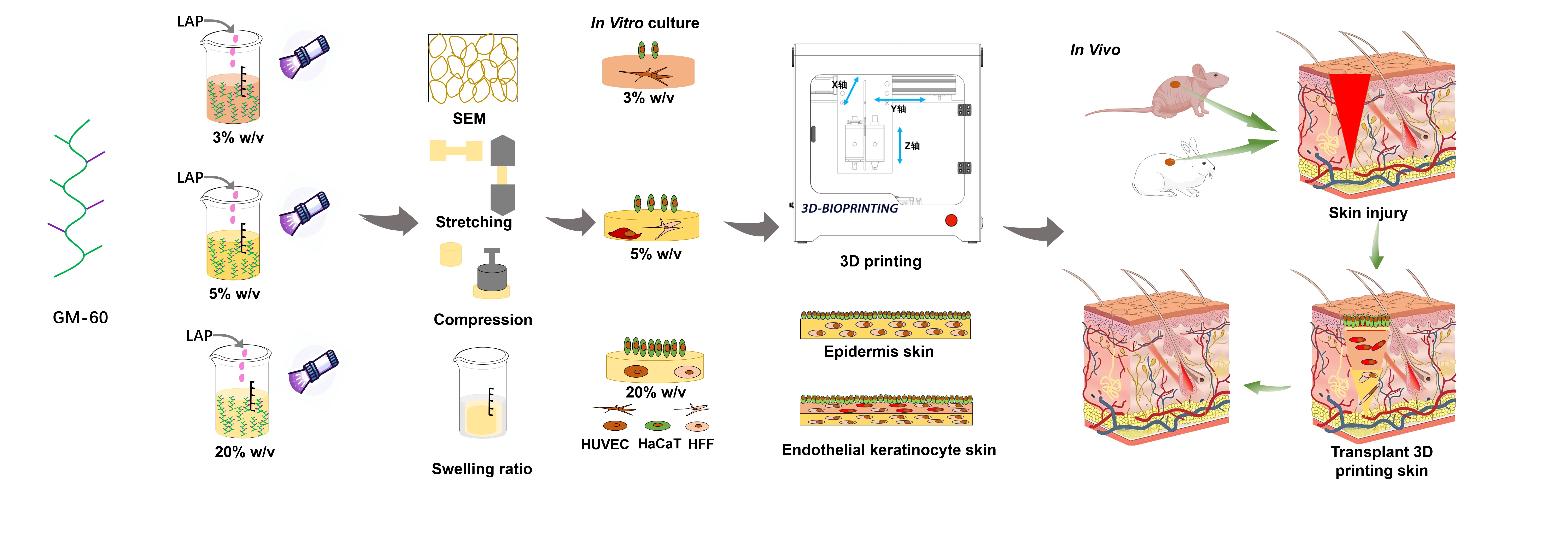
The skin is the largest organ of the human body and is the primary barrier against external stressors. However, in cases of severe skin damage or pathological conditions, the body’s natural physiological repair mechanisms are often insufficient to support effective skin tissue repair and regeneration. Bioprinting, a form of three-dimensional (3D) printing technology, utilizes various biomaterials and cells to construct complex 3D structures, offering the potential to overcome the limitations of conventional tissue-engineered skin and to develop functional skin substitutes. In this study, we developed a 3D bioprinter with excellent printing performance to fabricate vascularized skin substitutes. Through methacrylic anhydride-mediated modification of gelatin, we synthesized gelatin methacryloyl (GelMA) with varying degrees of substitution. The resulting GelMA hydrogel exhibited excellent mechanical properties, swelling ratio, porosity, and rheological properties. To create a hydrogel-multicellular composite bio-ink, we adjusted the concentration of the GelMA solution and co-cultured human immortalized epidermal cells, human foreskin fibroblasts, and human umbilical vein endothelial cells to optimize biological function. Importantly, by fine-tuning the printing parameters, the 3D extrusion-printed lines successfully fused into a continuous membrane, enhancing interlayer bonding and mechanical integrity. This process enabled the construction of a vascularized skin substitute with distinct reticular and papillary layers. In addition, the 3D-printed vascularized skin was implanted into skin defect models established in BALB/c nude mice and New Zealand rabbits to investigate its regenerative capabilities. These findings hold significant implications for the utilization of 3D-printed vascularized skin for improving skin injury repair, thereby advancing the field of skin tissue engineering.
- Lee SG, Lee S, Bae HK, et al. Evaluation of the therapeutic efficacy of human skin equivalents manufactured through droplet-based bioprinting/nebulization technology. Mol cell Toxicol. 2024;20(1):129-138. doi: 10.1007/s13273-023-00330-9
- Michael S, Sorg H, Peck CT, et al. Tissue engineered skin substitutes created by laser-assisted bioprinting form skin-like structures in the dorsal skin fold chamber in mice. PLoS ONE. 2013;8(3):e57741. doi: 10.1371/journal.pone.0057741
- Kafrashian Z, Brück S, Rogin P, et al. Segmented, side‐emitting hydrogel optical fibers for multimaterial extrusion printing. Adv Mater. 2025;37(4): e2309166. doi: 10.1002/adma.202309166
- Lin Z, Qiu X, Cai Z, et al. High internal phase emulsions gel ink for direct-ink-writing 3D printing of liquid metal. Nat Commun. 2024;15(1):4806. doi: 10.1038/s41467-024-48906-w
- Zhang C, Hua W, Mitchell K, Jin Y. Multiscale embedded printing of engineered human tissue and organ equivalents. Proc Natl Acad Sci USA. 2024;121(9): e2313464121. doi: 10.1073/pnas.2313464121
- Sobreiro‐Almeida R, Santos SC, Decarli MC, et al. Leveraging blood components for 3D printing applications through programmable ink engineering approaches. Adv Sci. 2024;11(47):2406569. doi: 10.1002/advs.202406569
- Dong T, Hu J, Dong Y, et al. Advanced biomedical and electronic dual-function skin patch created through microfluidic-regulated 3D bioprinting. Bioact Mater. 2024;40: 261-274. doi: 10.1016/j.bioactmat.2024.06.015
- Bebiano LB, Presa R, Vieira F, Lourenco BN, Pereira RF. Bioinspired and photo-clickable thiol-ene bioinks for the extrusion bioprinting of mechanically tunable 3D skin models. Biomimetics (Basel). 2024;9(4):228. doi: 10.3390/biomimetics9040228
- Gu Z, Fu J, Lin H, He Y. Development of 3D bioprinting: from printing methods to biomedical applications. Asian J Pharm Sci. 2020;15(5): 529-557. doi: 10.1016/j.ajps.2019.11.003
- Selvam SP, Ayyappan S, Jamir SI, Sellappan LK, Manoharan S. Recent advancements of hydroxyapatite and polyethylene glycol (PEG) composites for tissue engineering applications–a comprehensive review. Eur Polym J. 2024;215: 113226. doi: 10.1016/j.eurpolymj.2024.113226
- Taite LJ, Rowland ML, Ruffino KA, Smith BRE, Lawrence MB, West J L. Bioactive hydrogel substrates: probing leukocyte receptor–ligand interactions in parallel plate flow chamber studies. Ann Biomed Eng. 2006;34: 1705-1711. doi: 10.1007/s10439-006-9173-x
- Coudane J, Nottelet B, Mouton J, Garric X, Berghe HVD. Poly (ε-caprolactone)-based graft copolymers: synthesis methods and applications in the biomedical field: a review. Molecules. 2022;27(21):7339. doi: 10.3390/molecules27217339
- Milosevic M, Stojanovic DB, Simic V, et al. Preparation and modeling of three‐layered PCL/PLGA/PCL fibrous scaffolds for prolonged drug release. Sci Rep. 2020;10(1):11126. doi: 10.1038/s41598-020-68117-9
- Chen Y, Li J, Lu J, Ding M, Chen Y. Synthesis and properties of Poly (vinyl alcohol) hydrogels with high strength and toughness. Polym Test. 2022;108:107516. doi: 10.1016/j.polymertesting.2022.107516
- Zhao X, Lang Q, Yildirimer L, et al. Photocrosslinkable gelatin hydrogel for epidermal tissue engineering. Adv Health Mater. 2016;5(1):108-118. doi: 10.1002/adhm.201500005
- Xu W, Molino BZ, Cheng F, et al. On low-concentration inks formulated by nanocellulose assisted with gelatin methacrylate (GelMA) for 3D printing toward wound healing application. ACS Appl Mater Interfaces. 2019;11(9):8838-8848. doi: 10.1021/acsami.8b21268
- Mazio C, Casale C, Imparato G, et al. Pre-vascularized dermis model for fast and functional anastomosis with host vasculature. Biomaterials. 2019;192:159-170. doi: 10.1016/j.biomaterials.2018.11.018
- Xu H, Liu Z, Wei Y, et al. Complexation‐induced resolution enhancement pleiotropic small diameter vascular constructs with superior antibacterial and angiogenesis properties. Adv Healthc Mater. 2023;12(29):2301809. doi: 10.1002/adhm.202301809
- Wan H, Cao Y, Lo LW, Zhao J, Sepúlveda N, Wang C. Flexible carbon nanotube synaptic transistor for neurological electronic skin applications. ACS Nano. 2020;14(8):10402-10412. doi: 10.1021/acsnano.0c04259
- Vidal SEL, Tamamoto KA, Nguyen H, Abbott RD, Cairns DM, Kaplan DL. 3D biomaterial matrix to support long term, full thickness, immuno-competent human skin equivalents with nervous system components. Biomaterials. 2019;198:194-203. doi: 10.1016/j.biomaterials.2018.04.044
- Xiong M, Yang X, Shi Z, et al. Programmable artificial skins accomplish antiscar healing with multiple appendage regeneration. Adv Mater. 2024;36(50): 2407322. doi: 10.1002/adma.202407322
- Xia Y, Yan S, Wei H, et al. Multifunctional porous bilayer artificial skin for enhanced wound healing. ACS Appl Mater Interfaces. 2024;16(27):34578-34590. doi: 10.1021/acsami.4c05074
- Ma J, Qin C, Wu J, et al. 3D multicellular micropatterning biomaterials for hair regeneration and vascularization. Mater Horiz. 2023;10(9):3773-3784. doi: 10.1039/d3mh00528c
- Motter Catarino C, Cigaran Schuck D, Dechiario L, Karande P. Incorporation of hair follicles in 3D bioprinted models of human skin. Sci Adv. 2023;9(41):eadg0297. doi: 10.1126/sciadv.adg0297
- Chen H, Ma X, Gao T, et al. Robot-assisted in situ bioprinting of gelatin methacrylate hydrogels with stem cells induces hair follicle-inclusive skin regeneration. Biomed Pharmacother. 2023;158:114140. doi: 10.1016/j.biopha.2022.114140
- Zhao W, Chen H, Zhang Y, et al. Adaptive multi‐degree‐of‐freedom in situ bioprinting robot for hair‐follicle‐inclusive skin repair: a preliminary study conducted in mice. Bioeng Transl Med. 2022;7(3):e10303. doi: 10.1002/btm2.10303
- Dai LG, Dai NT, Chen TY, Kang LY, Hsu SH. A bioprinted vascularized skin substitute with fibroblasts, keratinocytes, and endothelial progenitor cells for skin wound healing. Bioprinting. 2022;28:e00237. doi: 10.1016/j.bprint.2022.e00237
- Ma J, Qin C, Wu J, et al. 3D printing of strontium silicate microcylinder‐containing multicellular biomaterial inks for vascularized skin regeneration. Adv Healthc Mater. 2021;10(16):2100523. doi: 10.1002/adhm.202100523
- Barros NR, Kim HJ, Gouidie MJ, et al. Biofabrication of endothelial cell, dermal fibroblast, and multilayered keratinocyte layers for skin tissue engineering. Biofabrication. 2021;13(3):035030. doi: 10.1088/1758-5090/aba503
- Xiang P, Yan L, Ge L, He X, Du N, Liu X. Development of a radial-flux machine with multi-shaped magnet rotor and non-ferromagnetic yoke for low torque ripple and rotor mass. IEEE Trans Ind Appl. 61;2025:2897-2910. doi: 10.1109/TIA.2025.3532558
- Xiang P, Yan L, Guo Y, He X, Gerada C, Chen IM. A concentrated-flux-type pm machine with irregular magnets and iron poles. IEEE/ASME Trans Mech. 2023;29(1): 691-702. doi: 10.1109/TMECH.2023.3293505
- Guo J, Gu H, Yin S, et al. Hepatocyte-derived Igκ promotes HCC progression by stabilizing electron transfer flavoprotein subunit α to facilitate fatty acid β-oxidation. J Exp Clin Cancer Res. 2024;43(1):280. doi: 10.1186/s13046-024-03203-8
- Liu C, Qin W, Wang Y, et al. 3D printed gelatin/sodium alginate hydrogel scaffolds doped with nano-attapulgite for bone tissue repair. Int J Nanomed. 2021;16:8417-8432. doi: 10.2147/IJN.S339500
- Pierce MC, Strasswimmer J, Hyle Park B, Cense B, De Boer JF. Birefringence measurements in human skin using polarization-sensitive optical coherence tomography. J Biomed Opt. 2004;9(2):287-291. doi: 10.1117/1.1645797
- Wang Y, Liu Y, Chen S, et al. Enhancing bone regeneration through 3D printed biphasic calcium phosphate scaffolds featuring graded pore sizes. Bioact Mater. 2024;46:21–36. doi: 10.1016/j.bioactmat.2024.11.024
- Kim BS, Yang WK, Jeong SK, et al. 3D cell printing of in vitro stabilized skin model and in vivo pre-vascularized skin patch using tissue-specific extracellular matrix bioink: a step towards advanced skin tissue engineering. Biomaterials. 2018;168:38-53. doi: 10.1016/j.biomaterials.2018.03.040
- Won-woo C, Minjun A, Byoung SK, Dong-Woo C. Blood‐lymphatic integrated system with heterogeneous melanoma spheroids via in‐bath three‐dimensional bioprinting for modelling of combinational targeted therapy. Adv Sci. 2022;9(29):2202093. doi: 10.1002/advs.202202093
- Song J, Liu T, Liao Z, et al. Digital light processing bioprinting neural systems with porous hydrogel in structure and function for disease models. Cell Rep Phys Sci. 2024;5(12):102311. doi: 10.1016/j.xcrp.2024.102311
- Zandi N, Daniele M, Brown A. Advances in fibrin-based bioprinting for skin tissue regeneration: exploring design, and innovative approaches. Biomed Mater Devices. 2025;3:330-348. doi: 10.1007/s44174-024-00198-w
- Fauzi MB, Rashidbenam Z, Bin Saim A, Binti Hj Idrus R. Preliminary study of in vitro three-dimensional skin model using an ovine collagen type i sponge seeded with co-culture skin cells: submerged versus air-liquid interface conditions. Polymers. 2020;12(12): 2784. doi: 10.3390/polym12122784
- Monsuur HN, Boink MA, Weijers EM, et al. Methods to study differences in cell mobility during skin wound healing in vitro. J Biomech. 2016;49(8):1381-1387. doi: 10.1016/j.jbiomech.2016.01.040
- Petry L, Kippenberger S, Meissner M, et al. Directing adipose‐derived stem cells into keratinocyte‐like cells: impact of medium composition and culture condition. J Eur Acad Dermatol Venereol. 2018;32(11):2010-2019. doi: 10.1111/jdv.15010
- Colin E, Plyer A, Golzio M, Meyer N, Faver G, Orlik X. Imaging of the skin microvascularization using spatially depolarized dynamic speckle. J Biomed Opt. 2022;27(4):046003. doi: 10.1117/1.Jbo.27.4.046003
- Hu X, Wang L, Deng J, et al. Dietary nitrate accelerates the healing of infected skin wounds in mice by increasing microvascular density. Biochem Biophys Res Commun. 2023;686:149176. doi: 10.1016/j.bbrc.2023.149176
- Fu T, Sullivan DP, Gonzalez AM, et al. Mechanotransduction via endothelial adhesion molecule CD31 initiates transmigration and reveals a role for VEGFR2 in diapedesis. Immunity. 2023;56(10):2311-2324.e6. doi: 10.1016/j.immuni.2023.08.001
- Park H, Collignon AM, Lepry WC, et al. Acellular dense collagen-S53P4 bioactive glass hybrid gel scaffolds form more bone than stem cell delivered constructs. Mater Sci Eng C Mater Biol Appl. 2021;120:111743. doi: 10.1016/j.msec.2020.111743
- Trappmann B, Gautrot JE, Connelly JT, et al. Extracellular-matrix tethering regulates stem-cell fate. Nat Mater. 2012;11(7): 642-649. doi: 10.1038/nmat3339
- Singh A, Dalal N, Tayalia P. An interplay of matrix stiffness, dimensionality and adhesivity on cellular behavior. Biomed Mater. 2023;18(2): 025010. doi: 10.1088/1748-605X/acb7c0
- Trujillo S, Gonzalez-Garcia C, Rico P, et al. Engineered 3D hydrogels with full-length fibronectin that sequester and present growth factors. Biomaterials. 2020; 252:120104. doi: 10.1016/j.biomaterials.2020.120104
- Ito M, Hiramatsu H, Kobayashi K, et al. NOD/SCID/γ c null mouse: an excellent recipient mouse model for engraftment of human cells. Blood. 2002;100(9):3175-3182. doi: 10.1182/blood-2001-12-0207
- Albanna M, Binder KW, Murphy SV, et al. In situ bioprinting of autologous skin cells accelerates wound healing of extensive excisional full-thickness wounds. Sci Rep. 2019;9(1):1856. doi: 10.1038/s41598-018-38366-w
- Wei Q, Su J, Meng S, et al. MiR‐17‐5p‐engineered sEVs encapsulated in GelMA hydrogel facilitated diabetic wound healing by targeting PTEN and p21. Adv Sci (Weinh). 2024;11(13):2307761. doi: 10.1002/advs.202307761
- Zhang G, Zhang Z, Cao G, et al. Engineered dermis loaded with confining forces promotes full-thickness wound healing by enhancing vascularisation and epithelialisation. Acta Biomater. 2023; 170: 464-478. doi: 10.1016/j.actbio.2023.08.049
- Jin T, Fu Z, Zhou L, et al. GelMA loaded with platelet lysate promotes skin regeneration and angiogenesis in pressure ulcers by activating STAT3. Sci Rep. 2024; 14(1): 18345. doi: 10.1038/s41598-024-67304-2
- Chen L, Ye JL, Gao C, Deng F, Liu W, Zhang Q. Design and fabrication of gelatin-based hydrogel loaded with modified amniotic extracellular matrix for enhanced wound healing. Heliyon. 2023;9(10):e20521. doi: 10.1016/j.heliyon.2023.e20521
- Hao X, Luo J, Huang Y, et al. Injectable antiswelling and high-strength bioactive hydrogels with a wet adhesion and rapid gelling process to promote sutureless wound closure and scar-free repair of infectious wounds. ACS Nano. 2023;17(21): 22015-22034. doi: 10.1021/acsnano.3c08625