

3D-printed GelMA/SA/CMCS hydrogel scaffolds containing Cynomorium songaricum polysaccharide for critical bone defect repair

Critical bone defect repair remains a major challenge in orthopedics. Cynomorium songaricum polysaccharide (CSP), derived from the traditional medicinal plant Cynomorium songaricum Rupr. in China, demonstrates excellent anti-inflammatory and osteogenic properties. Given these promising biological activities, we developed a novel therapeutic approach using a hydrogel composite scaffold incorporating CSP (GAC-C) for treating critical-sized bone defects. The composite scaffold was fabricated by embedding CSP into a methacrylated gelatin (GelMA)/sodium alginate (SA)/carboxymethyl chitosan (CMCS) blend via three-dimensional (3D) printing technology. The structural, mechanical, and biological properties of GAC-C were characterized, and osteogenic performance was evaluated both in vitro with rat bone marrow stromal cells (rBMSCs) and in vivo using a critical-sized bone defect model. Results indicated that the GAC-C scaffold demonstrated excellent biocompatibility, promoted osteogenic differentiation of rBMSCs, and enhanced bone integration and repair. Thus, the GAC-C scaffold has the potential for effectively repairing criticalsized bone defects.
1. Introduction
Critical bone defects remain a major challenge in orthopedics, as they exceed the natural bone healing capacity of our body. This often necessitates the use of bone repair materials to fill the defects and promote bone regeneration.1,2 Despite being considered the “gold standard” for treating bone defects, autologous bone grafting has several drawbacks, including limited donor availability and risks of secondary injury and infection.3 Given these limitations, bone tissue engineering scaffolds emerge as a promising alternative to address the challenges posed by bone defects.4–6 These scaffolds replicate the natural bone architecture, providing mechanical support to the defect site, serving as a foundation for new bone growth, and fostering a good environment for cells and growth factors to function.7 In recent years, hydrogel scaffolds, known for their excellent biological properties, have found wide applications in the biomedical field. In our study, a blend of gelatin methacryloyl (GelMA), sodium alginate (SA), and carboxymethyl chitosan (CMCS) was used as the three-dimensional (3D) printing ink. GelMA, a widely used hydrogel in biomedicine, is a double-bond modified gelatin that crosslinks and solidifies under ultraviolet (UV) light with a photoinitiator. The resulting scaffold combines the characteristics of both natural and synthetic biomaterials.8,9 GelMA hydrogels, due to their relatively low mechanical strength, are limited in their application for bone regeneration and tissue engineering, particularly for loadbearing applications. To address this limitation, researchers have explored various functionalization strategies to enhance both the mechanical and biological properties of GelMA hydrogels. For instance, incorporating materials like reduced graphene oxide (rGO) into GelMA hydrogels can improve their compressive strength and rheological properties, thereby promoting the adhesion and osteogenic differentiation of bone marrow stromal cells (BMSCs).10
In the field of bone tissue engineering, traditional growth factors such as bone morphogenetic protein-2 (BMP-2) have been widely used to promote bone regeneration. However, their high cost and dose-dependent side effects have restricted their broad clinical application. The high dose requirement of BMP-2 not only increases treatment costs but also increases the risk of severe side effects, such as ectopic ossification and inflammatory responses.11
Sodium alginate (SA), derived from brown algae, provides good biocompatibility and biodegradability. In aqueous environments, it spontaneously forms a 3D hydrogel network via hydrogen bonds.12,13 Physical blending of SA with other materials enhances cell adhesion and improves the mechanical properties of hydrogel scaffolds without chemical modification.14
Carboxymethyl chitosan (CMCS), a water-soluble derivative of chitosan, promotes the adhesion and proliferation of rat BMSCs (rBMSCs).15 The amino and carboxyl groups in CMCS enable gel formation with SA, enhancing the mechanical strength and mineralization of the composite hydrogel.16
Research has demonstrated that a developed UV-crosslinking system utilizing GelMA enables precise modulation of hydrogel stiffness across a broad elastic modulus range (0.5–30 kPa) by modulating UV intensity and exposure duration. This system demonstrated enhanced osteogenic differentiation of human mesenchymal stem cells (hMSCs) when cultured on high-stiffness hydrogels.17 Complementarily, SA/CMCS composite hydrogel microspheres fabricated through Ca²+-crosslinking exhibit good swelling capacity and biocompatibility. Incorporation of CMCS substantially increases microsphere diameter, internal porosity, and mechanical integrity. Notably, CMCS modification enhances BMP-2 loading/release efficiency while demonstrating superior cytocompatibility in vitro, effectively promoting MC3T3-E1 cell biomineralization and alkaline phosphatase (ALP) activity.18 The combination of these materials leverages their individual advantages and synergistic effects to enhance the biocompatibility and mechanical properties of the hydrogel scaffold, improving its performance in bone repair. The use of hydrogel materials for critical bone defect repair has gained significant attention due to their biocompatibility, tunable mechanical properties, and ability to mimic the extracellular matrix (ECM).19,20 However, several challenges persist in this field. Many types of hydrogels often exhibit insufficient mechanical strength for load-bearing applications, limited osteogenic induction capabilities, and difficulties in achieving controlled degradation rates that match bone regeneration timelines.9,21,22 The compressive modulus of traditional hydrogels is generally low, making it difficult for them to maintain structural integrity in dynamic stress environments, which increases the risk of early scaffold collapse. By introducing reinforcement mechanisms, the mechanical properties of hydrogels can be improved. For instance, Mpuhwe et al.23 introduced CeO2 nanoparticles into GelMA/PEGDA hydrogels, resulting in relatively enhanced mechanical properties of the scaffolds. Additionally, dual-crosslinked hydrogels are also a strategy to enhance the mechanical properties of hydrogels. For instance, in one study, a dual-crosslinked hydrogel was designed using photo-crosslinked GelMA and thiolated chitosan; the results indicated that this hydrogel had good mechanical properties and moisture retention ability and demonstrated a promoting effect on wound healing in vivo.24 However, the study focused on skin wound repair rather than bone repair. Therefore, if the hydrogels are to be used for bone defects, the problem of poor mechanical properties needs to be addressed. These limitations highlight the need for innovative composite hydrogels, like the GelMA/SA/CMCS (GAC) system, which combines the benefits of synthetic and natural polymers to effectively enhance mechanical strength and cell adhesion and can be tailored to match the bone regeneration cycle.
However, the osteogenic potential of these hydrogels is limited. Therefore, incorporating osteogenic active drugs is essential to enhance the osteogenic performance of the composite materials. Cynomorium songaricum (CS), a parasitic herb, is mainly found in the deserts and saline-alkali regions of northwest China.25 The stem of CS is a traditional Chinese medicinal material, rich in components like polysaccharides, flavonoids, and triterpenoids. It is commonly used to tonify the kidneys, replenish essence, enrich the blood, and promote defecation. CS polysaccharide (CSP), a water-soluble polysaccharide extracted from CS, mainly consists of arabinose (Ara), glucose (Glu), and galactose (Gal).26 CSP exhibits antioxidant, anti-inflammatory, anti-tumor, and anti-osteoporosis properties. Firstly, CSP enhances osteoblast differentiation and proliferation via activation of the PI3K/AKT signaling pathway, thereby augmenting bone tissue regenerative potential. This mechanism parallels the osteogenic effects of curculigoside on adipose-derived stem cells (ADSCs), where curculigoside promotes osteogenic marker expression through PI3K/ AKT signaling and demonstrates bone-loss protection in vivo.27 Furthermore, CSP exerts potent anti-inflammatory activity critical for bone repair processes. By regulating inflammation-associated signaling cascades, CSP effectively suppresses pro-inflammatory cytokine release, optimizing the regenerative microenvironment. This anti-inflammatory action aligns with the therapeutic mechanism of berberine-loaded thermosensitive hydrogels in periodontitis treatment. Berberine modulates PI3K/ AKT signaling to reduce inflammatory factor levels while upregulating osteogenesis-related markers.28 Notably, anti-osteoclastogenic properties of CSP share conceptual similarities with catalpol, which inhibits NF-κB and AKT pathways through PTEN activation, thereby suppressing osteoclastogenesis and preserving bone integrity.29 Additionally, CSP increases the OPG/RANKL expression ratio, inhibits osteoclast activity, regulates osteocalcin levels to reduce bone turnover, and restores the balance between bone formation and resorption.30 Water-soluble drugs pose unique challenges in drug delivery due to their rapid diffusion and potential for burst release, requiring the use of a carrier to manage these issues.31 In summary, CSP inclusion may provide other critical biological benefits, such as promoting cell adhesion, proliferation, and osteogenic differentiation.
In this study, we fabricated a bioactive hydrogel scaffold using 3D printing technology, aiming to promote the repair of critical bone defects (Scheme 1). This scaffold provides mechanical support at the defect site and stimulates the formation of new bone, offering a potential solution for repairing critical bone defects.

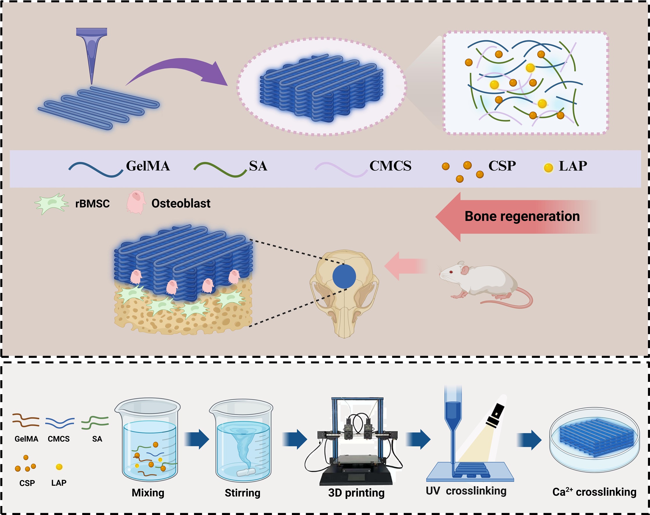
Scheme 1. Schematic diagram of the construction of GelMA/SA/CMCS composite hydrogel scaffolds and osteogenesis induction. Abbreviations: CMCS, carboxymethyl chitosan; CSP, cynomorium songaricum polysaccharide; GelMA, gelatin methacryloyl; LAP, lithium phenyl (2,4,6-trimethylbenzoyl) phosphinate; SA, sodium alginate; UV, ultraviolet.
2. Materials and methods
2.1. Materials
The chemicals used in this study were: GelMA (Engineering For Life, China), lithium phenyl (2,4,6-trimethylbenzoyl) phosphinate (LAP; Engineering For Life, China), SA (Macklin Inc., China), CMCS (Aladdin Chemical Inc., China), Cell Cytotoxicity Kit-8 (CCK-8) reagent (Beyotime, China), ALP color development kit (Beyotime, China), live/dead staining kit (Beijing Sora Biotechnology Co., Ltd., China), Alizarin Red S staining solution (Beijing Sora Biotechnology Co., Ltd., China), calcium chloride (Beijing Sora Biotechnology Co., Ltd., China), Hiscript II Q RT SuperMix Kit (Vazyme, China), BeyoFastTM SYBR Green qPCR Mix (Beyotime, China), CSP (Chengdu Pusi Biotechnology Co., Ltd., China), minimum essential medium eagle with alpha modification (α-MEM; Gibco, China), fetal bovine serum (FBS; Bio Channel, China), and penicillin-streptomycin (PS; Gibco, China).
2.2. Preparation of composite scaffolds
The GelMA/SA/CMCS composite was prepared using GelMA (4% w/v), SA (5% w/v), and CMCS (4% w/v). The GelMA/SA/CMCS-CSP composite was prepared by adding CSP (100 μg/mL) to the same components. GelMA was dissolved in phosphate-buffered saline (PBS; pH 7.4) at 70°С; LAP (0.25% w/v), SA, and CMCS were then added and stirred to form the printing ink.
GelMA/SA/CMCS and GelMA/SA/CMCS-CSP scaffolds were fabricated using a 3D printer (TL-H2 Pro, Changchun Qingken Trading Co., Ltd, China). The printing conditions were as follows: printing was conducted at room temperature, the inner diameter of the printing nozzle was 0.75 mm, the extrusion speed was 0.2 mL/min, and the nozzle movement speed was 50%. After the scaffolds were printed, they were exposed to ultraviolet light for 10 s and then placed in a CaCl2 (2% w/v) solution for 10 min before being set aside for later use. The two types of scaffolds were named GAC and GAC-C, respectively, and the control group was named CON for ease of description in subsequent experiments. Sample scaffolds with a length × width × height of 12 × 12 × 5 mm3 were prepared for in vitro characterization, and sample scaffolds with a diameter × height of 10 × 2 mm2 were prepared for in vitro osteogenesis experiments. Sample scaffolds with a diameter × height of 6 × 2 mm2 were prepared for animal experiments.
2.3. Characterization of the composite scaffolds
2.3.1. Morphological characteristics of the composite scaffolds
The composite scaffold was freeze-dried in a Scientz-12N freeze-dryer (SCIENTZ; China) for 24 h, and its appearance was captured using a digital camera. The surface and cross-sectional morphology of GAC and GAC-C scaffolds were observed using a field emission scanning electron microscope (FESEM; SU-8010; Hitachi, Japan) (n = 3 per group).
2.3.2. Mechanical properties of the composite scaffolds
The scaffolds were constructed based on a previously published technique.32 The compressive and tensile moduli (MPa) of GAC and GAC-C scaffolds were determined using a universal material testing machine (Zwick/Roell Z020; Germany) in accordance with ISO-37 guidelines, with subsequent quantitative evaluation (n = 3 per group).
2.3.3. Porosity of the composite scaffolds
In accordance with previous studies, the porosity of the scaffolds was measured by ethanol immersion in a gravimetric bottle.33 The porosity of the GAC and GAC-C scaffolds was tested separately (n = 3 per group).
2.3.4. Swelling performance of the composite scaffolds
The swelling behavior of the GAC and GAC-C scaffolds was measured using the weight-based method.35 Hydrogel samples were immersed in 5 mL PBS at 37°C. At preset time points, samples were weighed to determine the swelling weight (Ws) and final dry weight (Wf). The swelling rate (SR; n = 3 per group) was then calculated as:
2.3.5. Degradation of composite scaffolds in vitro
The degradation of GAC and GAC-C scaffolds was investigated in PBS (pH 7.4). The hydrogel scaffolds were submerged in 5 mL PBS and agitated continuously at 37°C. Samples of the scaffolds were retrieved at preset time points and then subjected to freeze-drying and weighing. The dry mass of the hydrogel recorded on Day 0 served as the initial weight (Wd0), while the weight of the sample at each subsequent time point is denoted as Wdn. The degradation rate (DR; n = 3 per group) was calculated using the following formula:
2.4. Biocompatibility of composite scaffolds in vitro
2.4.1. Cell culture
In accordance with previously reported methods,34 rBMSCs extracted from the bone marrow of Sprague-Dawley rats (male, 3-week-old) were incubated in α-MEM, supplemented with 10% FBS and 1% PS at 37°C and 5% CO2.
2.4.2. CCK-8 cytotoxicity assay
To determine the optimum CSP concentration, we conducted CCK-8 cytotoxicity assays. The CSP powder was dissolved in PBS and diluted by α-MEM containing 10% FBS and 1% PS into concentration gradients of 0, 5, 10, 20, 50, 100, 200, 500, and 1000 μg/mL. rBMSC cells (5 × 10³, 100 µL) were inoculated into 96-well plates and cultured overnight in an incubator (37°С, 5% CO2). The media was then replaced with a cell culture medium containing CSP. On Days 1, 2, and 3 of culture, CCK-8 solution (10 µL) was introduced into each well and allowed to incubate for 2 h. Absorbance (optical density [OD] 450 nm) was measured using iMark (BioRad, United States of America [USA]).
Subsequently, CCK-8 assays were performed to evaluate the effects of the hydrogel composite scaffold on rBMSCs proliferation. The experiment was divided into three groups: CON, GAC, and GAC-C. First, the scaffold soaking solution was prepared, and each group of scaffolds was immersed in α-MEM medium for 24 h at 0.1 g/mL according to the national standard (GB/T 16898.6-2017). The soaking solution was collected and filtered through a 0.22 μm filtration membrane to remove bacteria for reserve use. Third-passage rBMSCs were then inoculated into 96-well plates at a cell density of 5 × 10³ cells/well and incubated overnight in cell incubators. On the next day, the medium of each group was replaced with extracts for cell culture. The OD values for each group (n = 4 per group at each time point) were read using an enzyme-labeled meter at a wavelength of 450 nm on Days 1, 3, and 5.
2.4.3. Live/dead staining experiments
The effects of scaffolds on cell viability were determined using live/dead tests. The scaffolds of each group were placed on 24-well plates, and the third-passage rBMSCs were inoculated on the surface of the scaffolds at a cell density of 5 × 10⁴ cells/mL. The cell suspension (400 μL) was added to each group of 24-well plates. At specific time points (Days 1, 3, and 5), the original medium was removed, and the scaffold was washed three times with PBS. A buffer containing 4 μM calcein AM and 4.5 μM propyl iodide was added to each well and incubated for 20 min. Full-spectrum laser confocal microscopy (Leica STELLARIS 8; Germany) was used to observe the cells.
2.4.4. Cytoskeleton actin filament staining
After the scaffold was co-cultured with cells for 24 h, the medium was removed, and the scaffold was washed with PBS. The surface cells on the scaffold were fixed with 4% paraformaldehyde. The membrane was digested with 0.1% Triton X-100 at room temperature for 10 min, and the scaffold was washed three times with PBS. Each well was added with 300 μl FITC-phalloidin (Beijing Sora Biotechnology Co., Ltd, China) and cultured at room temperature for 1 h in the dark. After incubation, the cells were washed three times with PBS for 5 min each time, and the nuclei were stained with DAPI for 10 min. After staining, the residual dyes were washed with PBS. Cell morphology was observed and photographed using fullspectrum laser confocal microscopy at 488 and 545 nm.
2.5. In vitro osteogenic differentiation
2.5.1. Alkaline phosphatase activity
The GAC and GAC-C scaffolds were placed in 24-well plates, and rBMSCs were inoculated on the scaffolds at a density of 2 × 10⁴ cells/well, which reached 80% confluence when added to α-MEM medium. After 7 days of incubation with osteogenic induction medium (complete medium containing 0.25 mM ascorbic acid, 10 mM β-glycerophosphate, and 10 nM dexamethasone), the osteogenic induction medium was removed; the cell scaffold was fixed with 4% paraformaldehyde for 15 min and then washed with sterile water. The ALP colordeveloping working solution was added and incubated for 30 min. The cells were rinsed repeatedly with sterile water to eliminate the dye solution. The cells were observed with a microscope and photographed (n = 3 per group).
2.5.2. Alizarin Red S staining
The GAC and GAC-C scaffolds were placed in 24-well plates, and rBMSCs were inoculated on the scaffolds at a density of 2 × 10⁴ cells/well, which reached 80% confluence when added to α-MEM medium. After 14 days of incubation with an osteogenic induction medium (complete medium containing 0.25 mM ascorbic acid, 10 mM beta-glycerophosphate, and 10 nM dexamethasone), the osteogenic differentiation medium was removed, and the scaffold was fixed with 4% paraformaldehyde for 15 min, then washed with sterile water. The fixed cells were added with 1% Alizarin Red S staining solution and incubated for 5 min. The cells were then rinsed repeatedly with sterile water to remove the stain. After being observed under a microscope and photographed, calcium precipitation was dissolved with 10% cetylpyridine chloride monohydrate (Aladdin Chemical Inc., China) at room temperature for 30 min. Subsequently, the OD of the dissolved solution was measured at a wavelength of 562 nm (n = 3 per group).
2.5.3. Quantitative real-time polymerase chain reaction
Rat BMSCs (rBMSCs) were inoculated on scaffolds of each group and cultured in an osteogenic induction medium for 7 days (n = 3 per group). Total RNA was isolated from cells using FastPure Cell/Tissue Total RNA Isolation Kit V2 (Vazyme, China). Following the adjustment of RNA concentration with nuclease-free water, cDNA synthesis was carried out in accordance with the reverse transcription kit’s guidelines. For polymerase chain reaction (PCR), BeyoFastTM SYBR Green qPCR Mix and 96-well PCR plate (Nest Biotechnology, China) were used. The expression level of the target gene was standardized relative to the housekeeping gene glyceraldehyde 3-phosphate dehydrogenase (GAPDH). The primers of the osteogenic-related genes are listed in Table S1, Supporting Information.
2.6. In vivo osteogenesis evaluation of composite scaffolds
2.6.1. Establishment of skull defect model and scaffold implantation
A total of 18 12-week-old female Sprague-Dawley rats were provided by The First Affiliated Hospital, Zhejiang University School of Medicine (China), and raised in public facilities at the Laboratory Animal Medical Center. The animal experiment program was approved by the Ethics Committee of the First Affiliated Hospital of the Zhejiang University School of Medicine.
In accordance with established protocols, a cranial bone defect was created in the rats to form the critical bone defect.35 As previously detailed, the rats were anesthetized and laid in a prone position for depilation and ensuing steps. A longitudinal incision was created on the surface of the skull, followed by the sequential incision of the skin, subcutaneous tissue, and the periosteum. A hollow drill was used to create a full-layer bone defect with a diameter of 6 mm in the middle of the skull. The defects were randomly allocated into the following three groups: (i) a vacancy loss group serving as the negative control (n = 3); (ii) a group with GAC stent implantation acting as the positive control (n = 3); and (iii) a group with GAC-C stent implantation as the experimental group (n = 3). The stents were subsequently implanted into the rats, and the incisions were closed using sterile silk sutures.
2.6.2. Micro-computed tomography analysis
The rats were killed after 4 and 8 weeks of feeding, and skull specimens were collected. The sample was subjected to micro-computed tomography (micro-CT) analysis (Skyscan1276; Bruker, Belgium). The image data were analyzed using evaluation software, and 3D imaging was performed after the stent was removed. Following the reconstruction process, a fixed volume of interest (VOI) was centered on the region of damage, and a structural analysis was conducted. In addition, bone volume/total volume (BV/TV), trabecular thickness (TH.Tb), and trabecular separation (TH.Sp) were quantified.
2.6.3. Histological and immunofluorometric analysis
The rats were killed at 4 and 8 weeks after implantation; skull specimens were collected and fixed in 4% paraformaldehyde for 72 h. The sample was then placed in a 10% EDTA (Servicebio, China) solution for decalcification. After decalcification, the skull specimens were embedded in paraffin wax, and the middle area was cut into continuous sections of 5 μm thickness. The sections were stained with hematoxylin-eosin (H&E) and Masson staining solution. In addition, immunofluorescence staining was performed with 1/100 diluted Runx 2 and Col 1 antibodies (Affinity Biosciences, China) to evaluate the expression of Runx 2 and Col 1 proteins under fluorescence microscopy. Image J was used for quantitative fluorescence analysis.
2.7. Statistical analysis
Data were collated and statistically analyzed using GraphPad Prism 8 software (GraphPad Software Inc, America). The data are expressed as mean ± standard deviation, with *p < 0.05, **p < 0.01, ***p < 0.001, and ****p < 0.0001 indicating statistically significant differences. A two-tailed t-test was used for pair-to-pair comparison, and univariate analysis of variance or multivariate analysis of variance was used for data comparison between three groups or more. All data were obtained from at least three measurements.
3. Results and discussion
3.1. Characterization of composite hydrogel scaffolds
The EDS (energy-dispersive spectroscopy) analysis reveals prominent peaks corresponding to carbon (C) and oxygen (O), indicating the formation of a carbon-oxygen backbone and cross-linked structures within the polymer chains during the synthesis process (Figure S1, Supporting Information). The GAC and GAC-C scaffolds had milky white surfaces with a uniform porous network and rough surfaces conducive to cell adhesion. The addition of CSP did not significantly alter the scaffold diameter or pore size. The porous design encourages the development of connective tissue, including blood vessels and new bone.36 The mechanical properties of the composite scaffold are illustrated in Figure 1F and G. The Young’s modulus and compressive modulus of the GAC scaffold were 0.41 ± 0.077 and 0.24 ± 0.072 MPa, respectively, while the GAC-C scaffold exhibited values of 0.39 ± 0.059 and 0.25 ± 0.065 MPa, respectively. No significant differences were observed between the two groups. These results indicate that CSP incorporation does not reduce scaffold mechanical properties. Moreover, Mao et al.37 reported that Young’s modulus and the compressive modulus of GelMA hydrogel are approximately 0.04 MPa. Additionally, Qiao et al.38 synthesized a photo-crosslinked osteogenic growth peptide (OGP) and generated GelMA-c-OGP through cocrosslinking with GelMA, with the mechanical compressive strength of GelMA-c-OGP reaching 0.09 MPa. GAC-C exhibited superior mechanical properties compared to these hydrogels. The porosity of GAC scaffolds was 64.94 ± 0.23%, while that of GAC-C scaffolds was 66.56 ± 0.79%. Both scaffolds exhibited high porosity with no significant differences, indicating that CSP did not affect the pore structure (Figure 1H). Jin et al.39 reported that porosity of approximately 70% effectively promotes osteogenesis. The swelling rate of the GAC stent was 48.65 ± 1.16%, while that of the GAC-C stent was 54.07 ± 3.45%. Although no significant difference was observed, CSP, a hydrophilic substance, may enhance the swelling performance of the scaffold (Figure 1I). The GAC-C scaffold displayed a slightly higher degradation rate than the GAC scaffold at Week 1, though the difference was minimal. After 4 weeks, the GAC-C scaffold exhibited a slower degradation rate than the GAC group. After 7 weeks, both scaffold groups reached similar degradation rates (Figure 1J). The photocrosslinked network of GelMA provides mechanical stability, while the Ca²+ ion-crosslinking of SA enhances injectability. The amino-carboxyl electrostatic interaction of CMCS imparts pH responsiveness. The synergy of these three components achieves a balance between insitu shaping and long-term stability for the scaffold. The regenerated bone gradually matured into bone tissue from 4 to 8 weeks post-surgery, sufficient for healing in our study.40 The pace of scaffold degradation is important if it matches the pace of new bone formation.41 In comparison to single crosslinked hydrogels (e.g., pure GelMA or SA scaffolds), our composite hydrogel scaffold exhibited a more suitable degradation rate that better aligns with the needs of bone regeneration.37,42

Figure 1. Morphology and physicochemical properties of composite hydrogel scaffolds (GAC and GAC-C). Top view (A); side view (B); freeze-dried state (C); GAC (D); GAC-C (E). (A–C) Hybrid scaffold macrostructure of the as-prepared, sectioned, and dried GAC scaffolds; Top view (A); side view (B); freeze-dried state (C). (D and E) SEM images of the scaffold surface appearance and cross-section; GAC scaffold(D); GAC-C scaffold(E). (F) Young’s modulus of the hydrogels. (G) Compressive modulus of the hydrogels. (H) Porosity ratio of the hydrogels. (I and J) Swelling (I) and degradation ratio (J) of the 3D-printed composite scaffolds. Scale bars: 1 cm (A–C); 1 mm (D1 and E1); 50 μm (D2 and E2); 25 μm (D3 and E3). All statistical data are represented as mean ± standard deviation (n = 3). Abbreviations: ns, non-significant; SEM, scanning electron microscope.
3.2. Biocompatibility evaluation of scaffolds in vitro
First, we assessed the appropriate concentration of CSP based on the CCK-8 cytotoxicity assay (Figure S2, Supporting Information). On Day 1, no significant difference was observed between the control and CSP groups. On Day 2, cell proliferation was significantly inhibited in the 1000 μg/mL group. On Day 3, cell proliferation was significantly inhibited in the 200–1000 μg/mL concentration group. Based on these results, the concentration of 100 μg/mL was selected for subsequent experiments.
In addition, we evaluated the in vitro biocompatibility of the composite scaffold. The CCK-8 proliferation assay displayed no significant difference in cell proliferation among groups after 3 days of rBMSC culture with the composite scaffold extract (Figure 3C). The OD value in the GAC-C group significantly increased by Day 5, indicating that hydrogel scaffolds and CSP synergistically promoted rBMSC cell proliferation. Live/dead staining of rBMSC cells cultured on the scaffold surface displayed normal growth and proliferation (Figure 2A–C). Over time, cell density on the scaffold surface continuously increased. These results demonstrate that all scaffolds exhibit good biocompatibility. FITC-phalloidin staining illustrated the morphological features of rBMSCs on different scaffolds over 24 h (Figure 2D). Cells in all groups displayed multidirectional morphology, indicating good adhesion and healthy growth on the scaffold surface. Good biocompatibility is essential for biomaterials to transition from laboratory research to patient care.19 Previous research has primarily focused on the loading of traditional growth factors (such as BMP-2 and VEGF) or antibiotics.43,44 Plant and animal-based natural extracts are known for their high efficiency and low toxicity.44,45 Moreover, CSP, a natural plant polysaccharide, has not been fully explored for its osteogenic and anti-inflammatory properties in bone repair. Further research on CSP could contribute to its clinical translation.

Figure 2. Biocompatibility of composite hydrogel scaffolds. (A–C) Live/dead staining images of rBMSCs cultured on scaffolds for 1 (A), 3 (B), and 5 days (C). (D) Phalloidin staining images of rBMSCs cultured on scaffolds for 24 h. Scale bars: 500 μm (n = 3). Abbreviations: DAPI, 4’,6-diamidino-2-phenylindole; h, hours; PI, propidium iodide; rBMSCs, rat bone marrow stromal cells.

Figure 3. Composite hydrogel scaffolds with CSP promoted osteogenic differentiation in vitro. (A and B) ALP (7 days; A) and Alizarin Red staining (14 days; B) of rBMSCs cultured on scaffolds of different groups. (C) CCK-8 analysis of different groups on Days 1, 3, and 5. (D) Quantification of Alizarin Red staining. (E–H) Osteogenesis-related gene expression on Day 7. Scale bars: 500 μm (general view; A and B); 100 μm (microscopic view; A and B). All statistical data are represented as mean ± standard deviation; *p < 0.05, **p < 0.01, ***p < 0.001, ****p < 0.0001; n = 3. Abbreviations: CCK-8, cell counting kit-8; CSP, cynomorium songaricum polysaccharide; OD, optical density; rBMSCs, rat bone marrow stromal cells.
3.3. In vitro osteogenic evaluation of composite scaffolds
Alkaline phosphatase (ALP) staining results indicated that after 7 days of co-culture with rBMSCs, the scaffold group exhibited higher calcium deposition than the control group, with the GAC-C group outperforming the GAC group (Figure 3A). On Day 14, Alizarin Red S staining revealed dense orange-red deposits in both groups compared to the control group, with the GAC-C group demonstrating more pronounced deposits, indicating significant osteogenic potential (Figure 3B). Quantitative analysis of the Alizarin Red S stains further confirmed these results (Figure 3D). ALP staining was employed to evaluate early osteogenic effects, while Alizarin Red S staining was used to evaluate later effects.46 To further assess the effect of CSP-containing scaffolds on osteogenic differentiation, we evaluated osteogenic markers (ALP, Runx 2, Col 1, and OCN) using RT-qPCR on Day 7. ALP is an early indicator of bone formation, OCN marks later stages of osteogenesis, and calcified nodules indicate the completion of the osteogenic process.47 The results revealed that the expression levels of Alp, Runx 2, Col 1, and OCN were significantly upregulated in the GAC-C group (Figure 3E–H). Runx 2 is a key transcription factor for osteogenic differentiation48; Col 1 also indicates later stages of osteogenic differentiation.49 The late osteogenic markers, OCN and Col 1, exhibited a significant increase in mRNA expression levels compared to the controls. Furthermore, the late-stage osteogenesis effects were significantly improved, likely due to prolonged drug release.
Studies have demonstrated that CS administration significantly reduced the expression of RANK and RANKL while increasing osteoprotegerin (OPG) expression.50 RANKL, by binding to RANK on osteoprogenitor cells, regulates the formation of osteoclasts and bone resorption.51 OPG negatively regulates the RANK/ RANKL pathway by binding to RANKL and preventing its interaction with RANK.52
3.4. In vivo bone regeneration evaluation of composite hydrogel scaffolds
To evaluate the bone regeneration capabilities of 3D-printed scaffolds in vivo, each scaffold group was implanted into a 6 mm diameter skull defect in the rats (Figure 4A). The skull defect model is commonly used in preclinical studies to assess the osteogenic capacity of biomaterials and therapeutic strategies, valued for its reproducibility and clinical relevance. This model ensures proper experimental control and offers valuable insights into the cellular and ECM components, functionality, and mineralization of the tested materials.53 Micro-CT scans were conducted at 4 and 8 weeks, and 3D reconstruction images were used to measure new bone formation at the defect site (Figure 4B). In the control group, which did not receive scaffold implantation, no significant new bone growth was observed, confirming the successful creation of the skull defect model. In contrast, both the GAC and GAC-C groups displayed new bone formation extending toward the defect over time, with the GAC-C group demonstrating more pronounced bone formation. To further validate these results, we performed quantitative analyses, including BV/TV, TH.Tb, and TH.Sp (Figure 4C–E). The results confirmed that the quantity of new bone tissue after 8 weeks was significantly higher than after 4 weeks. Specifically, the BV/TV in the GAC and GAC-C groups was higher than in the control group; TH.Tb in the GAC-C group surpassed that in the GAC and control groups; and TH.Sp in the scaffold groups was lower than that in the control group. The GAC-C scaffold outperformed the other groups in all parameters. Bone defects are often associated with early-stage inflammation, which can inhibit bone regeneration.54 Inflammatory responses can impair osteogenesis and delay healing, emphasizing the need to address inflammation for effective bone repair.55 CPS is known for promoting osteogenic differentiation and exerting anti-inflammatory effects, which are essential for bone defect repair.30 These anti-inflammatory and osteogenic-promoting effects accelerate the healing of bone defects.

Figure 4. Bone regeneration in the critical-sized skull defect model. (A) Digital images displaying the surgical procedure of implanting the scaffold into the cranial defect in Sprague-Dawley rats. (B) Representative 3D reconstruction micro-CT images displaying regenerated bone around defects. Areas in yellow represent new bones. (C–E) Quantitative analysis of microstructural parameters of regenerated bone tissues, including bone volume/total volume (BV/ TV), trabecular thickness (Tb.Th), and trabecular separation (Tb.Sp). Scale bar: 2 mm (B). All statistical data are represented as mean ± standard deviation; *p < 0.05, **p < 0.01, ***p < 0.001, and ****p < 0.0001; n = 3. Abbreviations: micro-CT, micro computed tomography; W, weeks.
3.5. Histological evaluation
Both H&E and Masson staining were performed on specimens from each group at 4 and 8 weeks postimplantation, with results presented in Figure 5A and B, respectively. The control group displayed minimal new bone tissue, while the GAC and GAC-C groups exhibited significant bone islands. Masson staining further confirmed that these bone islands were composed of mature or immature gelatinous tissue complexes, with the most prominent formations in the GAC-C group. Thus, the GAC-C scaffold demonstrates the greatest potential for bone regeneration.

Figure 5. Histological analysis of bone regeneration at 4 and 8 weeks post-surgery in the rat calvarial defect model. (A) H&E staining of coronal sections of the calvarial defects at 4 and 8 weeks post-surgery. (B) Masson’s trichrome staining of coronal sections of the calvarial defects at Weeks 4 and 8 post-surgery. Yellow arrowheads indicate new bone; black arrowheads indicate host bone. Scale bars: 2 mm (first row); 500 μm (third row) (n = 3). Abbreviations: H&E, Hematoxylin & eosin staining; W, weeks.
Runx 2 is a key transcription factor that governs the transformation of mesenchymal stem cells into osteoblasts, essential for bone formation. It acts as a master regulator in osteoblast differentiation, influencing multiple stages of bone development and maturation. The regulation of Runx 2 is complex, involving various signaling pathways and transcriptional networks. For example, Runx 2 activity is regulated by post-translational modifications like phosphorylation, acetylation, and ubiquitination, all essential for controlling bone homeostasis and osteoblast differentiation. Moreover, Runx 2 is regulated by microRNAs, which can either promote or inhibit osteogenesis by targeting its coactivators or corepressors, respectively.56 Col 1 is another vital component in bone formation, serving as the primary organic matrix that provides structural support for mineral deposition. The interaction between Runx 2 and Col 1 is significant in the context of osteoblast differentiation and bone matrix formation. Runx 2 directly influences the expression of Col 1, thereby facilitating the synthesis of the bone matrix. This relationship underscores the importance of Runx 2 in the transcriptional regulation of genes involved in osteogenesis.57 The immunofluorescence staining results of Runx 2 and Col 1 expression are presented in Figure 6A and B, respectively. Over time, the fluorescence intensity of Runx 2 and Col 1 gradually increased, with the most pronounced increase in the GAC-C group, indicating that this scaffold had the most significant osteogenic effect. Quantitative analysis of Runx 2 and Col 1 expression in all positively stained areas further confirmed these findings, suggesting that the GAC-C scaffold significantly enhanced both Runx 2 and Col 1 expression (Figure 6C and D).

Figure 6. Histological analysis of bone regeneration at 4 weeks after surgery in the rat calvarial defect model. (A) Immunofluorescence images of Runx 2 (red) and cell nuclei (blue) stained at Weeks 4 and 8. (B) Immunofluorescence images of Col 1 (red) and cell nuclei (blue) stained at Weeks 4 and 8. (C) Quantitative fluorescence analysis of Runx 2. (D) Quantitative fluorescence analysis of Col 1. Scale bars: 100 μm (A and B). All statistical data are represented as mean ± standard deviation; *p < 0.05, **p < 0.01, ***p < 0.001, and ****p < 0.0001; n = 3. Abbreviations: ns, non-significant; W, weeks.
4. Conclusion
In this study, we developed a novel 3D-printed hydrogel composite scaffold incorporating CSP to address criticalsized bone defects. The GAC-C scaffold demonstrated excellent structural, mechanical, and biological properties, including enhanced biocompatibility and the ability to promote osteogenic differentiation of rBMSCs. Moreover, in vivo studies revealed significant bone integration and repair, highlighting its potential as a therapeutic material for treating critical bone defects. Thus, the GAC-C scaffold presents a potential solution for bone defect repair, leveraging the osteogenic benefits of CSP in regenerative medicine.
1.Shineh G, Patel K, Mobaraki M, Tayebi L. Functional approaches in promoting vascularization and angiogenesis in bone critical-sized defects via delivery of cells, growth factors, drugs, and particles. J Funct Biomater. 2023;14(2):99.
doi: 10.3390/jfb14020099
2.Zhang L, Yang G, Johnson BN, Jia X. Three-dimensional (3D) printed scaffold and material selection for bone repair. Acta Biomater. 2019;84:16-33.
doi: 10.1016/j.actbio.2018.11.039
3.Huang Y, Zhang L, Ji Y, et al. A non-invasive smart scaffold for bone repair and monitoring. Bioact Mater. 2023;19:499-510.
doi: 10.1016/j.bioactmat.2022.04.034
4.Wang C, Huang W, Zhou Y, et al. 3D printing of bone tissue engineering scaffolds. Bioact Mater. 2020;5(1):82-91.
doi: 10.1016/j.bioactmat.2020.01.004
5.Yuan X, Zhu W, Yang Z, et al. Recent advances in 3D printing of smart scaffolds for bone tissue engineering and regeneration. Adv Mater. 2024;36(34):e2403641.
6.Fu JN, Wang X, Yang M, et al. Scaffold-based tissue engineering strategies for osteochondral repair. Front Bioeng Biotechnol. 2021;9:812383.
doi: 10.3389/fbioe.2021.812383
7.Koushik TM, Miller CM, Antunes E. Bone tissue engineering scaffolds: function of multi-material hierarchically structured scaffolds. Adv Healthc Mater. 2023;12(9):e2202766.
8.Klotz BJ, Gawlitta D, Rosenberg A, Malda J, Melchels FPW. Gelatin-methacryloyl hydrogels: towards biofabricationbased tissue repair. Trends Biotechnol. 2016;34(5):394-407.
doi: 10.1016/j.tibtech.2016.01.002
9.Zhou B, Jiang X, Zhou X, et al. GelMA-based bioactive hydrogel scaffolds with multiple bone defect repair functions: therapeutic strategies and recent advances. Biomater Res. 2023;27(1):86.
doi: 10.1186/s40824-023-00422-6
10.Zhang X, Zhang H, Zhang Y, et al. 3D printed reduced graphene oxide-GelMA hybrid hydrogel scaffolds for potential neuralized bone regeneration. J Mater Chem B. 2023;11(6): 1288-1301.
doi: 10.1039/d2tb01979e
11.Qi J, Wu H, Liu G. Novel strategies for spatiotemporal and controlled BMP-2 delivery in bone tissue engineering. Cell Transplant. 2024;33:9636897241276733.
doi: 10.1177/09636897241276733
12.Wang T, Yi W, Zhang Y, et al. Sodium alginate hydrogel containing platelet-rich plasma for wound healing. Colloids Surf B Biointerfaces. 2023;222:113096.
doi: 10.1016/j.colsurfb.2022.113096
13.Chen Q, Yang ZR, Du S, Chen S, Zhang L, Zhu J. Polyphenolsodium alginate supramolecular injectable hydrogel with antibacterial and anti-inflammatory capabilities for infected wound healing. Int J Biol Macromol. 2024;257(Pt 1):128636.
doi: 10.1016/j.ijbiomac.2023.128636
14.Hu X, Zhang Z, Wu H, et al. Progress in the application of 3D-printed sodium alginate-based hydrogel scaffolds in bone tissue repair. Biomater Adv. 2023;152:213501.
doi: 10.1016/j.bioadv.2023.213501
15.Wang Y, Zhou X, Jiang J, et al. Carboxymethyl chitosan-enhanced multi-level microstructured composite hydrogel scaffolds for bone defect repair. Carbohydr Polym. 2025;348(Pt B):122847.
doi: 10.1016/j.carbpol.2024.122847
16.Shi Z, Yang Q, Pang Y, et al. The osteogenesis and the biologic mechanism of thermo-responsive injectable hydrogel containing carboxymethyl chitosan/sodium alginate nanoparticles towards promoting osteal wound healing. Int J Biol Macromol. 2023;224:533-543.
doi: 10.1016/j.ijbiomac.2022.10.142
17.Paek K, Woo S, Song SJ, et al. A well plate-based GelMA photo-crosslinking system with tunable hydrogel mechanical properties to regulate the PTH-mediated osteogenic fate. Biofabrication. 2024;16(2):025022.
18.Liying Q, Yining Y, Yongjian S, et al. Incorporation of carboxymethyl chitosan (CMCS) for the modulation of physio-chemical characteristics and cell proliferation environment of the composite hydrogel microspheres. Biomed Mater. 2024;19(6):065003.
19.Naahidi S, Jafari M, Logan M, et al. Biocompatibility of hydrogel-based scaffolds for tissue engineering applications. Biotechnol Adv. 2017;35(5):530-544.
doi: 10.1016/j.biotechadv.2017.05.006
20.Jin J, Yang Y, Yang J, et al. Macrophage metabolic reprogramming-based diabetic infected bone defect/bone reconstruction though multi-function silk hydrogel with exosome release. Int J Biol Macromol. 2024;278(Pt 4):134830.
doi: 10.1016/j.ijbiomac.2024.134830
21.Amrita, Arora A, Sharma P, Katti DS. Pullulan-based composite scaffolds for bone tissue engineering: improved osteoconductivity by pore wall mineralization. Carbohydr Polym. 2015;123:180-189.
doi: 10.1016/j.carbpol.2015.01.038
22.Xue C, Chen L, Wang N, et al. Stimuli-responsive hydrogels for bone tissue engineering. Biomater Transl. 2024;5(3):257-273.
doi: 10.12336/biomatertransl.2024.03.004
23.Mpuhwe NA, Kim GN, Koh YH. Vat photopolymerization of CeO(2)-incorporated hydrogel scaffolds with antimicrobial efficacy. Materials (Basel). 2025;18(5):1125.
doi: 10.3390/ma18051125
24.Ji S, Zhao Y, Zhai X, et al. A dual-crosslinked hydrogel based on gelatin methacryloyl and sulfhydrylated chitosan for promoting wound healing. Int J Mol Sci. 2023;24(3):2447.
doi: 10.3390/ijms24032447
25.Meng HC, Wang S, Li Y, Kuang YY, Ma CM. Chemical constituents and pharmacologic actions of Cynomorium plants. Chin J Nat Med. 2013;11(4):321-329.
doi: 10.1016/S1875-5364(13)60049-7
26.Wang J, Zhang J, Zhao B, Wu Y, Wang C, Wang Y. Structural features and hypoglycaemic effects of Cynomorium songaricum polysaccharides on STZ-induced rats. Food Chem. 2010;120(2):443-451.
doi: 10.1016/j.foodchem.2009.10.034
27.You WL, Xu ZL. Curculigoside promotes osteogenic differentiation of ADSCs to prevent ovariectomized-induced osteoporosis. J Orthop Surg Res. 2021;16(1):279.
doi: 10.1186/s13018-021-02389-3
28.Wang C, Liu C, Liang C, et al. Role of berberine thermosensitive hydrogel in periodontitis via PI3K/AKT pathway in vitro. Int J Mol Sci. 2023;24(7):6364.
doi: 10.3390/ijms25105104
29.Meng J, Zhang W, Wang C, et al. Catalpol suppresses osteoclastogenesis and attenuates osteoclast-derived bone resorption by modulating PTEN activity. Biochem Pharmacol. 2020;171:113715.
doi: 10.1016/j.bcp.2019.113715
30.Zhang J, Chen X, Han L, et al. Research progress in traditional applications, phytochemistry, pharmacology, and safety evaluation of Cynomorium songaricum. Molecules. 2024;29(5):941.
doi: 10.3390/molecules29050941
31.Negut I, Bita B, Groza A. Polymeric coatings and antimicrobial peptides as efficient systems for treating implantable medical devices associated-infections. Polymers. 2022;14(8):1611.
32.Lai Y, Li Y, Cao H, et al. Osteogenic magnesium incorporated into PLGA/TCP porous scaffold by 3D printing for repairing challenging bone defect. Biomaterials. 2019;197: 207-219.
doi: 10.1016/j.biomaterials.2019.01.013
33.Lai Y, Cao H, Wang X, et al. Porous composite scaffold incorporating osteogenic phytomolecule icariin for promoting skeletal regeneration in challenging osteonecrotic bone in rabbits. Biomaterials. 2018;153:1-13.
doi: 10.1016/j.biomaterials.2017.10.025
34.Fang J, Zhao X, Li S, et al. Protective mechanism of artemisinin on rat bone marrow-derived mesenchymal stem cells against apoptosis induced by hydrogen peroxide via activation of c-Raf-Erk1/2-p90(rsk)-CREB pathway. Stem Cell Res Ther. 2019;10(1):312.
doi: 10.1186/s13287-019-1419-2
35.Cui Z, Zhou L, Huang J, et al. Dual-model biomanufacturing of porous biomimetic scaffolds with concentrated growth factors and embedded endothelial vascular channels for bone defect regeneration. Chem Eng J. 2024;483:148933.
doi: 10.1016/j.cej.2024.148933
36.Eufinger H, Rasche C, Lehmbrock J, et al. Performance of functionally graded implants of polylactides and calcium phosphate/calcium carbonate in an ovine model for computer assisted craniectomy and cranioplasty. Biomaterials. 2007;28(3):475-485.
doi: 10.1016/j.biomaterials.2006.08.055
37.Mao Q, Wang Y, Li Y, et al. Fabrication of liver microtissue with liver decellularized extracellular matrix (dECM) bioink by digital light processing (DLP) bioprinting. Mater Sci Eng: C. 2020;109:110625.
doi: https://doi.org/10.1016/j.msec.2020.110625
38.Qiao Y, Liu X, Zhou X, et al. Gelatin templated polypeptide co-cross-linked hydrogel for bone regeneration. Adv Healthc Mater. 2020;9(1):e1901239.
39.Jin J, Wang D, Qian H, et al. Precision pore structure optimization of additive manufacturing porous tantalum scaffolds for bone regeneration: a proof-of-concept study. Biomaterials. 2025;313:122756.
doi: 10.1016/j.biomaterials.2024.122756
40.Ma D, Wang J, Zheng M, et al. Degradation behavior of ZE21C magnesium alloy suture anchors and their effect on ligament-bone junction repair. Bioact Mater. 2023;26:128-141.
doi: 10.1016/j.bioactmat.2023.02.021
41.Zhou K, Yu P, Shi X, et al. Hierarchically porous hydroxyapatite hybrid scaffold incorporated with reduced graphene oxide for rapid bone ingrowth and repair. ACS Nano. 2019;13(8):9595-9606.
42.Zhang X, Li Y, Ma Z, He D, Li H. Modulating degradation of sodium alginate/bioglass hydrogel for improving tissue infiltration and promoting wound healing. Bioact Mater. 2021;6(11):3692-3704.
doi: 10.1016/j.bioactmat.2021.03.038
43.Trujillo S, Gonzalez-Garcia C, Rico P, et al. Engineered 3D hydrogels with full-length fibronectin that sequester and present growth factors. Biomaterials. 2020;252:120104.
doi: 10.1016/j.biomaterials.2020.120104
44.Jiang Q, Zhou L, Yang Y, et al. Injectable NGF-loaded double crosslinked collagen/hyaluronic acid hydrogels for irregular bone defect repair via neuro-guided osteogenic process. Chem Eng J. 2024;497:154627.
doi: 10.1016/j.cej.2024.154627
45.Huang W, Liu C, Liu F, Liu Z, Lai G, Yi J. Hinokiflavone induces apoptosis and inhibits migration of breast cancer cells via EMT signalling pathway. Cell Biochem Funct. 2020;38(3):249-256.
doi: 10.1002/cbf.3443
46.Zhou Y, Liu X, She H, Wang R, Bai F, Xiang B. A silk fibroin/ chitosan/nanohydroxyapatite biomimetic bone scaffold combined with autologous concentrated growth factor promotes the proliferation and osteogenic differentiation of BMSCs and repair of critical bone defects. Regen Ther. 2022;21:307-321.
doi: 10.1016/j.reth.2022.08.006
47.Hu H, Wang D, Li L, Yin H, He G, Zhang Y. Role of microRNA-335 carried by bone marrow mesenchymal stem cells-derived extracellular vesicles in bone fracture recovery. Cell Death Dis. 2021;12(2):156.
doi: 10.1038/s41419-021-03430-3
48.Zhong X, Xiu LL, Wei GH, et al. Bezafibrate enhances proliferation and differentiation of osteoblastic MC3T3-E1 cells via AMPK and eNOS activation. Acta Pharmacol Sin. 2011;32(5):591-600.
doi: 10.1038/aps.2011.15
49.Wang J, Sun Y, Liu Y, et al. Effects of platelet-rich fibrin on osteogenic differentiation of Schneiderian membrane derived mesenchymal stem cells and bone formation in maxillary sinus. Cell Commun Signal. 2022;20(1):88.
doi: 10.1186/s12964-022-00844-0
50.Ma X, Liu J, Yang L, Zhang B, Dong Y, Zhao Q. Cynomorium songaricum prevents bone resorption in ovariectomized rats through RANKL/RANK/TRAF6 mediated suppression of PI3K/AKT and NF-κB pathways. Life Sci. 2018;209:140-148.
doi: https://doi.org/10.1016/j.lfs.2018.08.008
51.Fili S, Karalaki M, Schaller B. Therapeutic implications of osteoprotegerin. Cancer Cell Int. 2009;9:26.
52.Yan K, Wu C, Ye Y, et al. A20 inhibits osteoclastogenesis via TRAF6-dependent autophagy in human periodontal ligament cells under hypoxia. Cell Prolif. 2020;53(3): e12778.
doi: 10.1111/cpr.12778
53.Spicer PP, Kretlow JD, Young S, Jansen JA, Kasper FK, Mikos AG. Evaluation of bone regeneration using the rat critical size calvarial defect. Nat Protoc. 2012;7(10):1918-1929.
54.Luo T, Tan B, Zhu L, Wang Y, Liao J. A review on the design of hydrogels with different stiffness and their effects on tissue repair. Front Bioeng Biotechnol. 2022;10:817391.
doi: 10.3389/fbioe.2022.817391
55.Xu FF, Zhu H, Li XM, et al. Intercellular adhesion molecule-1 inhibits osteogenic differentiation of mesenchymal stem cells and impairs bio-scaffold-mediated bone regeneration in vivo. Tissue Eng Part A. 2014;20(19-20):2768-2782.
doi: 10.1089/ten.TEA.2014.0007
56.Gargalionis AN, Adamopoulos C, Vottis CT, Papavassiliou AG, Basdra EK. Runx2 and polycystins in bone mechanotransduction: challenges for therapeutic opportunities. Int J Mol Sci. 2024;25(10):5291.
doi: 10.3390/ijms25105291
57.Blair HC, Larrouture QC, Li Y, et al. Osteoblast differentiation and bone matrix formation in vivo and in vitro. Tissue Eng Part B Rev. 2017;23(3):268-280.