

3D-bioprinted model of adult neural stem cell microenvironment in Alzheimer’s disease

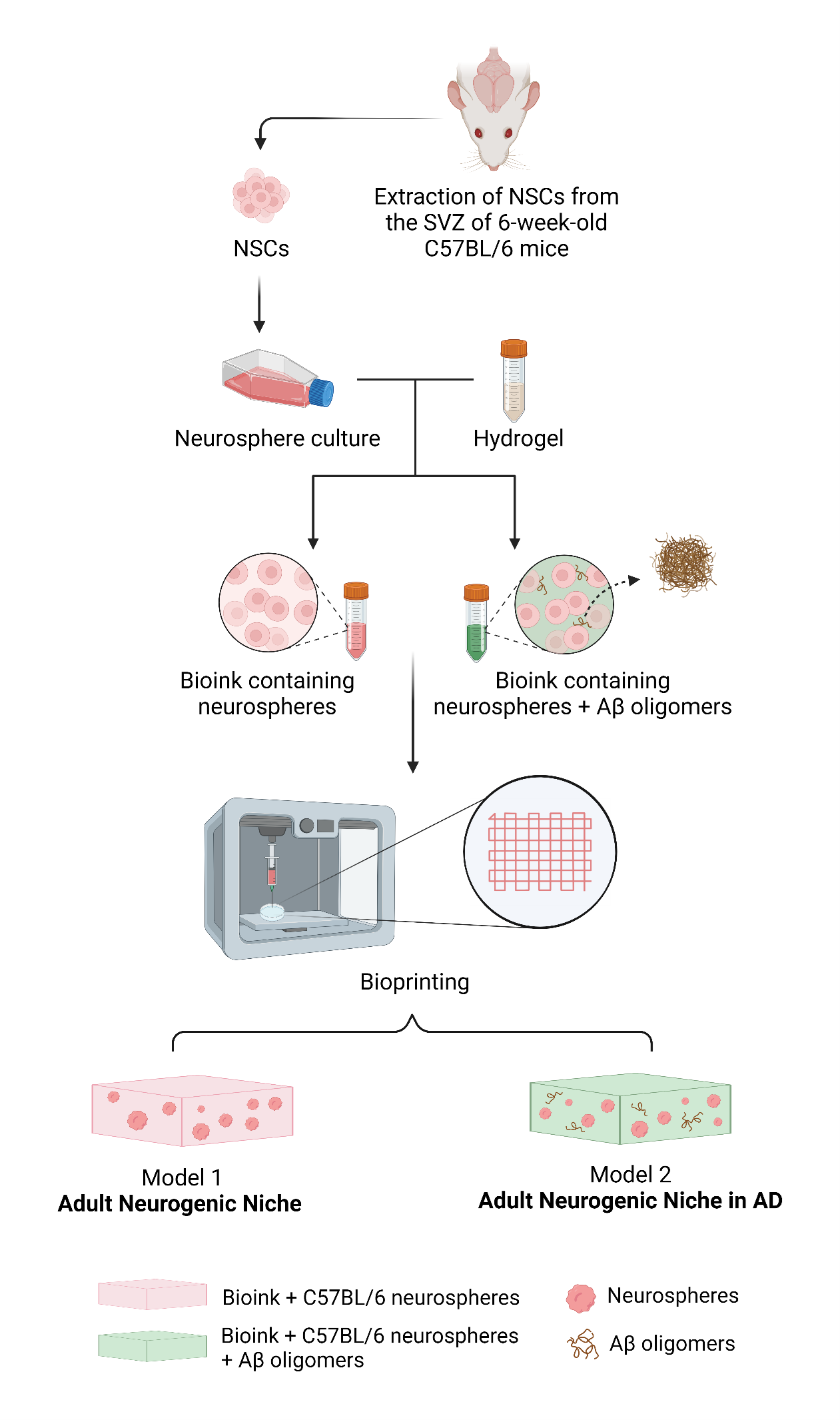
Neurogenesis plays a major role in neuroplasticity and memory. In adult human and mouse brains, neural stem cells (NSCs) are mainly distributed in two extensively characterized neurogenic niches: the subgranular zone (SGZ) of the hippocampus and the subventricular zone (SVZ) of the lateral ventricles. Impaired neurogenesis is one of the consequences of Alzheimer’s disease (AD), contributing to cognitive decline and progressive memory loss. Developing new in vitro models that resemble this three-dimensional (3D) structure is fundamental for enhancing our understanding of the SVZ neurogenic niche dynamics in AD. Herein, we produced and characterized a 3D-bioprinted model of the adult SVZ neurogenic niche containing amyloid β (Aβ) oligomers, mimicking the NSC microenvironment in AD. In this model, Aβ oligomers induce oxidative stress and reduce the proliferative potential of NSCs, while stimulating neuronal differentiation. We hypothesize that these events are an early attempt of adult NSCs to compensate for neuronal death in AD pathogenesis. Our 3D model simulates the NSC niche physiology, reproducing an early response of NSCs in AD, strengthening the importance of studying the potential of neurogenesis in neurodegeneration.
- Moreno-Jiménez EP, Flor-García M, Terreros-Roncal J, et al. Adult hippocampal neurogenesis is abundant in neurologically healthy subjects and drops sharply in patients with Alzheimer’s disease. Nat Med. 2019;25(4):554-560. doi: 10.1038/s41591-019-0375-9
- Selkoe DJ. Alzheimer’s disease: genes, proteins, and therapy. J Physiol Rev. 2001;81(2):741-766. doi: 10.1152/physrev.2001.81.2.741
- Li Puma DD, Piacentini R, Grassi C. Does impairment of adult neurogenesis contribute to pathophysiology of Alzheimer’s disease? A still open question. J Front Mol Neurosci. 2021;13:578211. doi: 10.3389/fnmol.2020.578211
- Kim HS, Shin SM, Kim S, Nam Y, Yoo A, Moon M. Relationship between adult subventricular neurogenesis and Alzheimer’s disease: pathologic roles and therapeutic implications. Front Aging Neurosci. 2022;14:1002281. doi: 10.3389/fnagi.2022.1002281
- Selkoe DJ. Alzheimer’s disease. Cold Spring Harb Perspect Biol. 2011;3:a004457. doi: 10.1101/cshperspect.a004457
- Culig L, Chu X, Bohr VA. Neurogenesis in aging and age-related neurodegenerative diseases. Ageing Res Rev. 2022;78:101636. doi: 10.1016/j.arr.2022.101636
- Scopa C, Marrocco F, Latina V, et al. Impaired adult neurogenesis is an early event in Alzheimer’s disease neurodegeneration, mediated by intracellular Aβ oligomers. Cell Death Differ. 2020;27(3):934-948. doi: 10.1038/s41418-019-0409-3
- Salles GN, Calió ML, Afewerki S, et al. Prolonged drug-releasing fibers attenuate Alzheimer’s disease-like pathogenesis. ACS Appl Mater Interfaces. 2018;10(43):36693-36702. doi: 10.1021/acsami.8b12649
- Salles GN, Calió ML, Hölscher C, Pacheco-Soares C, Porcionatto M, Lobo AO. Neuroprotective and restorative properties of the GLP-1/GIP dual agonist DA-JC1 compared with a GLP-1 single agonist in Alzheimer’s disease. Neuropharmacology. 2020;162:107813. doi: 10.1016/j.neuropharm.2019.107813
- Calió ML, Mosini AC, Marinho DS, et al. Leptin enhances adult neurogenesis and reduces pathological features in a transgenic mouse model of Alzheimer’s disease. Neurobiol. Dis. 2021;148:105219. doi: 10.1016/j.nbd.2020.105219
- Diaz Brinton R, Ming Wang J. Therapeutic potential of neurogenesis for prevention and recovery from Alzheimer’s disease: allopregnanolone as a proof of concept neurogenic agent. Curr Alzheimer Res. 2006;3(3):185-190. doi: 10.2174/156720506777632817
- Sotthibundhu A, Li Q-X, Thangnipon W, Coulson EJ. Aβ1–42 stimulates adult SVZ neurogenesis through the p75 neurotrophin receptor. Neurobiol Aging. 2009;30(12):1975-1985. doi: 10.1016/j.neurobiolaging.2008.02.004
- Bernabeu-Zornoza A, Coronel R, Palmer C, Monteagudo M, Zambrano A, Liste I. Physiological and pathological effects of amyloid-β species in neural stem cell biology. Neural Regen Res. 2019;14(12):2035-2042. doi: 10.4103/1673-5374.262571
- Walus K, Beyer S, Willerth SM. Three-dimensional bioprinting healthy and diseased models of the brain tissue using stem cells. Curr Opin Biomed Eng. 2020; 14:25-33. doi: 10.1016/j.cobme.2020.03.002
- de Melo BAG, Jodat YA, Cruz EM, Benincasa JC, Shin SR, Porcionatto MA. Strategies to use fibrinogen as bioink for 3D bioprinting fibrin-based soft and hard tissues. Acta Biomater. 2020;117:60-76. doi: 10.1016/j.actbio.2020.09.024
- de Melo BAG, Benincasa JC, Cruz EM, Maricato JT, Porcionatto MA. 3D culture models to study SARS-CoV-2 infectivity and antiviral candidates: From spheroids to bioprinting. Biomed J. 2021;44(1):31-42. doi: 10.1016/j.bj.2020.11.009
- Cruz EM, Machado LS, Zamproni LN, et al. A gelatin methacrylate-based hydrogel as a potential bioink for 3D bioprinting and neuronal differentiation. Pharmaceutics. 2023;15(2):627. doi: 10.3390/pharmaceutics15020627
- Knowlton S, Anand S, Shah T, Tasoglu S. Bioprinting for neural tissue engineering. Trends Neurosci. 2018; 41(1):31-46. doi: 10.1016/j.tins.2017.11.001
- Cadena M, Ning L, King A, et al. 3D bioprinting of neural tissues. Adv Healthc Mater. 2021;10(15):2001600. doi: 10.1002/adhm.202001600
- Parra-Cantu C, Li W, Quiñones-Hinojosa A, Zhang YS. 3D bioprinting of Glioblastoma model. J 3D Print Med. 2020;4(2):113-125. doi: 10.2217/3dp-2019-0027
- Gao T, Gillispie GJ, Copus JS, et al. Optimization of gelatin– alginate composite bioink printability using rheological parameters: A systematic approach. Biofabrication. 2018;10(3):034106. doi: 10.1088/1758-5090/aacdc7
- Ioannidis K, Angelopoulos I, Gakis G, et al. 3D reconstitution of the neural stem cell niche: connecting the dots. Front Bioeng Biotechnol. 2021;9:705470. doi: 10.3389/fbioe.2021.705470
- Centeno EGZ, Cimarosti H, Bithell A. 2D versus 3D human induced pluripotent stem cell-derived cultures for neurodegenerative disease modelling. Mol Neurodegener. 2018;13:1-15. doi: 10.1186/s13024-018-0258-4
- Ioannidis K, Danalatos RI, Champeris Tsaniras S, et al. A custom ultra-low-cost 3D bioprinter supports cell growth and differentiation. Front Bioeng Biotechnol. 2020;8:580889. doi: 10.3389/fbioe.2020.580889
- Benwood C, Walters-Shumka J, Scheck K, Willerth SM. 3D bioprinting patient-derived induced pluripotent stem cell models of Alzheimer’s disease using a smart bioink. Bioelectron Med. 2023;9(1):10. doi: 10.1186/s42234-023-00112-7
- Bovi dos Santos G, de Lima-Vasconcellos TH, Móvio MI, Birbrair A, Del Debbio CB, Kihara AH. New perspectives in stem cell transplantation and associated therapies to treat retinal diseases: from gene editing to 3D bioprinting. Stem Cell Rev Rep. 2024;20(3):722-737. doi: 10.1007/s12015-024-10689-4
- Romariz SAA, Sanabria V, da Silva KR, et al. High concentrations of cannabidiol induce neurotoxicity in neurosphere culture system. Neurotox Res. 2024; 42(1):14. doi: 10.1007/s12640-024-00692-5
- Fantini V, Bordoni M, Scocozza F, et al. Bioink composition and printing parameters for 3D modeling neural tissue. Cells. 2019;8(8):830. doi: 10.3390/cells8080830
- Zhou X, Cui H, Nowicki M, et al. Three-dimensional-bioprinted dopamine-based matrix for promoting neural regeneration. ACS Appl Mater Interfaces. 2018;10(10):8993-9001. doi: 10.1021/acsami.7b18197
- Joung D, Truong V, Neitzke CC, et al. 3D printed stem-cell derived neural progenitors generate spinal cord scaffolds. Adv Funct Mater. 2018;28(39):1801850. doi: 10.1002/adfm.201801850
- Suslov ON, Kukekov VG, Ignatova TN, Steindler DA. Neural stem cell heterogeneity demonstrated by molecular phenotyping of clonal neurospheres. Proc Natl Acad Sci USA. 2002;99(22):14506-14511. doi: 10.1073/pnas.212525299
- Othman SA, Soon CF, Ma NL, et al. Alginate-gelatin bioink for bioprinting of hela spheroids in alginate-gelatin hexagon shaped scaffolds. Polym Bull. 2021;78:6115-6135. doi: 10.1007/s00289-020-03421-y
- Li Z, Huang S, Liu Y, et al. Tuning alginate-gelatin bioink properties by varying solvent and their impact on stem cell behavior. Sci Rep. 2018;8(1):8020. doi: 10.1038/s41598-018-26407-3
- Cheng L, Yao B, Hu T, et al. Properties of an alginate-gelatin-based bioink and its potential impact on cell migration, proliferation, and differentiation. Int J Biol Macromol. 2019;135:1107-1113. doi: 10.1016/j.ijbiomac.2019.06.017
- Giuseppe MD, Law N, Webb B, et al. Mechanical behaviour of alginate-gelatin hydrogels for 3D bioprinting. J Mech Behav Biomed Mater. 2018;79:150-157. doi: 10.1016/j.jmbbm.2017.12.018
- Łabowska MB, Cierluk K, Jankowska AM, Kulbacka J, Detyna J, Michalak I. A review on the adaption of alginate-gelatin hydrogels for 3D cultures and bioprinting. Materials (Basel, Switzerland). 2021;14(4):858. doi: 10.3390/ma14040858
- Morgan C, Inestrosa NC. Interactions of laminin with the amyloid ß peptide: implications for Alzheimer’s disease. Braz J Med Biol Res. 2001;34:597-601. doi: 10.1590/S0100-879X2001000500006
- Bronfman FC, Garrido J, Alvarez A, Morgan C, Inestrosa NC. Laminin inhibits amyloid-beta-peptide fibrillation. Neurosci Lett. 1996;218(3):201-203. doi: 10.1016/s0304-3940(96)13147-5
- Rodin S, Kozin SA, Kechko OI, Mitkevich VA, Makarov AA. Aberrant interactions between amyloid-beta and alpha5 laminins as possible driver of neuronal disfunction in Alzheimer’s disease. Biochimie. 2020;174: 44-48. doi: 10.1016/j.biochi.2020.04.011
- Zhang Z, Wang J, Song Y, Wang Z, Dong M, Liu L. Disassembly of Alzheimer’s amyloid fibrils by functional upconversion nanoparticles under near-infrared light irradiation. Colloids Surf B Biointerfaces. 2019;181: 341-348. doi: 10.1016/j.colsurfb.2019.05.053
- Almenar-Queralt A, Falzone TL, Shen Z, et al. UV irradiation accelerates amyloid precursor protein (APP) processing and disrupts APP axonal transport. J Neurosci. 2014;34(9):3320-3339. doi: 10.1523/jneurosci.1503-13.2014
- Measey TJ, Gai F. Light-triggered disassembly of amyloid fibrils. Langmuir. 2012;28(34):12588-12592. doi: 10.1021/la302626d
- Gómez-Guillén MC, Giménez B, López-Caballero MEa, Montero MP. Functional and bioactive properties of collagen and gelatin from alternative sources: a review. Food Hydrocolloids. 2011;25(8):1813-1827. doi: 10.1016/j.foodhyd.2011.02.007
- Mancha Sánchez E, Gómez-Blanco JC, López Nieto E, et al. Hydrogels for bioprinting: a systematic review of hydrogels synthesis, bioprinting parameters, and bioprinted structures behavior. Front Bioeng Biotechnol. 2020;8:776. doi: 10.3389/fbioe.2020.00776
- Lee KY, Mooney DJ. Alginate: properties and biomedical applications. Prog Polym Sci. 2012;37(1):106-126. doi: 10.1016/j.progpolymsci.2011.06.003
- Ishiwata R, Iwasa M. Cellular inertia. Sci Rep. 2021;11(1):23799. doi: 10.1038/s41598-021-02384-y
- Derkach SR, Voron’ko NG, Kuchina YA, Kolotova DS. Modified fish gelatin as an alternative to mammalian gelatin in modern food technologies. Polymers. 2020;12(12):3051. doi: 10.3390/polym12123051
- Kokol V, Pottathara YB, Mihelčič M, Perše LS. Rheological properties of gelatine hydrogels affected by flow-and horizontally-induced cooling rates during 3D cryo-printing. Colloids Surf A Physicochem Eng Asp. 2021;616:126356. doi: 10.1016/j.colsurfa.2021.126356
- Liu S, Yang H, Chen D, et al. Three-dimensional bioprinting sodium alginate/gelatin scaffold combined with neural stem cells and oligodendrocytes markedly promoting nerve regeneration after spinal cord injury. Regen Biomater. 2022;9:rbac038. doi: 10.1093/rb/rbac038
- Kaliampakou C, Lagopati N, Pavlatou EA, Charitidis CA. Alginate–gelatin hydrogel scaffolds; an optimization of post-printing treatment for enhanced degradation and swelling behavior. Gels. 2023;9(11):857. doi: 10.3390/gels9110857
- Freeman FE, Kelly DJ. Tuning alginate bioink stiffness and composition for controlled growth factor delivery and to spatially direct MSC fate within bioprinted tissues. Sci Rep. 2017;7(1):17042. doi: 10.1038/s41598-017-17286-1
- Chung JHY, Naficy S, Yue Z, et al. Bio-ink properties and printability for extrusion printing living cells. Biomater Sci. 2013;1(7):763-773. doi: 10.1039/C3BM00012E
- Sonaye SY, Ertugral EG, Kothapalli CR, Sikder P. Extrusion 3D (bio) printing of alginate-gelatin-based composite scaffolds for skeletal muscle tissue engineering. Materials. 2022;15(22):7945. doi: 10.3390/ma15227945
- Hazur J, Detsch R, Karakaya E, et al. Improving alginate printability for biofabrication: establishment of a universal and homogeneous pre-crosslinking technique. Biofabrication. 2020;12(4):045004. doi: 10.1088/1758-5090/ab98e5
- Kim J, Choi YJ, Gal CW, Sung A, Park H, Yun HS. 142Development of an alginate-gelatin bioink enhancing osteogenic differentiation by gelatin release. Int J Bioprint. 2023;9(2):660. doi: 10.18063/ijb.v9i2.660
- Cooke ME, Rosenzweig DH. The rheology of direct and suspended extrusion bioprinting. APL Bioeng. 2021;5(1):011502. doi: 10.1063/5.0031475
- Chimene D, Kaunas R, Gaharwar AK. Hydrogel bioink reinforcement for additive manufacturing: a focused review of emerging strategies. Adv Mater. 2020;32(1):e1902026. doi: 10.1002/adma.201902026
- Mancha Sánchez E, Gómez-Blanco JC, López Nieto E, et al. Hydrogels for bioprinting: a systematic review of hydrogels synthesis, bioprinting parameters, and bioprinted structures behavior. Front Bioeng Biotechnol. 2020;8:776. doi: 10.3389/fbioe.2020.00776
- O’Connell C, Ren J, Pope L, et al. Characterizing bioinks for extrusion bioprinting: printability and rheology. Methods Mol Biol. 2020;2140:111-133. doi: 10.1007/978-1-0716-0520-2_7
- Semba JA, Mieloch AA, Tomaszewska E, Cywoniuk P, Rybka JD. Formulation and evaluation of a bioink composed of alginate, gelatin, and nanocellulose for meniscal tissue engineering. Int J Bioprint. 2023;9(1):621. doi: 10.18063/ijb.v9i1.621
- Schwab A, Levato R, D’Este M, Piluso S, Eglin D, Malda J. Printability and shape fidelity of bioinks in 3D bioprinting. Chem Rev. 2020;120(19):11028-11055. doi: 10.1021/acs.chemrev.0c00084
- Cui R, Li S, Li T, et al. Natural polymer derived hydrogel bioink with enhanced thixotropy improves printability and cellular preservation in 3D bioprinting. J Mater Chem B. 2023;11(17):3907-3918. doi: 10.1039/D2TB02786K
- Mouser VH, Melchels FP, Visser J, Dhert WJ, Gawlitta D, Malda J. Yield stress determines bioprintability of hydrogels based on gelatin-methacryloyl and gellan gum for cartilage bioprinting. Biofabrication. 2016;8(3):035003. doi: 10.1088/1758-5090/8/3/035003
- Venkata Krishna D, Ravi Sankar M. Persuasive factors on the bioink printability and cell viability in the extrusion-based 3D bioprinting for tissue regeneration applications. Eng Regener. 2023;4(4):396-410. doi: 10.1016/j.engreg.2023.07.002
- Herrada-Manchón H, Fernández MA, Aguilar E. Essential guide to hydrogel rheology in extrusion 3D printing: how to measure it and why it matters? Gels. 2023;9(7):517. doi: 10.3390/gels9070517
- Tuladhar S, Clark S, Habib A. Tuning shear thinning factors of 3D bio-printable hydrogels using short fiber. Materials (Basel, Switzerland). 2023;16(2):572. doi: 10.3390/ma16020572
- Malektaj H, Drozdov AD, deClaville Christiansen JJP. Mechanical properties of alginate hydrogels cross-linked with multivalent cations. Polymers (Basel). 2023;15(14):3012. doi: 10.3390/polym15143012
- Łabowska MB, Cierluk K, Jankowska AM, Kulbacka J, Detyna J, Michalak IJM. A review on the adaption of alginate-gelatin hydrogels for 3D cultures and bioprinting. Materials (Basel). 2021;14(4):858. doi: 10.3390/ma14040858
- Shams E, Barzad MS, Mohamadnia S, Tavakoli O, Mehrdadfar AJJoBA. A review on alginate-based bioinks, combination with other natural biomaterials and characteristics. J Biomater Appl. 2022;37(2):355-372. doi: 10.1177/08853282221085690
- Ioannidis K, Danalatos RI, Champeris Tsaniras S, et al. A custom ultra-low-cost 3D bioprinter supports cell growth and differentiation. Front Bioeng Biotechnol. 2020;8:580889. doi: 10.3389/fbioe.2020.580889
- Leonardo M, Prajatelistia E, Judawisastra HJB. Alginate-based bioink for organoid 3D bioprinting: a review. Bioprinting. 2022;28:e00246. doi: 10.1016/j.bprint.2022.e00246
- Mondal A, Gebeyehu A, Miranda M, et al. Characterization and printability of Sodium alginate-gelatin hydrogel for bioprinting NSCLC co-culture. Sci Rep. 2019; 9(1):19914. doi: 10.1038/s41598-019-55034-9
- Hiller T, Berg J, Elomaa L, et al. Generation of a 3D liver model comprising human extracellular matrix in an alginate/ gelatin-based bioink by extrusion bioprinting for infection and transduction studies. Int J Mol Sci. 2018;19(10):3129. doi: 10.3390/ijms19103129
- Di Giuseppe M, Law N, Webb B, et al. Mechanical behaviour of alginate-gelatin hydrogels for 3D bioprinting. J Mech Behav Biomed Mater. 2018;79:150-157. doi: 10.1016/j.jmbbm.2017.12.018
- Freeman FE, Kelly DJJSr. Tuning alginate bioink stiffness and composition for controlled growth factor delivery and to spatially direct MSC fate within bioprinted tissues. Sci Rep. 2017;7(1):17042. doi: 10.1038/s41598-017-17286-1
- Chung JH, Naficy S, Yue Z, et al. Bio-ink properties and printability for extrusion printing living cells. Biomater Sci. 2013;1(7):763-773. doi: 10.1039/C3BM00012E
- Maihemuti A, Zhang H, Lin X, et al. 3D-printed fish gelatin scaffolds for cartilage tissue engineering. J Bioact Mater. 2023;26:77-87. doi: 10.1016/j.bioactmat.2023.02.007
- Derkach SR, Voron’ko NG, Sokolan NI, Kolotova DS, Kuchina YA. Interactions between gelatin and sodium alginate: UV and FTIR studies. J Dispers Sci Technol. 2020;41(5):690-698. doi: 10.1080/01932691.2019.1611437
- Costa HdS, Dias MR. Alginate/bioactive glass beads: synthesis, morphological and compositional changes caused by SBF immersion method. Mater Res. 2021;24(4): e20200587. doi: 10.1590/1980-5373-MR-2020-0587
- Vosough F, Barth A. Characterization of homogeneous and heterogeneous amyloid-β42 oligomer preparations with biochemical methods and infrared spectroscopy reveals a correlation between infrared spectrum and oligomer size. ACS Chem Neurosci. 2021;12(3):473-488. doi: 10.1021/acschemneuro.0c00642
- Fraser PE, Nguyen JT, Inouye H, et al. Fibril formation by primate, rodent, and Dutch-hemorrhagic analogues of Alzheimer amyloid beta-protein. Biochemistry. 1992;31(44):10716-10723. doi: 10.1021/bi00159a011
- Zandomeneghi G, Krebs MR, McCammon MG, Fändrich M. FTIR reveals structural differences between native beta-sheet proteins and amyloid fibrils. Protein Sci. 2004;13(12):3314-3321. doi: 10.1110/ps.041024904
- Sarroukh R, Goormaghtigh E, Ruysschaert J-M, Raussens V. ATR-FTIR: A “rejuvenated” tool to investigate amyloid proteins. Biochim Biophys Acta. 2013;1828(10):2328-2338. doi: 10.1016/j.bbamem.2013.04.012
- Yankner BA, Duffy LK, Kirschner DA. Neurotrophic and neurotoxic effects of amyloid beta protein: reversal by tachykinin neuropeptides. Science (New York, NY). 1990;250(4978):279-282. doi: 10.1126/science.2218531
- Mazur-Kolecka B, Golabek A, Nowicki K, Flory M, Frackowiak J. Amyloid-beta impairs development of neuronal progenitor cells by oxidative mechanisms. Neurobiol Aging. 2006;27(9):1181-1192. doi: 10.1016/j.neurobiolaging.2005.07.006
- Wang X, Sun X, Gan D, et al. Bioadhesive and conductive hydrogel-integrated brain-machine interfaces for conformal and immune-evasive contact with brain tissue. Matter. 2022;5(4):1204-1223. doi: 10.1016/j.matt.2022.01.012
- Pettikiriarachchi JTS, Parish CL, Shoichet MS, Forsythe JS, Nisbet DR. Biomaterials for brain tissue engineering. Aust J Chem. 2010;63(8):1143-1154. doi: 10.1071/CH10159
- Sadeghi A, Afshari E, Hashemi M, Kaplan D, Mozafari M. Brainy biomaterials: latest advances in smart biomaterials to develop the next generation of neural interfaces. Curr Opin Biomed Eng. 2023;25:100420. doi: 10.1016/j.cobme.2022.100420
- Bierman‐Duquette RD, Safarians G, Huang J, et al. Engineering tissues of the central nervous system: interfacing conductive biomaterials with neural stem/progenitor cells. Adv Healthc Mater. 2022;11(7):2101577. doi: 10.1002/adhm.202101577
- Modulevsky DJ, Cuerrier CM, Pelling AE. Biocompatibility of subcutaneously implanted plant-derived cellulose biomaterials. PloS One. 2016;11(6):e0157894. doi: 10.1371/journal.pone.0157894
- Sordini L, Garrudo FFF, Rodrigues CAV, et al. Effect of electrical stimulation conditions on neural stem cells differentiation on cross-linked PEDOT: PSS films. Front Bioeng Biotechnol. 2021;9:591838. doi: 10.3389/fbioe.2021.591838
- Kaur G, Adhikari R, Cass P, Bown M, Gunatillake P. Electrically conductive polymers and composites for biomedical applications. RSC Adv. 2015;5(47):37553-37567. doi: 10.1039/C5RA01851J
- Jensen JB, Parmar M. Strengths and limitations of the neurosphere culture system. Mol Neurobiol. 2006;34(3): 153-161. doi: 10.1385/mn:34:3:153
- Simpson LW, Szeto GL, Boukari H, Good TA, Leach JB. Collagen hydrogel confinement of Amyloid-β (Aβ) accelerates aggregation and reduces cytotoxic effects. Acta Biomater. 2020;112:164-173. doi: 10.1016/j.actbio.2020.05.030
- Li YE, Jodat YA, Samanipour R, et al. Toward a neurospheroid niche model: optimizing embedded 3D bioprinting for fabrication of neurospheroid brain-like co-culture constructs. Biofabrication. 2020;13:015014. doi: 10.1088/1758-5090/abc1be
- Sears NA, Seshadri DR, Dhavalikar PS, Cosgriff-Hernandez E. A review of three-dimensional printing in tissue engineering. Tissue Eng Part B Rev. 2016;22(4):298-310. doi: 10.1089/ten.TEB.2015.0464
- Pillat MM, Ayupe AC, Juvenal G, et al. Differentiated embryonic neurospheres from familial Alzheimer’s disease model show innate immune and glial cell responses. Stem Cell Rev Rep. 2023;19(6):1800-1811. doi: 10.1007/s12015-023-10542-0
- Gaugler J, James B, Johnson T, et al. Alzheimer’s disease facts and figures. Alzheimers Dement. 2022;18(4):700-789. doi: 10.1002/alz.12638
- Andrade-Guerrero J, Santiago-Balmaseda A, Jeronimo- Aguilar P, et al. Alzheimer’s disease: an updated overview of its genetics. Int J Mol Sci. 2023;24(4):3754. doi: 10.3390/ijms24043754
- Esteve D, Molina-Navarro MM, Giraldo E, et al. Adult neural stem cell migration is impaired in a mouse model of Alzheimer’s disease. Mol Neurobiol. 2022;59(2): 1168-1182. doi: 10.1007/s12035-021-02620-6
- Choi YJ, Park J, Lee SH. Size-controllable networked neurospheres as a 3D neuronal tissue model for Alzheimer’s disease studies. Biomaterials. 2013;34(12):2938-2946. doi: 10.1016/j.biomaterials.2013.01.038
- Bernabeu-Zornoza A, Coronel R, Palmer C, Martín A, López-Alonso V, Liste I. Neurogenesis is increased in human neural stem cells by Aβ40 peptide. Int J Mol Sci. 2022;23(10):5820. doi: 10.3390/ijms23105820
- Cheignon C, Tomas M, Bonnefont-Rousselot D, Faller P, Hureau C, Collin F. Oxidative stress and the amyloid beta peptide in Alzheimer’s disease. Redox Biol. 2018;14:450-464. doi: 10.1016/j.redox.2017.10.014
- Li F, Gong Q, Dong H, Shi J. Resveratrol, a neuroprotective supplement for Alzheimer’s disease. Curr Pharm Des. 2012;18(1):27-33. doi: 10.2174/138161212798919075
- Chiang MC, Nicol CJB, Lin CH, Chen SJ, Yen C, Huang RN. Nanogold induces anti-inflammation against oxidative stress induced in human neural stem cells exposed to amyloid-beta peptide. Neurochem Int. 2021;145:104992. doi: 10.1016/j.neuint.2021.104992
- Walton NM, Shin R, Tajinda K, et al. Adult neurogenesis transiently generates oxidative stress. PloS One. 2012;7(4):e35264. doi: 10.1371/journal.pone.0035264
- Pérez Estrada C, Covacu R, Sankavaram SR, Svensson M, Brundin L. Oxidative stress increases neurogenesis and oligodendrogenesis in adult neural progenitor cells. Stem Cells Dev. 2014;23(19):2311-2327. doi: 10.1089/scd.2013.0452
- Madhavan L, Ourednik V, Ourednik J. Grafted neural stem cells shield the host environment from oxidative stress. Ann NY Acad Sci. 2005;1049:185-188. doi: 10.1196/annals.1334.017
- Fonseca MB, Solá S, Xavier JM, Dionísio PA, Rodrigues CM. Amyloid β peptides promote autophagy-dependent differentiation of mouse neural stem cells: Aβ-mediated neural differentiation. Mol Neurobiol. 2013;48(3):829-840. doi: 10.1007/s12035-013-8471-1
- Vázquez P, Arroba AI, Cecconi F, de la Rosa EJ, Boya P, de Pablo F. Atg5 and Ambra1 differentially modulate neurogenesis in neural stem cells. Autophagy. 2012;8(2):187-199. doi: 10.4161/auto.8.2.18535
- López-Toledano MA, Shelanski ML. Neurogenic effect of beta-amyloid peptide in the development of neural stem cells. J Neurosc. 2004;24(23):5439-5444. doi: 10.1523/jneurosci.0974-04.2004
- Bernabeu-Zornoza A, Coronel R, Palmer C, et al. Aβ42 peptide promotes proliferation and gliogenesis in human neural stem cells. Mol Neurobiol. 2019;56(6):4023-4036. doi: 10.1007/s12035-018-1355-7
- Baglietto-Vargas D, Sánchez-Mejias E, Navarro V, et al. Dual roles of Aβ in proliferative processes in an amyloidogenic model of Alzheimer’s disease. Sci Rep. 2017;7(1):10085. doi: 10.1038/s41598-017-10353-7
- Micci MA, Krishnan B, Bishop E, et al. Hippocampal stem cells promotes synaptic resistance to the dysfunctional impact of amyloid beta oligomers via secreted exosomes. Mol Neurodegener. 2019;14(1):25. doi: 10.1186/s13024-019-0322-8
- Wander CM, Song J. The neurogenic niche in Alzheimer’s disease. Neurosci Lett. 2021;762:136109. doi: 10.1016/j.neulet.2021.136109
- Jin K, Peel AL, Mao XO, et al. Increased hippocampal neurogenesis in Alzheimer’s disease. Proc Natl Acad Sci USA. 2004;101(1):343-347. doi: 10.1073/pnas.2634794100
- Porayette P, Gallego MJ, Kaltcheva MM, Bowen RL, Vadakkadath Meethal S, Atwood CS. Differential processing of amyloid-beta precursor protein directs human embryonic stem cell proliferation and differentiation into neuronal precursor cells. J Biol Chem. 2009;284(35): 23806-23817. doi: 10.1074/jbc.M109.026328
- Matta R, Gonzalez AL. Engineered biomimetic neural stem cell niche. Curr Stem Cell Rep. 2019;5(3):109-114. doi: 10.1007/s40778-019-00161-2