

Design and fabrication of a biomimetic artificial ear with enhanced mechanical properties

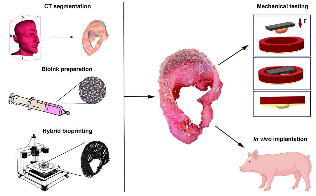
Microtia is a congenital malformation of the external part of the human ear. Recently, bioprinted auricles have been implanted in the first human patient. The remaining challenge in bioprinting of human ear is a post-implantation maintenance of bioprinted auricular construct size and shape. We hypothesize that the use of polylactide stiffeners will enable bioprinting of hybrid auricular constructs with stable post-implantation size and shape. Using the hybrid bioprinting method, auricular implants consisting of a custom-shaped polyurethane frame with polylactide stiffeners and filled with collagen hydrogel containing chondrocytes were printed. Mechanical testing of the implants was performed and it was shown that adding stiffeners to the frame increased the resistance of the structure to deformation. The implants were sutured under the temporal fascia in two mini-pigs for three months, after which a histologic and immunohistochemical study was performed. The formation of regenerated connective tissue with its own vascular network was observed, filling the entire volume of the implant. There was no evidence of inflammation or rejection. The implants maintained their size and shape after implantation. Thus, the in vivo evaluation of the auricular implant bioprinted by the described hybrid method gave satisfactory results in preclinical testing and the next logical step is a clinical translation.
- Reinisch JF, Tahiri Y, eds. Modern Microtia Reconstruction. Cham, Switzerland: Springer Nature Switzerland AG; 2019. doi: 10.1007/978-3-030-16387-7
- Huang Y, Zhao H, Wang Y, et al. The application and progress of tissue engineering and biomaterial scaffolds for total auricular reconstruction in microtia. Front Bioeng Biotechnol. 2023;11:1089031. doi: 10.3389/fbioe.2023.1089031
- Bhamare NC, Tardalkar KR, Kshersagar J, et al. Tissue engineered human ear pinna derived from decellularized goat ear cartilage: clinically useful and biocompatible auricle construct. Cell Tissue Bank. 2022;23(1):43-55. doi: 10.1007/s10561-021-09911-1
- Reighard CL, Hollister SJ, Zopf DA. Auricular reconstruction from rib to 3D printing. J 3D Print Med. 2018;2:35-41. doi: 10.2217/3dp-2017-0017
- Ali K, Trost J, Truong T, Harshbarger R. Total ear reconstruction using porous polyethylene. Semin Plast Surg. 2017;31(3):161-172. doi: 10.1055/s-0037-1604261
- Jiang C, Zhao C, Chen B, et al. Auricular reconstruction using Medpor combined with different hearing rehabilitation approaches for microtia. Acta Otolaryngol. 2021;141(6):572- 578. doi: 10.1080/00016489.2021.1900601
- Zhang B, Zeng X, Yang X. Clinical applications of ear reconstruction with Medpor [in Chinese]. Zhong Nan Da Xue Xue Bao Yi Xue Ban. 2019;44(5):562-570. doi: 10.11817/j.issn.1672-7347.2019.05.014
- Otto IA, Melchels FP, Zhao X, et al. Auricular reconstruction using biofabrication-based tissue engineering strategies. Biofabrication. 2015;7(3):032001. doi: 10.1088/1758-5090/7/3/032001
- Bichara DA, O’Sullivan NA, Pomerantseva I, et al. The tissue-engineered auricle: Past, present, and future. Tissue Eng Part B Rev. 2012;18(1):51-61. doi: 10.1089/ten.teb.2011.0326
- Bhamare N, Tardalkar K, Khadilkar A, Parulekar P, Joshi MG. Tissue engineering of human ear pinna. Cell Tissue Bank. 2022;23(3):441-457. doi: 10.1007/s10561-022-09991-7
- Lee JS, Kim BS, Seo D, Park JH, Cho DW. Three-dimensional cell printing of large-volume tissues: application to ear regeneration. Tissue Eng Part C Methods. 2017;23(3):136- 145. doi: 10.1089/ten.TEC.2016.0362
- Zhou G, Jiang H, Yin Z, et al. In Vitro regeneration of patient-specific ear-shaped cartilage and its first clinical application for auricular reconstruction. EBioMedicine. 2018;28:287-302. doi: 10.1016/j.ebiom.2018.01.011
- Su XH, Ye J, Lei C, et al. Secondary ear reconstruction based on the Nagata method after unsatisfactory microtia surgery outcomes. J Plast Reconstr Aesthet Surg. 2023;87:251-258. doi: 10.1016/j.bjps.2023.10.075
- Karalkin PA, Gryadunova AA, Pereira FDAS, et al. Morphological analysis of in vivo biocompatibility of printed auricle prosthesis. Morfologiia. 2017;152(6):61-66. doi: 10.17816/morph.398192
- Kasyanov VA, Pereira FDAS, Parfenov VA, et al. Development and implantation of a biocompatible auricular prosthesis. Biomed Eng. 2016;49(6):327-330. doi: 10.1007/s10527-016-9559-5
- Ovsianikov A, Yoo J, Mironov V, eds. 3D Printing and Biofabrication. Cham, Switzerland: Springer International Publishing AG; 2018. doi: 10.1007/978-3-319-45444-3
- Zopf DA, Flanagan CL, Mitsak AG, Brennan JR, Hollister SJ. Pore architecture effects on chondrogenic potential of patient-specific 3-dimensionally printed porous tissue bioscaffolds for auricular tissue engineering. Int J Pediatr Otorhinolaryngol. 2018;114:170-174. doi: 10.1016/j.ijporl.2018.07.033
- Visscher DO, Lee H, van Zuijlen PPM, et al. A photo-crosslinkable cartilage-derived extracellular matrix bioink for auricular cartilage tissue engineering. Acta Biomater. 2021;121:193-203. doi: 10.1016/j.actbio.2020.11.029
- Jia L, Hua Y, Zeng J, et al. Bioprinting and regeneration of auricular cartilage using a bioactive bioink based on microporous photocrosslinkable acellular cartilage matrix. Bioact Mater. 2022;16:66-81. doi: 10.1016/j.bioactmat.2022.02.032
- Wei Y, Li L, Xie C, et al. Current Status of Auricular Reconstruction Strategy Development. J Craniofac Surg. 2024;35(3):984-992. doi: 10.1097/SCS.0000000000009908
- Silva C, Pais A, Caldas GAR, Gouveia B, Alves JL, Belinha J. Study on 3D printing of gyroid-based structures for superior structural behavior. Prog Addit Manuf. 2021;6(4):689-703. doi: 10.1007/s40964-021-00191-5
- Fisch P, Kessler S, Ponta S, et al. Tissue Engineered Human Elastic Cartilage From Primary Auricular Chondrocytes for Ear Reconstruction. Adv Funct Mater. 2026;36:e30253. doi: 10.1002/adfm.202530253
- Brennan JR, Cornett A, Chang B, et al. Preclinical assessment of clinically streamlined, 3D-printed, biocompatible single-and two-stage tissue scaffolds for ear reconstruction. J Biomed Mater Res B Appl Biomater. 2021;109(3):394-400. doi: 10.1002/jbm.b.34707
- Cohen BP, Bernstein JL, Morrison KA, Spector JA, Bonassar LJ. Tissue engineering the human auricle by auricular chondrocyte-mesenchymal stem cell co-implantation. PLoS One. 2018;13(10):e0202356. doi: 10.1371/journal.pone.0202356
- Abdul Samat A, Abdul Hamid ZA, Jaafar M, Ong CC, Yahaya BH. Investigation of the In Vitro and In Vivo Biocompatibility of a Three-Dimensional Printed Thermoplastic Polyurethane/Polylactic Acid Blend for the Development of Tracheal Scaffolds. Bioengineering. 2023;10(4):394. doi: 10.3390/bioengineering10040394
- Joshi A, Choudhury S, Gugulothu SB, Visweswariah SS, Chatterjee K. Strategies to Promote Vascularization in 3D Printed Tissue Scaffolds: Trends and Challenges. Biomacromolecules. 2022;23(7):2730-2751. doi: 10.1021/acs.biomac.2c00423
- Später T, Menger MD, Laschke MW. Vascularization strategies for porous polyethylene implants. Tissue Eng Part B Rev. 2021;27(1):29-38. doi: 10.1089/ten.TEB.2020.0077
- Ma Y, Lloyd MS. Systematic Review of Medpor Versus Autologous Ear Reconstruction. J Craniofac Surg. 2022;33(2):602-606. doi: 10.1097/SCS.0000000000008130
- Hallmann R, Feinberg RN, Latker CH, Sasse J, Risau W. Regression of blood vessels precedes cartilage differentiation during chick limb development. Differentiation. 1987;34(2):98-105. doi: 10.1111/j.1432-0436.1987.tb00055.x
- Wu M, Zhang JD, Tang RN, et al. Elevated PTH induces endothelial-to-chondrogenic transition in aortic endothelial cells. Am J Physiol Renal Physiol. 2017;312(3):F436-F444. doi: 10.1152/ajprenal.00210.2016
- Zhang J, Wang L, Cao H, et al. Neurotrophin-3 acts on the endothelial-mesenchymal transition of heterotopic ossification in rats. J Cell Mol Med. 2019;23(4):2595-2609. doi: 10.1111/jcmm.14150
- Futrega K, Robey PG, Klein TJ, Crawford RW, Doran MR. A single day of TGF-β1 exposure activates chondrogenic and hypertrophic differentiation pathways in bone marrow-derived stromal cells. Commun Biol. 2021;4(1):29. doi: 10.1038/s42003-020-01520-0
- Wang W, Rigueur D, Lyons KM. TGFβ signaling in cartilage development and maintenance. Birth Defects Res C Embryo Today. 2014;102(1):37-51. doi: 10.1002/bdrc.21058
- Finestone SA, Ortiz-Ocasio LS, Shetty A, et al. Rib Autograft Versus Porous Polyethylene Implant Outcomes in Microtia Reconstruction: A Meta-Analysis and Systematic Review. Cleft Palate Craniofac J. 2026;63(6):1667-1676. doi: 10.1177/10556656251349274