

Three-dimensional-printed porous titanium scaffolds outperform biphasic calcium phosphate ceramics for load-bearing critical-sized bone defect repair

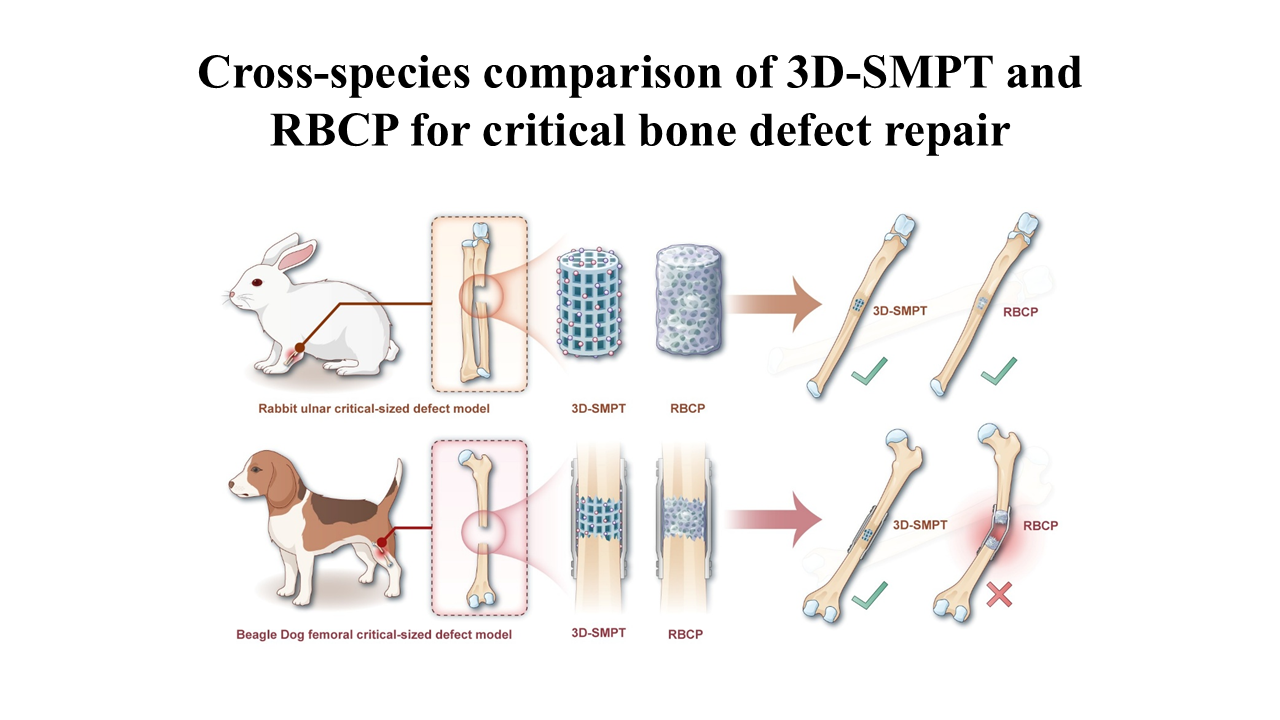
Repairing critical-sized, load-bearing bone defects remains a formidable clinical challenge, primarily due to the mechanical fragility of traditional bioceramics. This study systematically compared the bone regenerative efficacy of surface-microstructured three-dimensional (3D)-printed porous titanium (3D-SMPT) and reinforced biphasic calcium phosphate (RBCP) ceramics. In vitro evaluations demonstrated that a biomimetic coating effectively overcame titanium’s bioinertness, thereby endowing 3D-SMPT with excellent biocompatibility and osteoinductivity comparable to those of RBCP. In vivo cross-species assessments revealed significant biomechanical differences: in a low-load rabbit ulnar model, both scaffolds exhibited equivalent osteogenesis. However, in a true weight-bearing beagle femoral model, RBCP suffered severe structural collapse due to inherent brittleness. In stark contrast, leveraging its cortical bone-matched compressive strength (83.14 MPa, approximately 10-fold higher than that of RBCP at 7.68 MPa) and interconnected porosity, 3D-SMPT maintained long-term mechanical stability, effectively mitigating stress shielding to facilitate massive mature bone ingrowth and robust osseointegration. In conclusion, 3D-SMPT achieves a perfect integration of bioactivity and load-bearing stability, overcoming the inherent fragility of traditional ceramics and offering a highly promising clinical alternative for the repair of massive load-bearing bone defects.
- Wu AM, Bisignano C, James SL, et al. Global, regional, and national burden of bone fractures in 204 countries and territories, 1990-2019: a systematic analysis from the Global Burden of Disease Study 2019. Lancet Healthy Longev. 2021;2(9):e580-e592. doi: 10.1016/s2666-7568(21)00172-0
- Migliorini F, La Padula G, Torsiello E, Spiezia F, Oliva F, Maffulli N. Strategies for large bone defect reconstruction after trauma, infections or tumour excision: a comprehensive review of the literature. Eur J Med Res. 2021;26(1):118. doi: 10.1186/s40001-021-00593-9
- Taiwo BS, Okoye PC, Rayner A, Guryel E, Robertson A. Minding the gap: management of bone defects after trauma. Orthop Trauma. 2025;39(5):295-301. doi: 10.1016/j.mporth.2025.08.005
- Arealis G, Nikolaou VS. Bone printing: new frontiers in the treatment of bone defects. Injury. 2015;46:S20-S22. doi: 10.1016/S0020-1383(15)30050-4
- Hao S, Wang F, Huang J, et al. Periosteum Organoid: Biomimetic Design Inspired From the Bone Healing Process. Exploration. 2025;5(6):20240298. doi: 10.1002/EXP.20240298
- Smith CA, Richardson SM, Eagle MJ, Rooney P, Board T, Hoyland JA. The use of a novel bone allograft wash process to generate a biocompatible, mechanically stable and osteoinductive biological scaffold for use in bone tissue engineering. J Tissue Eng Regen Med. 2015;9(5):595-604. doi: 10.1002/term.1934
- Dumic-Cule I, Pecina M, Jelic M, et al. Biological aspects of segmental bone defects management. Int Orthop. 2015;39(5):1005-1011. doi: 10.1007/s00264-015-2728-4
- Hofmann GH, Sarsour R, Deursen Wv, et al. Immune rejection of orthopedic tissue allograft scoping review: Are we missing a cause of graft/procedural failure? Current concepts. J ISAKOS. 2025;15:101002. doi: 10.1016/j.jisako.2025.101002
- Gao J, Wang H, Li M, et al. DLP-printed GelMA-PMAA scaffold for bone regeneration through endochondral ossification. Int J Bioprint. 2023;9(5):754. doi: 10.18063/ijb.754
- Alonzo M, Primo FA, Kumar SA, et al. Bone tissue engineering techniques, advances and scaffolds for treatment of bone defects. Curr Opin Biomed Eng. 2021;17:100248. doi: 10.1016/j.cobme.2020.100248
- Chen Y, Hu H, Wang D, et al. 2D copper nanozyme patches facilitate bone regeneration via interfacial modulation of osteoclast-osteoblast dynamics. BMEMat. 2026. doi: 10.1002/bmm2.70067
- Tao Z, Yuan Z, Zhou D, et al. Fabrication of magnesium-doped porous polylactic acid microsphere for bone regeneration. Biomater Transl. 2023;4(4):280-290. doi: 10.12336/biomatertransl.2023.04.007
- Qu Y, Wang Yn, Kong W, et al. 3D-printed nano-hydroxyapatite/polylactic acid scaffold with simvastatin-loaded hydroxyethyl methacrylate/sulfobetaine methacrylate hydrogel for accelerated bone repair. Int J Bioprint. 2026;12(1):025490505. doi: 10.36922/ijb025490505
- Hou X, Zhang L, Zhou Z, et al. Calcium Phosphate- Based Biomaterials for Bone Repair. J Funct Biomater. 2022;13(4):187. doi: 10.3390/jfb13040187
- Wang P, Zhao L, Liu J, Weir MD, Zhou X, Xu HHK. Bone tissue engineering via nanostructured calcium phosphate biomaterials and stem cells. Bone Res. 2014;2(1):14017. doi: 10.1038/boneres.2014.17
- Jansen JA. Editorial: Calcium Phosphate Ceramics: The Living Bridge Between Biology and Biomaterials. Tissue Eng Part C Methods. 2025;31(11):381-383. doi: 10.1177/19373384251397945
- Lu J, Yu H, Chen C. Biological properties of calcium phosphate biomaterials for bone repair: a review. RSC Adv. 2018;8(4):2015-2033. doi: 10.1039/C7RA11278E
- Tang H, Zhao G, Lv Y, et al. Immunomodulatory 3D-printed hydroxyapatite/tricalcium phosphate/polycaprolactone scaffolds promote bone regeneration via macrophage polarization. Int J Bioprint. 2026;12(2):026060049. doi: 10.36922/ijb026060049
- Tang Z, Tan Y, Ni Y, et al. Comparison of ectopic bone formation process induced by four calcium phosphate ceramics in mice. Mater Sci Eng C Mater Biol Appl. 2017;70(Pt 2):1000-1010. doi: 10.1016/j.msec.2016.06.097
- Hu X, Zhang Z, Ning L, et al. Three-dimensional-printed bionic dual crosslinked drug-loaded hydrogel composite scaffolds for large bone defect repair. Int J Bioprint. 2025;12(1):348-370. doi: 10.36922/ijb025380391
- Stempels HW, Lehr AM, Delawi D, et al. Efficacy of Biphasic Calcium Phosphate Ceramic With a Needle-Shaped Surface Topography Versus Autograft in Instrumented Posterolateral Spinal Fusion. Spine. 2024;49(19):1323-1331. doi: 10.1097/brs.0000000000005075
- Zhu Y, Zhang K, Zhao R, et al. Bone regeneration with micro/nano hybrid-structured biphasic calcium phosphate bioceramics at segmental bone defect and the induced immunoregulation of MSCs. Biomaterials. 2017;147:133- 144. doi: 10.1016/j.biomaterials.2017.09.018
- Yang J, Chen HJ, Zhu XD, et al. Enhanced repair of a critical-sized segmental bone defect in rabbit femur by surface microstructured porous titanium. J Mater Sci Mater Med. 2014;25(7):1747-1756. doi: 10.1007/s10856-014-5202-8
- Gavalda-Diaz O, Saiz E, Chevalier J, Bouville F. Toughening of ceramics and ceramic composites through microstructure engineering: A review. Int Mater Rev. 2025;70(1):3-30. doi: 10.1177/09506608241308337
- Cai W, Huo Y, Liu Y, et al. Biomechanics in bone regeneration and mechanobiology in osteoblasts: Fundamental concepts and recent progress. EngMedicine. 2025;2(1):100057. doi: 10.1016/j.engmed.2025.100057
- Thanigaivel S, Priya AK, Balakrishnan D, Dutta K, Rajendran S, Soto-Moscoso M. Insight on recent development in metallic biomaterials: Strategies involving synthesis, types and surface modification for advanced therapeutic and biomedical applications. Biochem Eng J. 2022;187:108522. doi: 10.1016/j.bej.2022.108522
- Liu S, Manshaii F, Chen J, et al. Unleashing the Potential of Electroactive Hybrid Biomaterials and Self-Powered Systems for Bone Therapeutics. Nano-Micro Lett. 2024;17(1):44. doi: 10.1007/s40820-024-01536-9
- Abd-Elaziem W, Darwish MA, Hamada A, Daoush WM. Titanium-Based alloys and composites for orthopedic implants Applications: A comprehensive review. Mater Des. 2024;241:112850. doi: 10.1016/j.matdes.2024.112850
- Chen C, Xie Z, Yang S, et al. Machine Learning Approach to Investigating Macrophage Polarization on Various Titanium Surface Characteristics. BME Front. 2025;6:0100. doi: 10.34133/bmef.0100
- Fan S, Li S, Wu Y, et al. Customized 3D-printed heterogeneous porous titanium scaffolds for bone tissue engineering. MedComm Biomater Appl. 2024;3(2):e80. doi: 10.1002/mba2.80
- Yuan B, Liu P, Zhao R, et al. Functionalized 3D-printed porous titanium scaffold induces in situ vascularized bone regeneration by orchestrating bone microenvironment. J Mater Sci Technol. 2023;153:92-105. doi: 10.1016/j.jmst.2022.12.033
- Takemoto M, Fujibayashi S, Neo M, Suzuki J, Kokubo T, Nakamura T. Mechanical properties and osteoconductivity of porous bioactive titanium. Biomaterials. 2005;26(30):6014- 6023. doi: 10.1016/j.biomaterials.2005.03.019
- Dunand DC. Processing of Titanium Foams. Adv Eng Mater. 2004;6(6):369-376. doi: 10.1002/adem.200405576
- Hao P, Liu J, Zhang C, Lyu L. Design of the Elastic Modulus of porous lattice structures composed of cells with continuously variable cross section carrying structures. Clin Biomech. 2024;119:106330. doi: 10.1016/j.clinbiomech.2024.106330
- Luo B, Miu L, Luo Y. Titanium alloys for biomedical applications: a review on additive manufacturing process and surface modification technology. Int J Adv Manuf Technol. 2025;137(7-8):3215-3227. doi: 10.1007/s00170-025-15287-3
- Wang XX, Hayakawa S, Tsuru K, Osaka A. Improvement of bioactivity of H2O2/TaCl5-treated titanium after subsequent heat treatments. J Biomed Mater Res. 2000;52(1):171-176. doi: 10.1002/1097-4636(200010)52:1<171::AID-JBM22>3.0.CO;2-O
- Yang J, Wang J, Yuan T, et al. The enhanced effect of surface microstructured porous titanium on adhesion and osteoblastic differentiation of mesenchymal stem cells. J Mater Sci Mater Med. 2013;24(9):2235-2246. doi: 10.1007/s10856-013-4976-4
- Liu X, Gao J, Liu J, et al. Three-Dimensional-Printed Spherical Hollow Structural Scaffolds for Guiding Critical-Sized Bone Regeneration. ACS Biomater Sci Eng. 2024;10(4):2581-2594. doi: 10.1021/acsbiomaterials.3c01956
- Liu X, Chang Z, Li Z, et al. Hollow spherical mineralized scaffold integrated with a bone marrow mesenchymal stem cell-laden three-dimensional delivery system for regeneration of critical-sized bone defects. Int J Bioprint. 2026;12(2):026050046. doi: 10.36922/ijb026050046
- Qing F, Wang Z, Hong Y, et al. Selective effects of hydroxyapatite nanoparticles on osteosarcoma cells and osteoblasts. J Mater Sci Mater Med. 2012;23(9):2245-2251. doi: 10.1007/s10856-012-4703-6
- Heise T, Sawyer AY, Hirai T, Schaible S, Sy H, Wickramasekara S. Report on investigation of ISO 10993–12 extraction conditions. Regul Toxicol Pharmacol. 2022;131:105164. doi: 10.1016/j.yrtph.2022.105164
- Kovrlija I, Menshikh K, Abreu H, et al. Corrigendum to “Challenging applicability of ISO 10993-5 for calcium phosphate biomaterials evaluation: Towards more accurate in vitro cytotoxicity assessment” [Biomater. Adv. 160 (2024) 213866]. Biomater Adv. 2025;177:214386. doi: 10.1016/j.bioadv.2025.214386
- Yu D, Shen W, Dai J, Zhu H. Treatment of large bone defects in load-bearing bone: traditional and novel bone grafts. J Zhejiang Univ Sci B. 2025;26(5):421-447. doi: 10.1631/jzus.B2300669
- Reichert JC, Saifzadeh S, Wullschleger ME, et al. The challenge of establishing preclinical models for segmental bone defect research. Biomaterials. 2009;30(12):2149-2163. doi: 10.1016/j.biomaterials.2008.12.050
- Jiang S, Wang M, He J. A review of biomimetic scaffolds for bone regeneration: Toward a cell-free strategy. Bioeng Transl Med. 2021;6(2):e10206. doi: 10.1002/btm2.10206
- Todd EA, Mirsky NA, Silva BLG, et al. Functional Scaffolds for Bone Tissue Regeneration: A Comprehensive Review of Materials, Methods, and Future Directions. J Funct Biomater. 2024;15(10):280. doi: 10.3390/jfb15100280
- Chen Q, Gu J, Zhang H, et al. Promoting implant osseointegration via the osteoblast-selective β-amino acid polymer strategy. Nat Commun. 2025;16(1). doi: 10.1038/s41467-025-58394-1
- Cheng X, Yang X, Liu C, et al. Stabilization of Apatite Coatings on PPENK Surfaces by Mechanical Interlocking to Promote Bioactivity and Osseointegration In Vivo. ACS Appl Mater Interfaces. 2023;15(1):697-710. doi: 10.1021/acsami.2c20633
- Chang L, Chen P, Mokudai T, Kawashita M, Mizoguchi I, Kanetaka H. Enhancing Titanium Osteoconductivity by Alkali-Hot Water Treatment. ACS Omega. 2024;9(44):44568- 44576. doi: 10.1021/acsomega.4c06702
- Tuikampee S, Chaijareenont P, Rungsiyakull P, Yavirach A. Titanium Surface Modification Techniques to Enhance Osteoblasts and Bone Formation for Dental Implants: A Narrative Review on Current Advances. Metals. 2024;14(5):515. doi: 10.3390/met14050515
- Ruan D, Wu C, Deng S, Zhang Y, Guan G. The Anatase Phase of Nanotopography Titania with Higher Roughness Has Better Biocompatibility in Osteoblast Cell Morphology and Proliferation. Biomed Res Int. 2020;2020(1):8032718. doi: 10.1155/2020/8032718
- Arcos D, Vallet-Regí M. Substituted hydroxyapatite coatings of bone implants. J Mater Chem B. 2020;8(9):1781-1800. doi: 10.1039/c9tb02710f
- Liu W, Cheong N, He Z, Zhang T. Application of Hydroxyapatite Composites in Bone Tissue Engineering: A Review. J Funct Biomater. 2025;16(4):127. doi: 10.3390/jfb16040127
- Zhang W, Wang G, Liu Y, et al. The synergistic effect of hierarchical micro/nano-topography and bioactive ions for enhanced osseointegration. Biomaterials. 2013;34(13):3184- 3195. doi: 10.1016/j.biomaterials.2013.01.008
- Duan G, Chang D, Zhang C, et al. Biofunctionalisation strategies of material surface and the inspired biological effects for bone repair. Biosurf Biotribol. 2024;10(2):17-41. doi: 10.1049/bsb2.12081
- Ng WL, An J, Chua CK. Process, Material, and Regulatory Considerations for 3D Printed Medical Devices and Tissue Constructs. Engineering. 2024;36:146-166. doi: 10.1016/j.eng.2024.01.028
- Guo W, Li P, Wei Y, et al. Ionic substitution through bredigite doping for microstructure and performance adjustment in DLP 3D-printed TPMS porous HA bone scaffolds. Virtual Phys Prototyp. 2024;19(1):e2423840. doi: 10.1080/17452759.2024.2423840
- Xu Z, Qi X, Bao M, et al. Biomineralization inspired 3D printed bioactive glass nanocomposite scaffolds orchestrate diabetic bone regeneration by remodeling micromilieu. Bioact Mater. 2023;25:239-255. doi: 10.1016/j.bioactmat.2023.01.024
- Bergoglio M, Najmi Z, Cochis A, Miola M, Vernè E, Sangermano M. Silanized and Cu-doped bioactive glass as filler for biobased photocurable 3D printed scaffolds. Mater Today Chem. 2025;44:102559. doi: 10.1016/j.mtchem.2025.102559
- Ganeshkumar S, Rahman HA, Gowtham TM, Adithya T, Suyambulinagm I, Maniraj J. Multifunctional Polymer Composites: Design, Properties, and Emerging Applications—A Critical Review. In: Springer Proceedings in Materials. Singapore: Springer Nature Singapore; 2024:637- 649. doi: 10.1007/978-981-97-7071-7_45
- Pu Z, Gong W, Wu H, et al. Innovative applications and future challenges of shape memory scaffolds for functional reconstruction in diseases of the musculoskeletal system. Colloids Surf B Biointerfaces. 2026;261:115426. doi: 10.1016/j.colsurfb.2026.115426
- Dong D, Su H, Li X, et al. Microstructures and mechanical properties of biphasic calcium phosphate bioceramics fabricated by SLA 3D printing. J Manuf Process. 2022;81:433- 443. doi: 10.1016/j.jmapro.2022.07.016
- Miri Z, Haugen HJ, Loca D, et al. Review on the strategies to improve the mechanical strength of highly porous bone bioceramic scaffolds. J Eur Ceram Soc. 2024;44(1):23-42. doi: 10.1016/j.jeurceramsoc.2023.09.003
- Rohlmann A, Zilch H, Bergmann G, Kölbel R. Material properties of femoral cancellous bone in axial loading. Part I: Time independent properties. Arch Orthop Trauma Surg. 1980;97(2):95-102. doi: 10.1007/bf00450930
- Wu Y, Li M, Su H, Chen H, Zhu Y. Up-to-date progress in bioprinting of bone tissue. Int J Bioprint. 2023;9(1):628. doi: 10.18063/ijb.v9i1.628
- Bose S, Roy M, Bandyopadhyay A. Recent advances in bone tissue engineering scaffolds. Trends Biotechnol. 2012;30(10):546-554. doi: 10.1016/j.tibtech.2012.07.005