

One-step decellularization of porcine uterine tissue for developing alginate–decellularized uterine ECM hydrogel for uterine tissue engineering

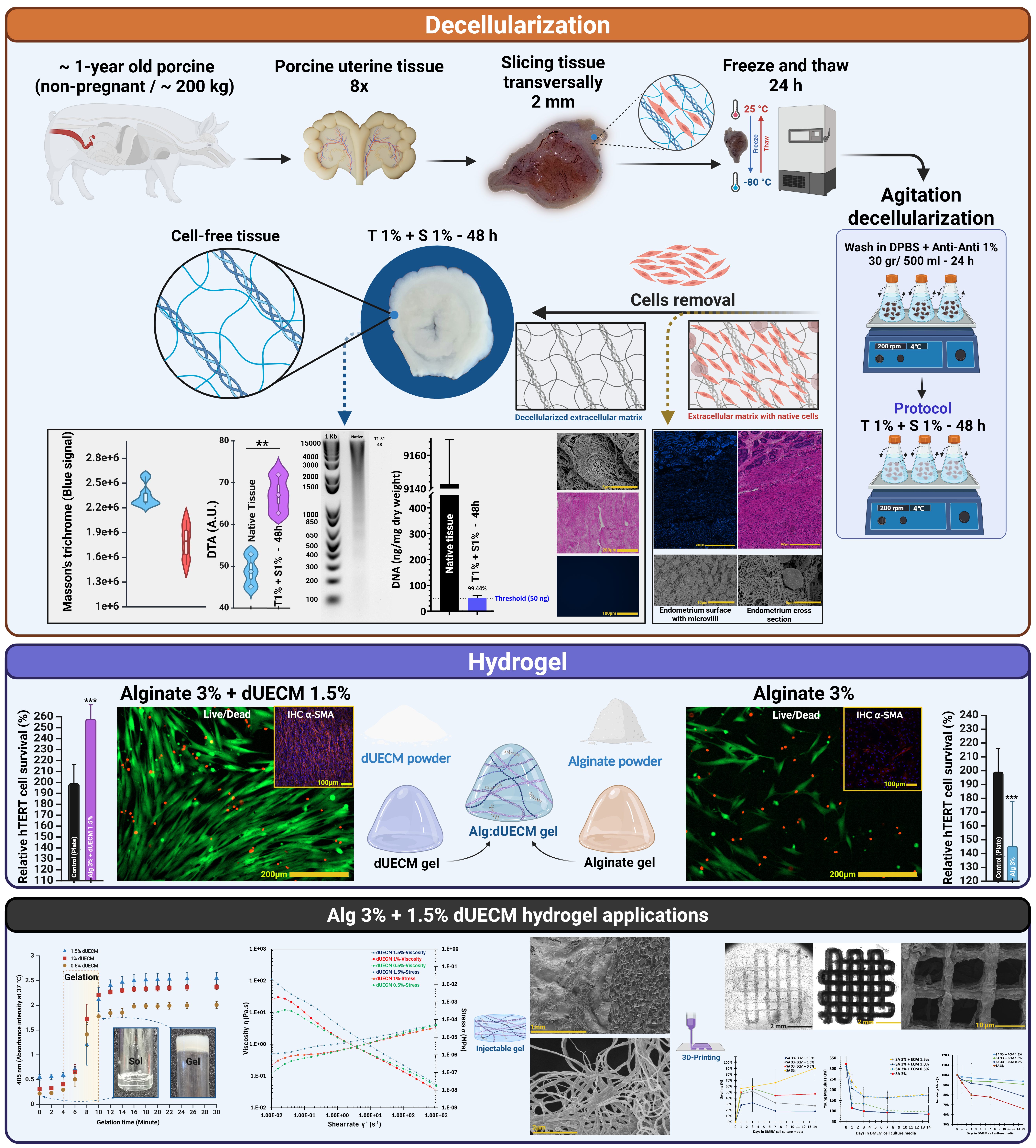
Decellularized uterine extracellular matrix (dUECM) is promising for uterine tissue engineering because of its inherent bioactivity and structural complexity. However, transforming dUECM into porous, functional 3D constructs remains a significant challenging. This study aimed to: (1) synthesize dUECM using a modified decellularization protocol and formulate it into a hydrogel ink, and (2) to fabricate 3D-printed constructs from this ink to assess their capacity to support human uterine myometrial cell growth in vitro. Porcine uterine tissues were decellularized using 1% Triton™ X-100 with varying concentrations of sodium dodecyl sulfate (SDS) (0.1–1.5%) for 48–72 h. The resulting dUECM was characterized using DNA and glycosaminoglycan (GAG) quantification, Picrosirius Red-polarized light microscopy, routine histology, scanning electron microscopy (SEM), Fourier-transform infrared spectroscopy (FTIR), Raman spectroscopy, and thermogravimetric analysis (TGA). To prepare the ink, dUECM powder was enzymatically digested with pepsin and subsequently blended with 2% and 3% alginate to obtain a printable hydrogel formulation. Constructs were fabricated using extrusion-based 3D printing and assessed for filament fidelity, swelling, degradation behavior, and mechanical properties. Biocompatibility was evaluated using hTERT-HM myometrial cells through MTT metabolic assays, Live/Dead staining, and immunohistochemical α-SMA staining. The optimal protocol (1% Triton™ X-100 + 1% SDS for 48 h) reduced DNA to 51.3 ± 9 ng/mg while retaining a high level of GAGs (54.9 ± 7.6 μg/mg). Preservation of the ECM structure was confirmed by spectroscopy analyses. The 3% Alg + 1.5% dUECM hydrogel exhibited suitable printability (1.5 ± 0.2), swelling capacity (47 ± 12%), degradation resistance (94 ± 18% mass retention), and mechanical strength (decreasing from 323 kPa to 175 kPa over 14 days), along with high viability and proliferation (258 ± 13%). The developed dUECM-based hydrogel supports 3D bioprinting with strong mechanical and biological performance, offering a promising platform for uterine tissue engineering.
- Ana Pilar B, Jiangfeng Y, Ann-Beth M, João Paulo S, Jun Z. Trends and projections of caesarean section rates: global and regional estimates. BMJ Glob Health. 2021;6(6):e005671. doi: 10.1136/bmjgh-2021-005671
- Hanuman S, Pande G, Nune M. Current status and challenges in uterine myometrial tissue engineering. Bioengineered. 2023;14(1):2251847. doi: 10.1080/21655979.2023.2251847
- Tahermanesh K, Hanjani S, Saadat Mostafavi SR, et al. Hourglass cesarean scar: A neglected external niche in association with the internal niche. Int J Gynaecol Obstet. 2022;157(2):478-480. doi: 10.1002/ijgo.14052
- Tahermanesh K, Allahqoli L, Bozorgmehr B, et al. Comprehensive evaluation of residual myometrial thickness of the cesarean scar in uterine samples after hysterectomy. Fertil Steril. 2026;125(4):717-719. doi: 10.1016/j.fertnstert.2025.10.011
- Tahermanesh K, Allahqoli L, Karimzadeh A, et al. Cesarean scar niche: comparison of transvaginal ultrasound findings and uterine tissue samples after hysterectomy. Am J Obstet Gynecol. 2025;232(4):373.e1-373.e10. doi: 10.1016/j.ajog.2024.10.010
- Francés-Herrero E, Lopez R, Hellström M, et al. Bioengineering trends in female reproduction: a systematic review. Hum Reprod Update. 2022;28(6):798-837. doi: 10.1093/humupd/dmac025
- Fazel Anvari Yazdi A, Tahermanesh K, Ejlali M, et al. Comparative analysis of porcine-uterine decellularization for bioactive-molecule preservation and DNA removal. Front Bioeng Biotechnol. 2024;12:1418034. doi: 10.3389/fbioe.2024.1418034
- Chen DX. Extrusion bioprinting of scaffolds: an introduction. In: Extrusion Bioprinting of Scaffolds for Tissue Engineering. Cham, Switzerland: Springer; 2024:1-15. doi: 10.1007/978-3-031-72471-8_1
- Chen XB, Fazel Anvari-Yazdi A, Duan X, et al. Biomaterials / bioinks and extrusion bioprinting. Bioact Mater. 2023;28:511-536. doi: 10.1016/j.bioactmat.2023.06.006
- Ahn J, Sen T, Lee D, et al. Uterus-Derived Decellularized Extracellular Matrix-Mediated Endometrial Regeneration and Fertility Enhancement. Adv Funct Mater. 2023;33(34):2214291. doi: 10.1002/adfm.202214291
- Cha E, Choi YS, Lee MJ, et al. Uterus-Mimetic Extracellular Microenvironment for Engineering Female Reproductive System. Adv Funct Mater. 2025;35(6):2415149. doi: 10.1002/adfm.202415149
- Hynes RO, Naba A. Overview of the matrisome— an inventory of extracellular matrix constituents and functions. Cold Spring Harb Perspect Biol. 2012;4(1):a004903. doi: 10.1101/cshperspect.a004903
- Kasravi M, Ahmadi A, Babajani A, et al. Immunogenicity of decellularized extracellular matrix scaffolds: a bottleneck in tissue engineering and regenerative medicine. Biomater Res. 2023;27(1):10. doi: 10.1186/s40824-023-00348-z
- Fazel Anvari-Yazdi A, Badea I, Chen X. Biomaterials in Postoperative Adhesion Barriers and Uterine Tissue Engineering. Gels. 2025;11(6):441. doi: 10.3390/gels11060441
- Yunoki S, Matsuda T. Simultaneous Processing of Fibril Formation and Cross-Linking Improves Mechanical Properties of Collagen. Biomacromolecules. 2008;9(3):879- 885. doi: 10.1021/bm7012058
- McInnes AD, Moser MA, Chen X. Preparation and use of decellularized extracellular matrix for tissue engineering. J Funct Biomater. 2022;13(4):240. doi: 10.3390/jfb13040240
- Choudhury D, Yee M, Sheng ZLJ, Amirul A, Naing MW. Decellularization systems and devices: State-of-the-art. Acta Biomater. 2020;115:51-59. doi: 10.1016/j.actbio.2020.07.060
- Wilson GJ, Courtman DW, Klement P, Michael Lee J, Yeger H. Acellular matrix: A biomaterials approach for coronary artery bypass and heart valve replacement. Ann Thorac Surg. 1995;60:S353-S358. doi: 10.1016/0003-4975(95)98967-Y
- Cartmell JS, Dunn MG. Effect of chemical treatments on tendon cellularity and mechanical properties. J Biomed Mater Res. 2000;49(1):134-140. doi: 10.1002/(SICI)1097-4636(200001)49:1<134::AID-JBM17>3.0.CO;2-D
- Gilbert TW, Sellaro TL, Badylak SF. Decellularization of tissues and organs. Biomaterials. 2006;27(19):3675-3683. doi: 10.1016/j.biomaterials.2006.02.014
- Mendibil U, Ruiz-Hernandez R, Retegi-Carrion S, Garcia- Urquia N, Olalde-Graells B, Abarrategi A. Tissue-specific decellularization methods: rationale and strategies to achieve regenerative compounds. Int J Mol Sci. 2020;21(15):5447. doi: 10.3390/ijms21155447
- Crapo PM, Gilbert TW, Badylak SF. An overview of tissue and whole organ decellularization processes. Biomaterials. 2011;32(12):3233-3243. doi: 10.1016/j.biomaterials.2011.01.057
- Santoso EG, Yoshida K, Hirota Y, et al. Application of detergents or high hydrostatic pressure as decellularization processes in uterine tissues and their subsequent effects on in vivo uterine regeneration in murine models. PLoS One. 2014;9(7):e103201. doi: 10.1371/journal.pone.0103201
- Campo H, Baptista PM, López-Pérez N, Faus A, Cervelló I, Simón C. De-and recellularization of the pig uterus: a bioengineering pilot study. Biol Reprod. 2017;96(1):34-45. doi: 10.1095/biolre/bio143396
- Sargazi Z, Zavareh S, Jafarabadi M, Salehnia M. An efficient protocol for decellularization of the human endometrial fragments for clinical usage. Prog Biomater. 2021;10(2):119- 130. doi: 10.1007/s40204-021-00156-5
- Almeida GHDR, Gibin MS, Rinaldi JdC, et al. Development and Biocompatibility Assessment of Decellularized Porcine Uterine Extracellular Matrix-Derived Grafts. Tissue Eng Part C Methods. 2024;30(12):569-589. doi: 10.1089/ten.tec.2024.0229
- Arezoo N, Mohammad H, Malihezaman M. Tissue engineering of mouse uterus using menstrual blood stem cells (MenSCs) and decellularized uterine scaffold. Stem Cell Res Ther. 2021;12:1-12. doi: 10.1186/s13287-021-02543-y
- Sehic E, Brännström M, Hellström M. Progress in Preclinical Research on Uterus Bioengineering That Utilizes Scaffolds Derived from Decellularized Uterine Tissue. Biomed Mater Devices. 2023;1(1):66-73. doi: 10.1007/s44174-022-00036-x
- Golebiowska AA, Intravaia JT, Sathe VM, Kumbar SG, Nukavarapu SP. Decellularized extracellular matrix biomaterials for regenerative therapies: Advances, challenges and clinical prospects. Bioact Mater. 2024;32:98-123. doi: 10.1016/j.bioactmat.2023.09.017
- Gorroñogoitia I, Urtaza U, Zubiarrain-Laserna A, Alonso- Varona A, Zaldua AM. A Study of the Printability of Alginate-Based Bioinks by 3D Bioprinting for Articular Cartilage Tissue Engineering. Polymers. 2022;14(2):354. doi: 10.3390/polym14020354
- Li N, Duan X, Ding XF, Zhu N, Chen X. Characterization of hydrogel-scaffold mechanical properties and microstructure by using synchrotron propagation-based imaging. J Mech Behav Biomed Mater. 2025;163:106844. doi: 10.1016/j.jmbbm.2024.106844
- Alizadeh Sardroud H, Rosa GDS, Dust W, et al. Comparison study on hyaline cartilage versus fibrocartilage formation in a pig model by using 3D-bioprinted hydrogel and hybrid constructs. Biofabrication. 2025;17(1):015014. doi: 10.1088/1758-5090/ad88a6
- Leonardo M, Prajatelistia E, Judawisastra H. Alginate-based bioink for organoid 3D bioprinting: A review. Bioprinting. 2022;28:e00246. doi: 10.1016/j.bprint.2022.e00246
- Heidari Kani M, Chan E-C, Young RC, Butler T, Smith R, Paul JW. 3D Cell Culturing and Possibilities for Myometrial Tissue Engineering. Ann Biomed Eng. 2017;45(7):1746- 1757. doi: 10.1007/s10439-016-1749-5
- Condon J, Yin S, Mayhew B, et al. Telomerase immortalization of human myometrial cells. Biol Reprod. 2002;67(2):506-514. doi: 10.1095/biolreprod67.2.506
- Ghorbani F, Ekhtiari M, Moeini Chaghervand B, Moradi L, Mohammadi B, Kajbafzadeh A-M. Detection of the residual concentration of sodium dodecyl sulfate in the decellularized whole rabbit kidney extracellular matrix. Cell Tissue Bank. 2022;23(1):119-128. doi: 10.1007/s10561-021-09921-z
- Mathapati S, Galla S, Sankaranarayanan K, Verma RS, Cherian KM, Guhathakurta S. Qualitative and quantitative detection of sodium deoxycholic acid in decellularized tissue. Indian J Thorac Cardiovasc Surg. 2010;26(2):129-131. doi: 10.1007/s12055-010-0016-x
- Zvarova B, Uhl FE, Uriarte JJ, et al. Residual Detergent Detection Method for Nondestructive Cytocompatibility Evaluation of Decellularized Whole Lung Scaffolds. Tissue Eng Part C Methods. 2016;22(5):418-428. doi: 10.1089/ten.tec.2015.0439
- Mehdizadeh Kashi A, Tahemanesh K, Chaichian S, Joghataei MT, Moradi F, Tavangar SM. How to prepare biological samples and live tissues for scanning electron microscopy (SEM). Galen Med J. 2014;3(2):63-80. doi: 10.31661/gmj.v3i2.267
- Mehdizadehkashi A, Tahermanesh K, Anvari-Yazdi AF, et al. Ultrastructural investigation of pelvic peritoneum in patients with chronic pelvic pain and subtle endometriosis in association with chromoendoscopy. J Minim Invasive Gynecol. 2017;24(1):114-123. doi: 10.1016/j.jmig.2016.09.013
- de Campos Vidal B, Mello MLS. Collagen type I amide I band infrared spectroscopy. Micron. 2011;42(3):283-289. doi: 10.1016/j.micron.2010.09.010
- Voytik-Harbin SL, Brightman AO, Waisner BZ, Robinson JP, Lamar CH. Small intestinal submucosa: A tissue-derived extracellular matrix that promotes tissue-specific growth and differentiation of cells in vitro. Tissue Eng. 1998;4(2):157- 174. doi: 10.1089/ten.1998.4.157
- Pati F, Jang J, Ha D-H, et al. Printing three-dimensional tissue analogues with decellularized extracellular matrix bioink. Nat Commun. 2014;5(1):3935. doi: 10.1038/ncomms4935
- Ketabat F, Maris T, Duan X, et al. Optimization of 3D printing and in vitro characterization of alginate/gelatin lattice and angular scaffolds for potential cardiac tissue engineering. Front Bioeng Biotechnol. 2023;11:1161804. doi: 10.3389/fbioe.2023.1161804
- Carney SA, Tahara H, Swartz CD, et al. Immortalization of Human Uterine Leiomyoma and Myometrial Cell Lines After Induction of Telomerase Activity: Molecular and Phenotypic Characteristics. Lab Invest. 2002;82(6):719-728. doi: 10.1097/01.LAB.0000017499.51216.3E
- Scholze M, Singh A, Lozano PF, et al. Utilization of 3D printing technology to facilitate and standardize soft tissue testing. Sci Rep. 2018;8(1):11340. doi: 10.1038/s41598-018-29583-4
- Fadare O, Roma AA. Normal Histology of the Uterine Corpus. In: Atlas of Uterine Pathology. Cham, Switzerland: Springer International Publishing; 2019:1-22. doi: 10.1007/978-3-030-17931-1_1
- Júnior ZSS, Botta SB, Ana PA, et al. Effect of papain-based gel on type I collagen-spectroscopy applied for microstructural analysis. Sci Rep. 2015;5(1):11448. doi: 10.1038/srep11448
- Hopkins J, Brenner L, Tumosa C. Variation of the amide I and amide II peak absorbance ratio in human hair as measured by Fourier transform infrared spectroscopy. Forensic Sci Int. 1991;50(1):61-65. doi: 10.1016/0379-0738(91)90134-5
- Cárcamo JJ, Aliaga AE, Clavijo RE, Brañes MR, Campos- Vallette MM. Raman study of the shockwave effect on collagens. Spectrochim Acta A Mol Biomol Spectrosc. 2012;86:360-365. doi: 10.1016/j.saa.2011.10.049
- France CAM, Thomas DB, Doney CR, Madden O. FT-Raman spectroscopy as a method for screening collagen diagenesis in bone. J Archaeol Sci. 2014;42:346-355. doi: 10.1016/j.jas.2013.11.020
- Cárcamo-Vega JJ, Brañes MR, Loske AM, Campos-Vallette MM. The influence of the number of shock waves and the energy flux density on the Raman spectrum of collagen type I from rat. Shock Waves. 2020;30(2):201-214. doi: 10.1007/s00193-019-00920-4
- Bergholt MS, Serio A, Albro MB. Raman Spectroscopy: Guiding Light for the Extracellular Matrix. Front Bioeng Biotechnol. 2019;7. doi: 10.3389/fbioe.2019.00303
- Takahashi Y, Sugano N, Takao M, Sakai T, Nishii T, Pezzotti G. Raman spectroscopy investigation of load-assisted microstructural alterations in human knee cartilage: Preliminary study into diagnostic potential for osteoarthritis. J Mech Behav Biomed Mater. 2014;31:77-85. doi: 10.1016/j.jmbbm.2013.02.014
- Unal M, Akkus O. Raman spectral classification of mineral-and collagen-bound water’s associations to elastic and post-yield mechanical properties of cortical bone. Bone. 2015;81:315-326. doi: 10.1016/j.bone.2015.07.024
- Dong R, Yan X, Pang X, Liu S. Temperature-dependent Raman spectra of collagen and DNA. Spectrochim Acta A Mol Biomol Spectrosc. 2004;60(3):557-561. doi: 10.1016/s1386-1425(03)00262-2
- Xiao J. Raman Detection of Collagen Biomarkers. In: Collagen Mimetic Peptides and Their Biophysical Characterization. Singapore: Springer Nature Singapore; 2024:175-189. doi: 10.1007/978-981-10-1941-8_10
- McColl IH, Blanch EW, Gill AC, et al. A new perspective on β-sheet structures using vibrational Raman optical activity: from poly (L-lysine) to the prion protein. J Am Chem Soc. 2003;125(33):10019-10026. doi: 10.1021/ja021464v
- Madzharova F, Heiner Z, Kneipp J. Surface enhanced hyper-Raman scattering of the amino acids tryptophan, histidine, phenylalanine, and tyrosine. J Phys Chem C. 2017;121(2):1235-1242. doi: 10.1021/acs.jpcc.6b10905
- Mitra S, Werling K, Berquist EJ, Lambrecht DS, Garrett- Roe S. CH mode mixing determines the band shape of the carboxylate symmetric stretch in Apo-EDTA, Ca2+–EDTA, and Mg2+–EDTA. J Phys Chem A. 2021;125(22):4867-4881. doi: 10.1021/acs.jpca.1c03061
- Chamberlain LJ, Yannas IV, Hsu HP, Strichartz G, Spector M. Collagen-GAG Substrate Enhances the Quality of Nerve Regeneration through Collagen Tubes up to Level of Autograft. Exp Neurol. 1998;154(2):315-329. doi: 10.1006/exnr.1998.6955
- Fernández-Galiana Á, Bibikova O, Vilms Pedersen S, Stevens MM. Fundamentals and Applications of Raman- Based Techniques for the Design and Development of Active Biomedical Materials. Adv Mater. 2024;36(43):2210807. doi: 10.1002/adma.202210807
- Freitas A, Aroso M, Barros A, et al. Characterization of the Striatal Extracellular Matrix in a Mouse Model of Parkinson’s Disease. Antioxidants. 2021;10(7):1095. doi: 10.3390/antiox10071095
- Pecul M, Deillon C, Thorvaldsen AJ, Ruud K. The aqueous Raman optical activity spectra of 4(R)-hydroxyproline: theory and experiment. J Raman Spectrosc. 2010;41(10):1200- 1210. doi: 10.1002/jrs.2572
- Ye H, Rahul, Kruger U, et al. Burn-related Collagen Conformational Changes in ex vivo Porcine Skin using Raman Spectroscopy. Sci Rep. 2019;9(1):19138. doi: 10.1038/s41598-019-55012-1
- Movasaghi Z, Rehman S, Rehman IU. Raman Spectroscopy of Biological Tissues. Appl Spectrosc Rev. 2007;42(5):493- 541. doi: 10.1080/05704920701551530
- Owoyomi O, Jide I, Akanni M, Soriyan O, Morakinyo M. Interactions between sodium dodecylsulphate and Triton X-100: molecular properties and kinetics investigations. J Appl Sci. 2005;5:729-734. doi: 10.3923/jas.2005.729.734
- Bergström M, Eriksson JC. A Theoretical Analysis of Synergistic Effects in Mixed Surfactant Systems. Langmuir. 2000;16(18):7173-7181. doi: 10.1021/la000397k
- Denkova PS, Van Lokeren L, Willem R. Mixed Micelles of Triton X-100, Sodium Dodecyl Dioxyethylene Sulfate, and Synperonic L61 Investigated by NOESY and Diffusion Ordered NMR Spectroscopy. J Phys Chem B. 2009;113(19):6703-6709. doi: 10.1021/jp8104369
- Zheng MH, Chen J, Kirilak Y, Willers C, Xu J, Wood D. Porcine small intestine submucosa (SIS) is not an acellular collagenous matrix and contains porcine DNA: Possible implications in human implantation. J Biomed Mater Res B Appl Biomater. 2005;73B(1):61-67. doi: 10.1002/jbm.b.30170
- Tiemann TT, Padma AM, Sehic E, et al. Towards uterus tissue engineering: a comparative study of sheep uterus decellularisation. Mol Hum Reprod. 2020;26(3):167-178. doi: 10.1093/molehr/gaaa009
- Henderson PW, Nagineni VV, Harper A, et al. Development of an Acellular Bioengineered Matrix with a Dominant Vascular Pedicle. J Surg Res. 2010;164(1):1-5. doi: 10.1016/j.jss.2010.07.017
- Bruyneel AAN, Carr CA. Ambiguity in the Presentation of Decellularized Tissue Composition: The Need for Standardized Approaches. Artif Organs. 2017;41(8):778- 784. doi: 10.1111/aor.12838
- Sehic E, de Miguel-Gómez L, Thorén E, et al. Decellularization and enzymatic preconditioning of bovine uterus for improved recellularization. Transl Med Commun. 2024;9(1):16. doi: 10.1186/s41231-024-00175-x
- Daryabari SS, Fendereski K, Ghorbani F, et al. Whole-organ decellularization of the human uterus and in vivo application of the bio-scaffolds in animal models. J Assist Reprod Genet. 2022;39(6):1237-1247. doi: 10.1007/s10815-022-02492-2
- Kasturi M, Vasanthan KS. Effect of decellularization using sodium dodecyl sulfate on glycosaminoglycans content in the liver. Regen Med. 2023;18(7):527-530. doi: 10.2217/rme-2023-0050
- Martínez Cortizas A, López-Costas O. Linking structural and compositional changes in archaeological human bone collagen: an FTIR-ATR approach. Sci Rep. 2020;10(1):17888. doi: 10.1038/s41598-020-74993-y
- Frushour BG, Koenig JL. Raman scattering of collagen, gelatin, and elastin. Biopolymers. 1975;14(2):379-391. doi: 10.1002/bip.1975.360140211
- Martinez MG, Bullock AJ, MacNeil S, Rehman IU. Characterisation of structural changes in collagen with Raman spectroscopy. Appl Spectrosc Rev. 2019;54(6):509- 542. doi: 10.1080/05704928.2018.1506799
- Tahermanesh K, Karimzadeh A, Allahqoli L, et al. Assessing the Impact of Collagenase Clostridium histolyticum Injection on Biomechanical Properties of Uterine Fibroids and Adjacent Myometrial Tissue. Reprod Sci. 2026;33(2):391- 398. doi: 10.1007/s43032-025-02035-x
- Barros da Silva P, Zhao X, Bidarra SJ, et al. Tunable Hybrid Hydrogels of Alginate and Cell-Derived dECM to Study the Impact of Matrix Alterations on Epithelial-to-Mesenchymal Transition. Adv Healthc Mater. 2024;13(29):e2401032. doi: 10.1002/adhm.202401032
- Lin Y, Lin Y, Hu H, et al. Nature-Inspired Macromolecular Biocomposites Based on Decellularized Extracellular Matrix. Macromol Rapid Commun. 2025;46(14):2401049. doi: 10.1002/marc.202401049
- Pang X, Lin L, Tang B. Unraveling the role of Calcium ions in the mechanical properties of individual collagen fibrils. Sci Rep. 2017;7(1):46042. doi: 10.1038/srep46042
- Fazel Anvari-Yazdi A, MacPhee DJ, Badea I, Chen X. Gynecologic postoperative anti-adhesion barriers: From biomaterials to barrier development. Biomater Biosyst. 2025;19:100115. doi: 10.1016/j.bbiosy.2025.100115
- Kani MH. Exploring Novel Application of Tissue Engineering Strategies to Human Myometrium. Dissertation. Newcastle, Australia: University of Newcastle; 2017. https://hdl.handle.net/1959.13/1385416
- Platz J, Bonenfant NR, Uhl FE, et al. Comparative Decellularization and Recellularization of Wild-Type and Alpha 1,3 Galactosyltransferase Knockout Pig Lungs: A Model for Ex Vivo Xenogeneic Lung Bioengineering and Transplantation. Tissue Eng Part C Methods. 2016;22(8):725- 739. doi: 10.1089/ten.TEC.2016.0109
- Lu Y, Shao A, Shan Y, et al. A standardized quantitative method for detecting remnant alpha-Gal antigen in animal tissues or animal tissue-derived biomaterials and its application. Sci Rep. 2018;8(1):15424. doi: 10.1038/s41598-018-32959-1
- Gonçalves AC, Griffiths LG, Anthony RV, Orton EC. Decellularization of bovine pericardium for tissue-engineering by targeted removal of xenoantigens. J Heart Valve Dis. 2005;14(2):212-217.
- Gasek N, Dearborn J, Enes SR, et al. Comparative immunogenicity of decellularized wild type and alpha 1,3 galactosyltransferase knockout pig lungs. Biomaterials. 2021;276:121029. doi: 10.1016/j.biomaterials.2021.121029