

Automated multi-class wound assessment using dedicated instance segmentation models for boundary detection and classification

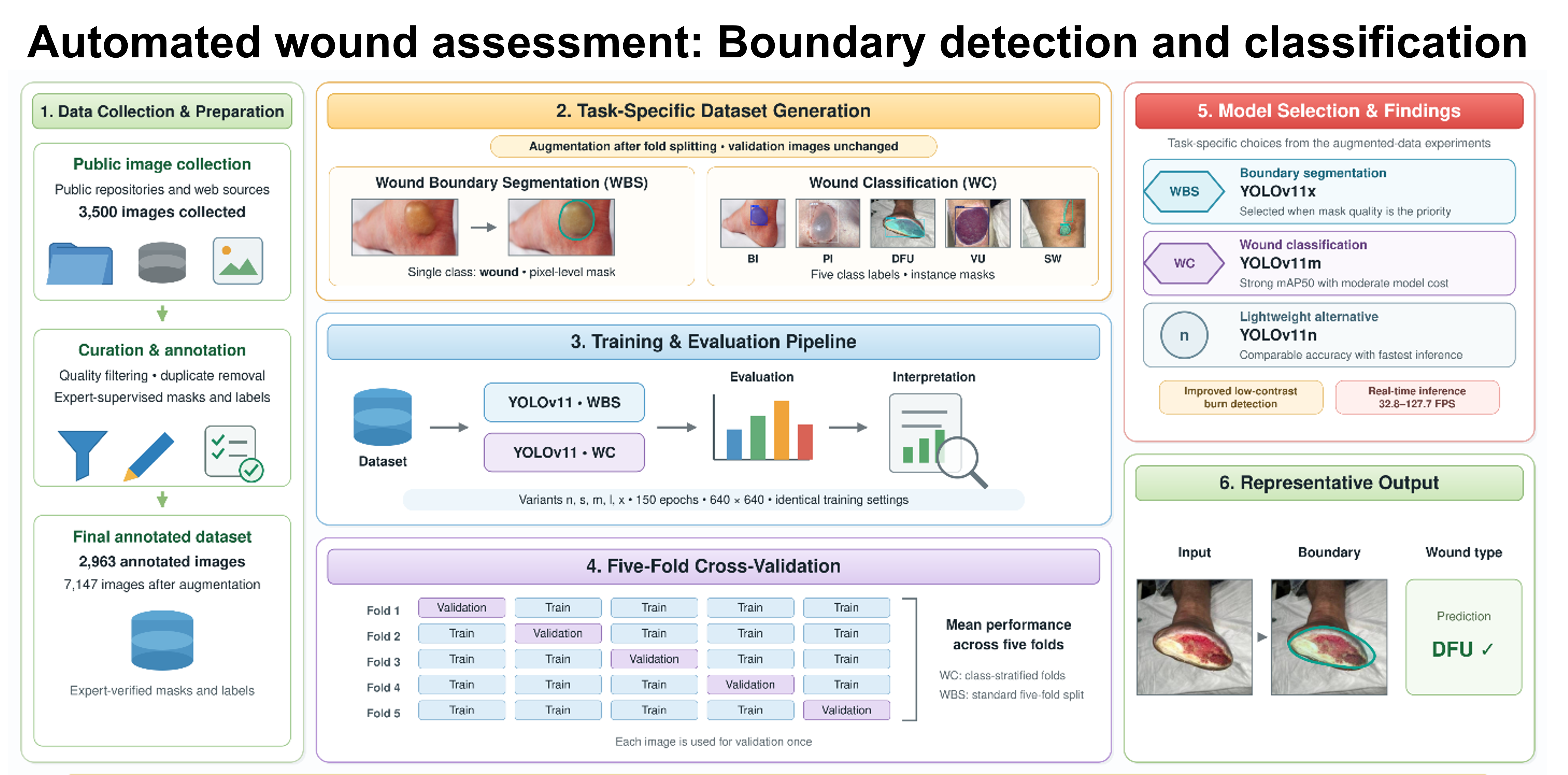
Accurate wound classification (WC) and boundary segmentation are essential for guiding clinical decisions in both chronic and acute wound management. However, most existing artificial intelligence (AI) models are limited, focusing on a narrow set of wound types, limited variations in wound severity, or a single task (segmentation or classification), which reduces their clinical applicability. This study presents two dedicated instance segmentation models based on You Only Look Once (YOLO)v11 that perform wound boundary segmentation (WBS) and WC across five clinically relevant wound types: burn injury (BI), pressure injury, diabetic foot ulcer, vascular ulcer, and surgical wound. A wound-type balanced dataset of 2,963 annotated images was created to train the models for both tasks, using five-fold cross-validation to ensure robust and unbiased evaluation. Models trained on the original, non-augmented dataset achieved consistent performance across folds, though BI detection accuracy was relatively low. Therefore, the dataset was augmented using rotation, flipping, and variations in brightness, saturation, and exposure to help the model learn more generalizable, invariant features. This augmentation significantly improved performance, particularly for visually subtle BI cases. Among the tested variants, YOLOv11x achieved the best WBS performance (F1-score: 0.9341; mean average precision [mAP]50: 0.9629). For WC, YOLOv11m achieved the highest mAP50 (0.9194) and mAP50–95 (0.6950), whereas YOLOv11l achieved the highest F1-score (0.8797). The lightweight YOLOv11n provided comparable accuracy at lower computational cost, making it suitable for resource-constrained deployments. Supported by confusion matrices and visual detection outputs, the results confirm the model’s robustness against complex backgrounds and high intra-class variability, demonstrating the potential of YOLOv11-based architectures for accurate, real-time wound analysis in both clinical and remote care settings.
- Falanga V, Isseroff RR, Soulika AM, et al. Chronic wounds. Nat Rev Dis Primers. 2022;8(1). doi: 10.1038/s41572-022-00377-3
- Frykberg RG, Banks J. Challenges in the Treatment of Chronic Wounds. Adv Wound Care. 2015;4(9):560-582. doi: 10.1089/wound.2015.0635
- Rostami B, Anisuzzaman DM, Wang C, Gopalakrishnan S, Niezgoda J, Yu Z. Multiclass wound image classification using an ensemble deep CNN-based classifier. Comput Biol Med. 2021;134:104536. doi: 10.1016/j.compbiomed.2021.104536
- Nussbaum SR, Carter MJ, Fife CE, et al. An Economic Evaluation of the Impact, Cost, and Medicare Policy Implications of Chronic Nonhealing Wounds. Value in Health. 2018;21(1):27-32. doi: 10.1016/j.jval.2017.07.007
- Yakupu A, Zhang J, Dong W, Song F, Dong J, Lu S. The epidemiological characteristic and trends of burns globally. BMC Public Health. 2022;22(1). doi: 10.1186/s12889-022-13887-2
- Carter JE, Amani H, Carter D, et al. Evaluating Real-World National and Regional Trends in Definitive Closure in U.S. Burn Care: A Survey of U.S. Burn Centers. J Burn Care Res. 2022;43(1):141-148. doi: 10.1093/jbcr/irab151
- Sen CK. Human Wounds and Its Burden: An Updated Compendium of Estimates. Adv Wound Care. 2019;8(2):39-48. doi: 10.1089/wound.2019.0946
- Odame P, Ahiamadzor MM, Derkyi NKB, et al. Multi-Wound Classification: Exploring Image Enhancement and Deep Learning Techniques. Eng Rep. 2025;7(1). doi: 10.1002/eng2.70001
- Alhababi M, Auner G, Malik H, Aljasem M, Aldoulah Z. Unified wound diagnostic framework for wound segmentation and classification. Mach Learn Appl. 2025;19:100616. doi: 10.1016/j.mlwa.2024.100616
- Maurya L, Mirza S. MCTFWC: a multiscale CNN-transformer fusion-based model for wound image classification. Signal Image Video Process. 2025;19(7). doi: 10.1007/s11760-025-04072-0
- Cassidy B, McBride C, Kendrick C, et al. An enhanced harmonic densely connected hybrid transformer network architecture for chronic wound segmentation utilising multi-colour space tensor merging. Comput Biol Med. 2025;192:110172. doi: 10.1016/j.compbiomed.2025.110172
- Aldoulah ZA, Malik H, Molyet R, Aljasem M. SwishRes-U-Net: A deep neural architecture for chronic wound segmentation. Biomed Signal Process Control. 2025;100:107048. doi: 10.1016/j.bspc.2024.107048
- Zahia S, Garcia Zapirain MB, Sevillano X, González A, Kim PJ, Elmaghraby A. Pressure injury image analysis with machine learning techniques: A systematic review on previous and possible future methods. Artif Intell Med. 2020;102:101742. doi: 10.1016/j.artmed.2019.101742
- Liu H, Sun W, Cai W, et al. Current status, challenges, and prospects of artificial intelligence applications in wound repair theranostics. Theranostics. 2025;15(5):1662-1688. doi: 10.7150/thno.105109
- Wu Y, Wu L, Yu M. The clinical value of intelligent wound measurement devices in patients with chronic wounds: A scoping review. Int Wound J. 2024;21(3). doi: 10.1111/iwj.14843
- Jørgensen LB, Sørensen JA, Jemec GBE, Yderstræde KB. Methods to assess area and volume of wounds – a systematic review. Int Wound J. 2016;13(4):540-553. doi: 10.1111/iwj.12472
- Liu TJ, Wang H, Christian M, Chang CW, Lai F, Tai HC. Automatic segmentation and measurement of pressure injuries using deep learning models and a LiDAR camera. Sci Rep. 2023;13(1).680. doi: 10.1038/s41598-022-26812-9
- Rippon MG, Fleming L, Chen T, Rogers AA, Ousey K. Artificial intelligence in wound care: diagnosis, assessment and treatment of hard-to-heal wounds: a narrative review. J Wound Care. 2024;33(4):229-242. doi: 10.12968/jowc.2024.33.4.229
- Anisuzzaman DM, Wang C, Rostami B, Gopalakrishnan S, Niezgoda J, Yu Z. Image-Based Artificial Intelligence in Wound Assessment: A Systematic Review. Adv Wound Care. 2022;11(12):687-709. doi: 10.1089/wound.2021.0091
- Anisuzzaman DM, Patel Y, Niezgoda JA, Gopalakrishnan S, Yu Z. A Mobile App for Wound Localization Using Deep Learning. IEEE Access. 2022;10:61398-61409. doi: 10.1109/ACCESS.2022.3179137
- Wongvibulsin S, Yan MJ, Pahalyants V, Murphy W, Daneshjou R, Rotemberg V. Current State of Dermatology Mobile Applications with Artificial Intelligence Features. JAMA Dermatol. 2024;160(6):646-650. doi: 10.1001/jamadermatol.2024.0468
- Sarp S, Kuzlu M, Pipattanasomporn M, Guler O. Simultaneous wound border segmentation and tissue classification using a conditional generative adversarial network. J Eng. 2021;2021(3):125-134. doi: 10.1049/tje2.12016
- Chang CW, Ho CY, Lai F, et al. Application of multiple deep learning models for automatic burn wound assessment. Burns. 2023;49(5):1039-1051. doi: 10.1016/j.burns.2022.07.006
- Chauhan J, Goyal P. Convolution neural network for effective burn region segmentation of color images. Burns. 2021;47(4):854-862. doi: 10.1016/j.burns.2020.08.016
- Niri R, Zahia S, Stefanelli A, et al. Wound Segmentation with U-Net Using a Dual Attention Mechanism and Transfer Learning. J Imaging Inform Med. 2025;38(5):3351-3365. doi: 10.1007/s10278-025-01386-w
- Shah SMAH, Rizwan A, Atteia G, Alabdulhafith M. CADFU for Dermatologists: A Novel Chronic Wounds & Ulcers Diagnosis System with DHuNeT (Dual-Phase Hyperactive UNet) and YOLOv8 Algorithm. Healthcare. 2023;11(21):2840. doi: 10.3390/healthcare11212840
- Pandey B, Joshi D, Arora AS, Upadhyay N, Chhabra HS. A Deep Learning Approach for Automated Detection and Segmentation of Pressure Ulcers Using Infrared-Based Thermal Imaging. IEEE Sens J. 2022;22(15):14762-14768. doi: 10.1109/JSEN.2022.3184105
- Li F, Zhang H, Xu S, et al. Exploration of machine learning models for surgical incision healing assessment based on thermal imaging: A feasibility study. Int Wound J. 2024;21(2). doi: 10.1111/iwj.14677
- Dhar MK, Wang C, Patel Y, et al. Wound Tissue Segmentation in Diabetic Foot Ulcer Images Using Deep Learning: A Pilot Study. arXiv. Preprint posted online 2024. doi: 10.48550/arXiv.2406.16012
- Scebba G, Zhang J, Catanzaro S, et al. Detect-and-segment: A deep learning approach to automate wound image segmentation. Inform Med Unlocked. 2022;29:100884. doi: 10.1016/j.imu.2022.100884
- Huang PH, Pan YH, Luo YS, et al. Development of a deep learning-based tool to assist wound classification. J Plast Reconstr Aesthetic Surg. 2023;79:89-97. doi: 10.1016/j.bjps.2023.01.030
- Moses O, Qureshi M, King ICC. Letter to the Editor regarding “Development of a deep learning-based tool to assist wound classification.” J Plast Reconstr Aesthet Surg. 2023;87:215-216. doi: 10.1016/j.bjps.2023.10.089
- Yadav DP, Aljrees T, Kumar D, Kumar A, Singh KU, Singh T. Spatial attention-based residual network for human burn identification and classification. Sci Rep. 2023;13(1). doi: 10.1038/s41598-023-39618-0
- Patel Y, Shah T, Dhar MK, et al. Integrated image and location analysis for wound classification: a deep learning approach. Sci Rep. 2024;14(1). doi: 10.1038/s41598-024-56626-w
- Anisuzzaman DM, Patel Y, Rostami B, Niezgoda J, Gopalakrishnan S, Yu Z. Multi-modal wound classification using wound image and location by deep neural network. Sci Rep. 2022;12(1). doi: 10.1038/s41598-022-21813-0
- Huang HN, Zhang T, Yang CT, et al. Image segmentation using transfer learning and Fast R-CNN for diabetic foot wound treatments. Front Public Health. 2022;10. doi: 10.3389/fpubh.2022.969846
- Ay B, Tasar B, Utlu Z, Ay K, Aydin G. Deep transfer learning-based visual classification of pressure injuries stages. Neural Comput Appl. 2022;34(18):16157-16168. doi: 10.1007/s00521-022-07274-6
- Ramachandram D, Ramirez-GarciaLuna JL, Fraser RDJ, Martínez-Jiménez MA, Arriaga-Caballero JE, Allport J. Fully Automated Wound Tissue Segmentation Using Deep Learning on Mobile Devices: Cohort Study. JMIR Mhealth Uhealth. 2022;10(4):e36977. doi: 10.2196/36977
- Tusar MH, Fayyazbakhsh F, Zendehdel N, et al. AI-Powered Image-Based Assessment of Pressure Injuries Using You Only Look Once (YOLO) Version 8 Models. Adv Wound Care. 2025;15(5). doi: 10.1089/wound.2024.0245
- Tusar MH, Fayyazbakhsh F, Leu M. Reducing Cross Class Confusion of Wounds Using a Two-Stage Hierarchical Deep Learning Approach for Pressure Injury Staging. In: BMES Annual Meeting. BMES; 2025. Available from: https://www.researchgate.net/publication/403529186_Reducing_Cross_Class_Confusion_of_Wounds_Using_a_Two-Stage_Hierarchical_Deep_Learning_Approach_for_Pressure_Injury_Staging_Introduction
- Huang ST, Chu YC, Liu LR, et al. Deep Learning-Based Clinical Wound Image Analysis Using a Mask R-CNN Architecture. J Med Biol Eng. 2023;43(4):417-426. doi: 10.1007/s40846-023-00802-2
- Bossuyt PM, Reitsma JB, Bruns DE, et al. STARD 2015: An updated list of essential items for reporting diagnostic accuracy studies. BMJ. 2015;351:h5527. doi: 10.1136/bmj.h5527
- Cohen JF, Korevaar DA, Altman DG, et al. STARD 2015 guidelines for reporting diagnostic accuracy studies: explanation and elaboration. BMJ Open. 2016;6(11):e012799. doi: 10.1136/bmjopen-2016-012799.
- Roboflow, version 1.0 [software]. Roboflow Inc.; 2022. https://roboflow.com
- Ultralytics. YOLOv11 Release Notes. 2024. https://github.com/ultralytics/assets/releases
- Cepeda S, Esteban-Sinovas O, Romero R, et al. Real-time brain tumor detection in intraoperative ultrasound: From model training to deployment in the operating room. Comput Biol Med. 2025;193:110481. doi: 10.1016/j.compbiomed.2025.110481
- Sapkota R, Flores-Calero M, Qureshi R, et al. YOLO advances to its genesis: a decadal and comprehensive review of the You Only Look Once (YOLO) series. Artif Intell Rev. 2025;58(9). doi: 10.1007/s10462-025-11253-3
- Khanam R, Hussain M. YOLOv11: An overview of the key architectural enhancements. arXiv. 2024. doi: 10.48550/arXiv.2410.17725
- Jocher G. YOLOv8 release v8.1.0. 2024. Accessed August 27, 2026. https://github.com/ultralytics/assets/releases.
- Terven J, Córdova-Esparza DM, Romero-González JA. A Comprehensive Review of YOLO Architectures in Computer Vision: From YOLOv1 to YOLOv8 and YOLO-NAS. Mach Learn Knowl Extr. 2023;5(4):1680-1716. doi: 10.3390/make5040083
- He L, Zhou Y, Liu L, Cao W, Ma J. Research on object detection and recognition in remote sensing images based on YOLOv11. Sci Rep. 2025;15(1):14032. doi: 10.1038/s41598-025-96314-x
- Sapkota R, Karkee M. Integrating YOLO11 and convolution block attention module for multi-season segmentation of tree trunks and branches in commercial apple orchards. arXiv. 2024. doi: 10.48550/arXiv.2412.05728
- Jegham N, Koh CY, Abdelatti M, Hendawi A. YOLO evolution: A comprehensive benchmark and architectural review of YOLOv12, YOLO11, and their previous versions. arXiv. 2024. doi: 10.48550/arXiv.2411.00201
- Rasheed AF, Zarkoosh M. YOLOv11 Optimization for Efficient Resource Utilization. arXiv. 2024. doi: 10.48550/arXiv.2412.14790
- Cino L, Distante C, Martella A, Mazzeo PL. Skin Lesion Classification Through Test Time Augmentation and Explainable Artificial Intelligence. J Imaging. 2025;11(1):15. doi: 10.3390/jimaging11010015
- Neuwieser H, Jami NVSJ, Meier RJ, et al. Interpreting Venous and Arterial Ulcer Images Through the Grad-CAM Lens: Insights and Implications in CNN-Based Wound Image Classification. Diagnostics. 2025;15(17):2184. doi: 10.3390/diagnostics15172184
- Lin TY, Maire M, Belongie S, et al. Microsoft COCO: Common Objects in Context. In: Computer Vision - ECCV 2014 (Lecture Notes in Computer Science). Springer International Publishing; 2014:740-755. doi: 10.1007/978-3-319-10602-1_48