

Microfluidic bioprinting of cell-laden gradients in RADA16-I self-assembling peptide hydrogels

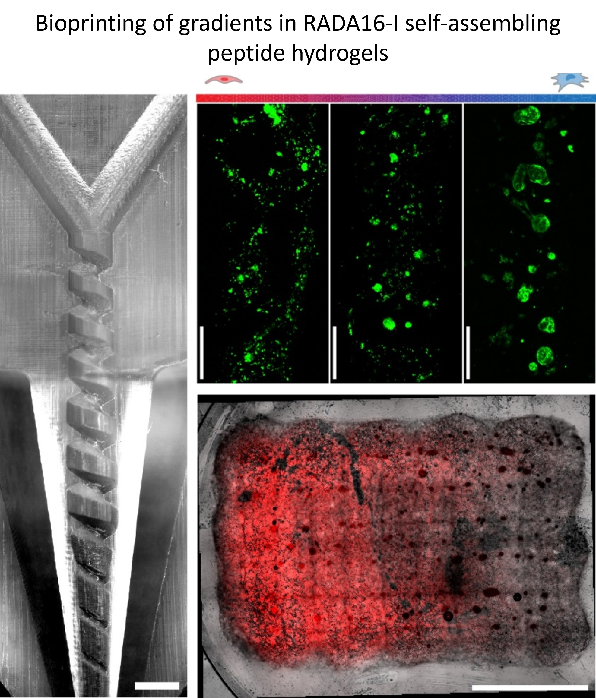
Reproducing the continuous compositional and cellular transitions found in native tissues remains a major challenge in extrusion-based bioprinting, which typically generates constructs composed of discrete regions and imposes stringent rheological requirements on bioinks. Microfluidic bioprinting offers new opportunities to reproduce tissue heterogeneity by enabling controlled mixing of biomaterials and cell populations during extrusion. In this study, we present a coaxial microfluidic bioprinting strategy for fabricating hydrogel scaffolds with continuous cellular gradients using low-viscosity self-assembling peptide bioinks. The system combines three independently controlled syringe pumps with a 3D-printed coaxial nozzle containing a screw-like passive mixer. Two low-viscosity RADA16-I peptide solutions containing different cell populations are mixed in situ, while a methylcellulose–alginate shell stabilizes the filament during printing and supports post-printing self-assembly of the core hydrogel. Computational simulations confirm efficient mixing within the nozzle, and fluorescence imaging demonstrates smooth compositional transitions along printed filaments. The system enables the fabrication of scaffolds containing co-culture gradients of endothelial cells and mesenchymal stem cells, which remain viable and display cell-type-specific organization. Overall, this approach enables the printing of soft peptide hydrogels and the fabrication of biomimetic constructs with continuous cellular transitions, highlighting its potential for tissue engineering and regenerative medicine.
- Mota C, Camarero-Espinosa S, Baker MB, Wieringa P, Moroni L. Bioprinting: From Tissue and Organ Development to in Vitro Models. Chem Rev. 2020;120(19):10547-10607. doi: 10.1021/acs.chemrev.9b00789
- Dai W, Wu T, Leng X, et al. Advances in biomechanical and biochemical engineering methods to stimulate meniscus tissue. Am J Transl Res. 2021;13(8):8540-8560.
- Mukund K, Subramaniam S. Skeletal muscle: A review of molecular structure and function, in health and disease. WIREs Mech Dis. 2019;12(1). doi: 10.1002/wsbm.1462
- Santos-Beato P, Midha S, Pitsillides AA, Miller A, Torii R, Kalaskar DM. Biofabrication of the osteochondral unit and its applications: Current and future directions for 3D bioprinting. J Tissue Eng. 2022;13. doi: 10.1177/20417314221133480
- Hama R, Reinhardt JW, Ulziibayar A, Watanabe T, Kelly J, Shinoka T. Recent Tissue Engineering Approaches to Mimicking the Extracellular Matrix Structure for Skin Regeneration. Biomimetics. 2023;8(1):130. doi: 10.3390/biomimetics8010130
- Fang L, Lin X, Xu R, et al. Advances in the Development of Gradient Scaffolds Made of Nano-Micromaterials for Musculoskeletal Tissue Regeneration. Nano-Micro Lett. 2024;17(1). doi: 10.1007/s40820-024-01581-4
- Niu X, Li N, Du Z, Li X. Integrated gradient tissue-engineered osteochondral scaffolds: Challenges, current efforts and future perspectives. Bioact Mater. 2023;20:574- 597. doi: 10.1016/j.bioactmat.2022.06.011
- Murphy SV, Atala A. 3D bioprinting of tissues and organs. Nat Biotechnol. 2014;32(8):773-785. doi: 10.1038/nbt.2958
- Zhang YS, Haghiashtiani G, Hübscher T, et al. 3D extrusion bioprinting. Nat Rev Methods Primers. 2021;1(1). doi: 10.1038/s43586-021-00073-8
- Jergitsch M, Mateos-Timoneda MA. 3D extrusion bioprinting: rational bioink design and advanced fabrication techniques. Trends Biotechnol. 2026;44(2):378-388. doi: 10.1016/j.tibtech.2025.06.008
- Cui X, Li J, Hartanto Y, et al. Advances in Extrusion 3D Bioprinting: A Focus on Multicomponent Hydrogel-Based Bioinks. Adv Healthc Mater. 2020;9(15). doi: 10.1002/adhm.201901648
- Levato R, Visser J, Planell JA, Engel E, Malda J, Mateos- Timoneda MA. Biofabrication of tissue constructs by 3D bioprinting of cell-laden microcarriers. Biofabrication. 2014;6(3):035020. doi: 10.1088/1758-5082/6/3/035020
- Kilian D, Ahlfeld T, Akkineni AR, Bernhardt A, Gelinsky M, Lode A. 3D Bioprinting of osteochondral tissue substitutes – in vitro-chondrogenesis in multi-layered mineralized constructs. Sci Rep. 2020;10(1). doi: 10.1038/s41598-020-65050-9
- Bittner SM, Smith BT, Diaz-Gomez L, et al. Fabrication and mechanical characterization of 3D printed vertical uniform and gradient scaffolds for bone and osteochondral tissue engineering. Acta Biomater. 2019;90:37-48. doi: 10.1016/j.actbio.2019.03.041
- Malda J, Visser J, Melchels FP, et al. 25th Anniversary Article: Engineering Hydrogels for Biofabrication. Adv Mater. 2013;25(36):5011-5028. doi: 10.1002/adma.201302042
- Ouyang L. Pushing the rheological and mechanical boundaries of extrusion-based 3D bioprinting. Trends Biotechnol. 2022;40(7):891-902. doi: 10.1016/j.tibtech.2022.01.001
- Colosi C, Shin SR, Manoharan V, et al. Microfluidic Bioprinting of Heterogeneous 3D Tissue Constructs Using Low-Viscosity Bioink. Adv Mater. 2015;28(4):677-684. doi: 10.1002/adma.201503310
- Susapto HH, Alhattab D, Abdelrahman S, et al. Ultrashort Peptide Bioinks Support Automated Printing of Large-Scale Constructs Assuring Long-Term Survival of Printed Tissue Constructs. Nano Lett. 2021;21(7):2719-2729. doi: 10.1021/acs.nanolett.0c04426
- Idaszek J, Costantini M, Karlsen TA, et al. 3D bioprinting of hydrogel constructs with cell and material gradients for the regeneration of full-thickness chondral defect using a microfluidic printing head. Biofabrication. 2019;11(4):044101. doi: 10.1088/1758-5090/ab2622
- Ravanbakhsh H, Karamzadeh V, Bao G, Mongeau L, Juncker D, Zhang YS. Emerging Technologies in Multi-Material Bioprinting. Adv Mater. 2021;33(49). doi: 10.1002/adma.202104730
- Jergitsch M, Soiunov R, Selinger F, et al. Fabrication and validation of an affordable DIY coaxial 3D extrusion bioprinter. Sci Rep. 2025;15(1). doi: 10.1038/s41598-025-06478-9
- Liu W, Zhong Z, Hu N, et al. Coaxial extrusion bioprinting of 3D microfibrous constructs with cell-favorable gelatin methacryloyl microenvironments. Biofabrication. 2018;10(2):024102. doi: 10.1088/1758-5090/aa9d44
- Jergitsch M, Perez-Amodio S, Delgado LM, Perez RA, Mateos-Timoneda MA. 3D coaxial bioprinting of RADA16-I self-assembling peptide hydrogel. Mater Today Bio. 2026;37:102900. doi: 10.1016/j.mtbio.2026.102900
- Schneider CA, Rasband WS, Eliceiri KW. NIH Image to ImageJ: 25 years of image analysis. Nat Methods. 2012;9(7):671-675. doi: 10.1038/nmeth.2089
- Stöber W, Fink A, Bohn E. Controlled growth of monodisperse silica spheres in the micron size range. J Colloid Interface Sci. 1968;26(1):62-69. doi: 10.1016/0021-9797(68)90272-5
- Dani F, Ahlfeld T, Albrecht F, et al. Homogeneous and Reproducible Mixing of Highly Viscous Biomaterial Inks and Cell Suspensions to Create Bioinks. Gels. 2021;7(4):227. doi: 10.3390/gels7040227
- Wang M, Li W, Mille LS, et al. Digital Light Processing Based Bioprinting with Composable Gradients. Adv Mater. 2021;34(1). doi: 10.1002/adma.202107038
- Shafiee S, Shariatzadeh S, Zafari A, Majd A, Niknejad H. Recent Advances on Cell-Based Co-Culture Strategies for Prevascularization in Tissue Engineering. Front Bioeng Biotechnol. 2021;9. doi: 10.3389/fbioe.2021.745314
- Mazloomnejad R, Babajani A, Kasravi M, et al. Angiogenesis and Re-endothelialization in decellularized scaffolds: Recent advances and current challenges in tissue engineering. Front Bioeng Biotechnol. 2023;11. doi: 10.3389/fbioe.2023.1103727
- Ouyang L, Yao R, Zhao Y, Sun W. Effect of bioink properties on printability and cell viability for 3D bioplotting of embryonic stem cells. Biofabrication. 2016;8(3):035020. doi: 10.1088/1758-5090/8/3/035020
- Ahlfeld T, Guduric V, Duin S, et al. Methylcellulose – a versatile printing material that enables biofabrication of tissue equivalents with high shape fidelity. Biomater Sci. 2020;8(8):2102-2110. doi: 10.1039/d0bm00027b
- Gonzalez La Corte S, Stevens CA, Cárcamo-Oyarce G, Ribbeck K, Wingreen NS, Datta SS. Morphogenesis of bacterial cables in polymeric environments. Sci Adv. 2025;11(3). doi: 10.1126/sciadv.adq7797