

Advances in three-dimensional bioprinted tumor organoids: From model construction to clinical translation

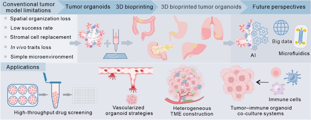
Traditional cancer research models face inherent limitations, as two-dimensional cell cultures fail to capture the complexity of tumor biology, while animal models are confounded by species-specific discrepancies. The integration of tumor organoids with three-dimensional (3D) bioprinting has recently emerged as a transformative strategy. This approach combines the histological and genetic fidelity of organoids with the spatial precision and structural controllability of 3D bioprinting, thereby enabling the fabrication of biomimetic tumor models. Such models more faithfully recapitulate critical features of the tumor microenvironment (TME), addressing major gaps in conventional experimental systems. This review systematically examines the principles, recent advances, and translational applications of 3D bioprinting-enabled tumor organoids, including the biological basis of organoids, key bioprinting strategies, and technical considerations. Major applications include constructing heterogeneous TMEs with immune interactions, engineering vascularized tumor structures, enabling high-throughput drug screening, validating bioprinted organoids using clinical samples, and advancing clinical translation, regulatory frameworks, and Good Manufacturing Practice-compliant manufacturing of tumor organoids. Despite substantial progress, several challenges remain, including limited printing resolution, bioink instability, difficulties in sustaining long-term cultures, and gaps in standardization. Nevertheless, integration with emerging technologies, such as microfluidics, artificial intelligence, big data analytics, and standardized biomanufacturing platforms, is anticipated to bridge the gap between basic tumor research and clinical translation. Ultimately, these synergistic advances may accelerate the development of personalized cancer therapies and improve patient outcomes.
- Kvåle G, Heuch I, Nilssen S. Parity in relation to mortality and cancer incidence: a prospective study of Norwegian women. Int J Epidemiol. 1994;23(4):691-699. doi: 10.1093/ije/23.4.691.
- Martin DB, Nelson PS. From genomics to proteomics: techniques and applications in cancer research. Trends Cell Biol. 2001;11(11):S60-S65. doi: 10.1016/s0962-8924(01)02123-7.
- Tang XY, Wu S, Wang D, et al. Human organoids in basic research and clinical applications. Signal Transduct Target Ther. 2022;7(1):168. doi: 10.1038/s41392-022-01024-9.
- Wang G, Mao X, Wang W, Wang X, Li S, Wang Z. Bioprinted research models of urological malignancy. Exploration. 2024;4(4):20230126.doi: 10.1002/EXP.20230126.
- Hwangbo H, Chae S, Kim W, Jo S, Kim GH. Tumor-on-a-chip models combined with mini-tissues or organoids for engineering tumor tissues. Theranostics. 2024;14(1):33-55. doi: 10.7150/thno.90093.
- Zhou Z, Pang Y, Ji J, et al. Harnessing 3D in vitro systems to model immune responses to solid tumours: a step towards improving and creating personalized immunotherapies. Nat Rev Immunol. 2024;24(1):18-32. doi: 10.1038/s41577-023-00896-4.
- Lv J, Du X, Wang M, Su J, Wei Y, Xu C. Construction of tumor organoids and their application to cancer research and therapy. Theranostics. 2024;14(3):1101-1125. doi: 10.7150/thno.91362.
- Ma W, Lu H, Xiao Y, Wu C. Advancing organoid development with 3D bioprinting. Organoid Res. 2025;1(1):025040004. doi: 10.36922/or025040004.
- Xu X, Li H, Chen J, et al. A universal strategy to construct high-performance homo- and heterogeneous microgel assembly bioinks. Small Methods. 2024;8(12):e2400223. doi: 10.1002/smtd.202400223.
- Wang X, Luo Y, Ma Y, Wang P, Yao R. Converging bioprinting and organoids to better recapitulate the tumor microenvironment. Trends Biotechnol. 2024;42(5):648-663. doi: 10.1016/j.tibtech.2023.11.006.
- An Y, He Y, Ge N, Guo J, Yang F, Sun S. Organoids to remodel SARS-CoV-2 research: updates, limitations and perspectives. Aging Dis. 2023;14(5):1677-1699. doi: 10.14336/ad.2023.0209.
- Sato T, Vries RG, Snippert HJ, et al. Single Lgr5 stem cells build crypt-villus structures in vitro without a mesenchymal niche. Nature. 2009;459(7244):262-265. doi: 10.1038/nature07935.
- Sachs N, de Ligt J, Kopper O, et al. A living biobank of breast cancer organoids captures disease heterogeneity. Cell. 2018;172(1-2):373-386.e310. doi: 10.1016/j.cell.2017.11.010.
- Drost J, van Jaarsveld RH, Ponsioen B, et al. Sequential cancer mutations in cultured human intestinal stem cells. Nature. 2015;521(7550):43-47. doi: 10.1038/nature14415.
- Viegas J, Sarmento B. Bridging the gap between testing and clinics exploring alternative pre-clinical models in melanoma research. Adv Drug Deliv Rev. 2024;208:115295. doi: 10.1016/j.addr.2024.115295.
- Mierke CT. Bioprinting of cells, organoids and organs-on-a-chip together with hydrogels improves structural and mechanical cues. Cells. 2024;13(19):1638. doi: 10.3390/cells13191638.
- Blandino G, Satchi-Fainaro R, Tinhofer I, et al. Cancer organoids as reliable disease models to drive clinical development of novel therapies. J Exp Clin Cancer Res. 2024;43(1):334. doi: 10.1186/s13046-024-03258-7.
- Fredrikson JP, Roth DM, Cosgrove JA, et al. Engineering neuronal networks in granular microgels to innervate bioprinted cancer organoids on-a-chip. Lab Chip. 2025;25(14):3467-3481. doi: 10.1039/d5lc00134j.
- Gu WJ, Liu XX, Shen YW, et al. TRIM4 enhances small-molecule-induced neddylated-degradation of CORO1A for triple negative breast cancer therapy. Theranostics. 2024;14(18):7023-7041. doi: 10.7150/thno.97662.
- Barretina J, Caponigro G, Stransky N, et al. The Cancer Cell Line Encyclopedia enables predictive modelling of anticancer drug sensitivity. Nature. 2012;483(7391):603-607. doi: 10.1038/nature11003.
- Dietlein F, Weghorn D, Taylor-Weiner A, et al. Identification of cancer driver genes based on nucleotide context. Nat Genet. 2020;52(2):208-218. doi: 10.1038/s41588-019-0572-y.
- Yu M, Ni M, Xu F, et al. NSUN6-mediated 5-methylcytosine modification of NDRG1 mRNA promotes radioresistance in cervical cancer. Mol Cancer. 2024;23(1):139. doi: 10.1186/s12943-024-02055-2.
- Lencioni G, Gregori A, Toledo B, et al. Unravelling the complexities of resistance mechanism in pancreatic cancer: insights from in vitro and ex-vivo model systems. Semin Cancer Biol. 2024;106-107:217-233. doi: 10.1016/j.semcancer.2024.09.002.
- Liu Y, Wu W, Cai C, et al. Patient-derived xenograft models in cancer therapy: technologies and applications. Signal Transduct Target Ther. 2023;8(1):160. doi: 10.1038/s41392-023-01419-2.
- Dogan E, Galifi CA, Cecen B, Shukla R, Wood TL, Miri AK. Extracellular matrix regulation of cell spheroid invasion in a 3D bioprinted solid tumor-on-a-chip. Acta Biomater. 2024;186:156-166. doi: 10.1016/j.actbio.2024.07.040.
- Kuwata T, Yanagihara K, Iino Y, et al. Establishment of novel gastric cancer patient-derived xenografts and cell lines: pathological comparison between primary tumor, patient-derived, and cell-line derived xenografts. Cells. 2019;8(6):585. doi: 10.3390/cells8060585.
- Lang Y, Lyu Y, Tan Y, Hu Z. Progress in construction of mouse models to investigate the pathogenesis and immune therapy of human hematological malignancy. Front Immunol. 2023;14:1195194. doi: 10.3389/fimmu.2023.1195194.
- Lallo A, Schenk MW, Frese KK, Blackhall F, Dive C. Circulating tumor cells and CDX models as a tool for preclinical drug development. Transl Lung Cancer Res. 2017;6(4):397-408. doi: 10.21037/tlcr.2017.08.01.
- Lancaster MA, Knoblich JA. Organogenesis in a dish: modeling development and disease using organoid technologies. Science. 2014;345(6194):1247125. doi: 10.1126/science.1247125.
- Huang MS, Christakopoulos F, Roth JG, Heilshorn SC. Organoid bioprinting: from cells to functional tissues. Nat Rev Bioeng. 2024;3(2):126-142. doi: 10.1038/s44222-024-00268-0.
- D’Antoni C, Mautone L, Sanchini C, et al. Unlocking neural function with 3D in vitro models: a technical review of self-assembled, guided, and bioprinted brain organoids and their applications in the study of neurodevelopmental and neurodegenerative disorders. Int J Mol Sci. 2023;24(13):10762. doi: 10.3390/ijms241310762.
- Puls TJ, Tan X, Husain M, Whittington CF, Fishel ML, Voytik-Harbin SL. Development of a novel 3D tumor-tissue invasion model for high-throughput, high-content phenotypic drug screening. Sci Rep. 2018;8(1):13039. doi: 10.1038/s41598-018-31138-6.
- Cao R, Li NT, Shing CB, Kutulakos Z, Tan CM, McGuigan AP. Reproducible manufacturing of SPOT as a high-throughput scaffold-based culture platform. J Vis Exp. 2025;221:e68405. doi: 10.3791/68405.
- Maloney E, Clark C, Sivakumar H, et al. Immersion bioprinting of tumor organoids in multi-well plates for increasing chemotherapy screening throughput. Micromachines. 2020;11(2):208. doi: 10.3390/mi11020208.
- Yang Q, Li M, Yang X, et al. Flourishing tumor organoids: history, emerging technology, and application. Bioeng Transl Med. 2023;8(5):e10559. doi: 10.1002/btm2.10559.
- van de Wetering M, Francies HE, Francis JM, et al. Prospective derivation of a living organoid biobank of colorectal cancer patients. Cell. 2015;161(4):933-945. doi: 10.1016/j.cell.2015.03.053.
- Vlachogiannis G, Hedayat S, Vatsiou A, et al. Patient-derived organoids model treatment response of metastatic gastrointestinal cancers. Science. 2018;359(6378):920-926. doi: 10.1126/science.aao2774.
- Xiang D, He A, Zhou R, et al. Building consensus on the application of organoid-based drug sensitivity testing in cancer precision medicine and drug development. Theranostics. 2024;14(8):3300-3316. doi: 10.7150/thno.96027.
- Xu X, Lv T, Yao Y, et al. Comprehensive dissection of rectal cancer organoids in responses to chemoradiation. Cell Rep Med. 2025;6(10):102397. doi: 10.1016/j.xcrm.2025.102397.
- Zhou Z, He J, Pang Y, Sun W. Reconstruction of tumor microenvironment via in vitro three-dimensional models. Biofabrication. 2023;15(3):032002. doi: 10.1088/1758-5090/acd1b8.
- Liu Q, Mille LS, Villalobos C, et al. 3D-bioprinted cholangiocarcinoma-on-a-chip model for evaluating drug responses. Bio-des Manuf. 2023;6(4):373-389. doi: 10.1007/s42242-022-00229-9.
- Yang J, Chen Z, Gao C, et al. A mechanical-assisted post-bioprinting strategy for challenging bone defects repair. Nat Commun. 2024;15(1):3565. doi: 10.1038/s41467-024-48023-8.
- Latimer JM, Maekawa S, Yao Y, Wu DT, Chen M, Giannobile WV. Regenerative medicine technologies to treat dental, oral, and craniofacial defects. Front Bioeng Biotechnol. 2021;9:704048. doi: 10.3389/fbioe.2021.704048.
- Cui X, Jiao J, Yang L, et al. Advanced tumor organoid bioprinting strategy for oncology research. Mater Today Bio. 2024;28:101198. doi: 10.1016/j.mtbio.2024.101198.
- Shi W, Mirza S, Kuss M, et al. Embedded bioprinting of breast tumor cells and organoids using low-concentration collagen-based bioinks. Adv Healthc Mater. 2023;12(26):e2300905. doi: 10.1002/adhm.202300905.
- Murphy JF, Lavelle M, Asciak L, et al. Biofabrication and biomanufacturing in Ireland and the UK. Biodes Manuf. 2024;7(6):825-856. doi: 10.1007/s42242-024-00316-z.
- Budharaju H, Sundaramurthi D, Sethuraman S. Embedded 3D bioprinting – an emerging strategy to fabricate biomimetic & large vascularized tissue constructs. Bioact Mater. 2024;32:356-384. doi: 10.1016/j.bioactmat.2023.10.012.
- Zhang X, Zhang X, Li Y, Zhang Y. Applications of light-based 3D bioprinting and photoactive biomaterials for tissue engineering. Materials. 2023;16(23):7461. doi: 10.3390/ma16237461.
- Zhao Y, Li Y, Mao S, Sun W, Yao R. The influence of printing parameters on cell survival rate and printability in microextrusion-based 3D cell printing technology. Biofabrication. 2015;7(4):045002. doi: 10.1088/1758-5090/7/4/045002.
- Blaeser A, Duarte Campos DF, Puster U, Richtering W, Stevens MM, Fischer H. Controlling shear stress in 3D bioprinting is a key factor to balance printing resolution and stem cell integrity. Adv Healthc Mater. 2016;5(3):326-333. doi: 10.1002/adhm.201500677.
- You S, Xiang Y, Hwang HH, et al. High cell density and high-resolution 3D bioprinting for fabricating vascularized tissues. Sci Adv. 2023;9(8):eade7923. doi: 10.1126/sciadv.ade7923.
- Utama RH, Atapattu L, O’Mahony AP, et al. A 3D bioprinter specifically designed for the high-throughput production of matrix-embedded multicellular spheroids. iScience. 2020;23(10):101621. doi: 10.1016/j.isci.2020.101621.
- Xu T, Jin J, Gregory C, Hickman JJ, Boland T. Inkjet printing of viable mammalian cells. Biomaterials. 2005;26(1):93-99. doi: 10.1016/j.biomaterials.2004.04.011.
- Mandrycky C, Wang Z, Kim K, Kim DH. 3D bioprinting for engineering complex tissues. Biotechnol Adv. 2016;34(4):422-434. doi: 10.1016/j.biotechadv.2015.12.011.
- Parodi I, Di Lisa D, Pastorino L, Scaglione S, Fato MM. 3D bioprinting as a powerful technique for recreating the tumor microenvironment. Gels. 2023;9(6):482. doi: 10.3390/gels9060482.
- Ma X, Qu X, Zhu W, et al. Deterministically patterned biomimetic human iPSC-derived hepatic model via rapid 3D bioprinting. Proc Natl Acad Sci U S A. 2016;113(8): 2206-2211. doi: 10.1073/pnas.1524510113.
- Melchels FP, Feijen J, Grijpma DW. A review on stereolithography and its applications in biomedical engineering. Biomaterials. 2010;31(24):6121-6130. doi: 10.1016/j.biomaterials.2010.04.050.
- Huang S, Wu Y, Zhao H, et al. Advancements in bone organoids: perspectives on construction methodologies and application strategies. J Adv Res. 2025;S2090-1232(25):00397-2. doi: 10.1016/j.jare.2025.06.011.
- Burns N, Rajesh A, Manjula-Basavanna A, Duraj-Thatte A. 3D extrusion bioprinting of microbial inks for biomedical applications. Adv Drug Deliv Rev. 2025;217:115505. doi: 10.1016/j.addr.2024.115505.
- Lee Y, Min J, Kim S, Park W, Ko J, Jeon NL. Recapitulating the cancer-immunity cycle on a chip. Adv Healthc Mater. 2025;14(1):e2401927. doi: 10.1002/adhm.202401927.
- Yang Q, Li M, Xiao Z, Feng Y, Lei L, Li S. A new perspective on precision medicine: the power of digital organoids. Biomater Res. 2025;29:0171. doi: 10.34133/bmr.0171.
- Daskalakis P, Kanakousaki E, Ntoulias C, et al. 3D bioprinting with high-viscosity bioinks: a custom-designed extrusion head for high-resolution cellulose acetate scaffolds. Int J Bioprinting. 2025;11(3):337-357. doi: 10.36922/ijb025060047.
- Barjuei ES, Shin J, Kim K, Lee J. Precision improvement of robotic bioprinting via vision-based tool path compensation. Sci Rep. 2024;14(1):17764. doi: 10.1038/s41598-024-68597-z.
- Cui X, Boland T, D’Lima DD, Lotz MK. Thermal inkjet printing in tissue engineering and regenerative medicine. Recent Pat Drug Deliv Formul. 2012;6(2):149-155. doi: 10.2174/187221112800672949.
- Ng WL, Yeong WY, Naing MW. Polyvinylpyrrolidone-based bio-ink improves cell viability and homogeneity during drop-on-demand printing. Materials. 2017;10(2):190. doi: 10.3390/ma10020190.
- Stepanov I, Gottshall NR, Ahmadianyazdi A, et al. Low-cost robotic manipulation of live microtissues for cancer drug testing. Sci Adv. 2025;11(20):eads1631. doi: 10.1126/sciadv.ads1631.
- Lee SY, Phuc HD, Um SH, Mongrain R, Yoon J-K, Bhang SH. Photocuring 3D printing technology as an advanced tool for promoting angiogenesis in hypoxia-related diseases. J Tissue Eng. 2024;15:1-21. doi: 10.1177/20417314241282476.
- Li Z, Chen L, Wu J, et al. A review of 3D bioprinting for organoids. Med Rev. 2025;5(4):318-338. doi: 10.1515/mr-2024-0089.
- Chansoria P, Rizzo R, Rütsche D Liu H, Delrot P, Zenobi- Wong M. Light from afield: fast, high-resolution, and layer-free deep vat 3D printing. Chem Rev. 2024;124(14): 8787-8822. doi: 10.1021/acs.chemrev.4c00134.
- Sabnis A, Rahimi M, Chapman C, Nguyen KT. Cytocompatibility studies of an in situ photopolymerized thermoresponsive hydrogel nanoparticle system using human aortic smooth muscle cells. J Biomed Mater Res A. 2009;91(1):52-59. doi: 10.1002/jbm.a.32194.
- Bhusal A, Dogan E, Nguyen HA, et al. Multi-material digital light processing bioprinting of hydrogel-based microfluidic chips. Biofabrication. 2021;14(1):014103. doi: 10.1088/1758-5090/ac2d78.
- Son J, Li S, Jeong W. Bioprinting vascularized constructs for clinical relevance: engineering hydrogel systems for biological maturity. Gels. 2025;11(8):636. doi: 10.3390/gels11080636.
- Paulsen SJ, Mitcham TM, Pan CS, et al. Projection-based stereolithography for direct 3D printing of heterogeneous ultrasound phantoms. PLoS One. 2021;16(12):e0260737. doi: 10.1371/journal.pone.0260737.
- Kim GT, Go HB, Yu JH, et al. Cytotoxicity, colour stability and dimensional accuracy of 3D printing resin with three different photoinitiators. Polymers. 2022;14(5):979. doi: 10.3390/polym14050979.
- Banigo AT, Nauta L, Zoetebier B, Karperien M. Hydrogel-based bioinks for coaxial and triaxial bioprinting: a review of material properties, printing techniques, and applications. Polymers. 2025;17(7):917. doi: 10.3390/polym17070917.
- Bhatt SS, Krishna Kumar J, Laya S, Thakur G, Nune M. Scaffold-mediated liver regeneration: a comprehensive exploration of current advances. J Tissue Eng. 2024;15:1-31. doi: 10.1177/20417314241286092.
- Han J, Jeong HJ, Choi J, et al. Bioprinted patient-derived organoid arrays capture intrinsic and extrinsic tumor features for advanced personalized medicine. Adv Sci. 2025;12(20):e2407871. doi: 10.1002/advs.202407871.
- Schütte M, Risch T, Abdavi-Azar N, et al. Molecular dissection of colorectal cancer in pre-clinical models identifies biomarkers predicting sensitivity to EGFR inhibitors. Nat Commun. 2017;8:14262. doi: 10.1038/ncomms14262.
- Wierzbicka A, Bartniak M, Waśko J, Kolesińska B, Grabarczyk J, Bociaga D. The impact of gelatin and fish collagen on alginate hydrogel properties: a comparative study. Gels. 2024;10(8):491. doi: 10.3390/gels10080491.
- Yuan W, Sun Y, Du B, et al. Double network hydrogel based on curdlan and gelatin methacryloyl (GelMA) with enhanced mechanical and tissue adhesive properties. nt J Biol Macromol. 2025;320(Pt 3):146080. doi: 10.1016/j.ijbiomac.2025.146080.
- Tanadchangsaeng N, Pasanaphong K, Tawonsawatruk T, et al. 3D bioprinting of fish skin-based gelatin methacryloyl (GelMA) bio-ink for use as a potential skin substitute. Sci Rep. 2024;14(1):23240. doi: 10.1038/s41598-024-73774-1.
- Janmohammadi M, Nazemi Z, Salehi AOM, et al. Cellulose-based composite scaffolds for bone tissue engineering and localized drug delivery. Bioact Mater. 2023;20:137-163. doi: 10.1016/j.bioactmat.2022.05.018.
- Paneer Selvam S, Ayyappan S, I Jamir S, Sellappan LK, Manoharan S. Recent advancements of hydroxyapatite and polyethylene glycol (PEG) composites for tissue engineering applications – a comprehensive review. Eur Polym J. 2024;215:113226. doi: 10.1016/j.eurpolymj.2024.113226.
- Wang M, Lin S, Liu M, et al. An injectable and rapidly degraded carboxymethyl chitosan/polyethylene glycol hydrogel for postoperative antiadhesion. Chem Eng J. 2023;463:142283. doi: 10.1016/j.cej.2023.142283.
- Li X, Ren J, Huang Y, Cheng L, Gu Z. Applications and recent advances in 3D bioprinting sustainable scaffolding techniques. Molecules. 2025;30(14):3027. doi: 10.3390/molecules30143027.
- Perry AC, Lan X, Ma Z, et al. Gelatin methacryloyl bioinks for bioprinting nasal cartilage: Balancing mechanical integrity and extracellular matrix formation. Int J Biol Macromol. 2025;311(Pt 1):143559. doi: 10.1016/j.ijbiomac.2025.143559.
- Yue K, Trujillo-de Santiago G, Alvarez MM, Tamayol A, Annabi N, Khademhosseini A. Synthesis, properties, and biomedical applications of gelatin methacryloyl (GelMA) hydrogels. Biomaterials. 2015;73:254-271. doi: 10.1016/j.biomaterials.2015.08.045.
- Urciuolo A, Giobbe GG, Dong Y, et al. Hydrogel-in-hydrogel live bioprinting for guidance and control of organoids and organotypic cultures. Nat Commun. 2023;14(1):3128. doi: 10.1038/s41467-023-37953-4.
- Korkeamäki JT, Rashad A, Ojansivu M, et al. Systematic development and bioprinting of novel nanostructured multi-material bioinks for bone tissue engineering. Biofabrication. 2025;17(2):025005. doi: 10.1088/1758-5090/ada63b.
- Ravanbakhsh H, Karamzadeh V, Bao G, Mongeau L, Juncker D, Zhang YS. Emerging technologies in multi-material bioprinting. Adv Mater. 2021;33(49):e2104730. doi: 10.1002/adma.202104730.
- Loebel C, Rodell CB, Chen MH, Burdick JA. Shear-thinning and self-healing hydrogels as injectable therapeutics and for 3D-printing. Nat Protoc. 2017;12(8):1521-1541. doi: 10.1038/nprot.2017.053.
- Kirillova A, Maxson R, Stoychev G, Gomillion CT, Ionov L. 4D biofabrication using shape-morphing hydrogels. Adv Mater. 2017;29(46):1703443. doi: 10.1002/adma.201703443.
- Cao P, Yang J, Gong J, et al. 4D printing of bilayer tubular structure with dual-stimuli responsive based on self-rolling behavior. J Appl Polym Sci. 2023;140(1):e53241. doi: 10.1002/app.53241.
- Jia Q, Wang A, Yuan Y, Zhu B, Long H. Heterogeneity of the tumor immune microenvironment and its clinical relevance. Exp Hematol Oncol. 2022;11(1):24. doi: 10.1186/s40164-022-00277-y.
- Meng F, Meyer CM, Joung D, Vallera DA, McAlpine MC, Panoskaltsis-Mortari A. 3D bioprinted in vitro metastatic models via reconstruction of tumor microenvironments. Adv Mater. 2019;31(10):e1806899. doi: 10.1002/adma.201806899.
- Yang H, Sun L, Liu M, Mao Y. Patient-derived organoids: a promising model for personalized cancer treatment. Gastroenterol Rep. 2018;6(4):243-245. doi: 10.1093/gastro/goy040.
- Alsaed B, Lin L, Son J, et al. Intratumor heterogeneity of EGFR expression mediates targeted therapy resistance and formation of drug tolerant microenvironment. Nat Commun. 2025;16(1):28. doi: 10.1038/s41467-024-55378-5.
- Wang J, Tao X, Zhu J, et al. Tumor organoid-immune co-culture models: exploring a new perspective of tumor immunity. Cell Death Discov. 2025;11(1):195. doi: 10.1038/s41420-025-02407-x.
- Zhang L, Liao W, Chen S, et al. Towards a new 3Rs era in the construction of 3D cell culture models simulating tumor microenvironment. Front Oncol. 2023;13:1146477. doi: 10.3389/fonc.2023.1146477.
- Miao Y, Pek NM, Tan C, et al. Co-development of mesoderm and endoderm enables organotypic vascularization in lung and gut organoids. Cell. 2025;188(16):4295-4313. doi: 10.1016/j.cell.2025.05.041.
- Velasco-Rodriguez B, Diaz-Vidal T, Rosales-Rivera LC, et al. Hybrid methacrylated gelatin and hyaluronic acid hydrogel scaffolds. Preparation and systematic characterization for prospective tissue engineering applications. Int J Mol Sci. 2021;22(13):6758. doi: 10.3390/ijms22136758.
- Tang M, Xie Q, Gimple RC, et al. Three-dimensional bioprinted glioblastoma microenvironments model cellular dependencies and immune interactions. Cell Res. 2020;30(10):833-853. doi: 10.1038/s41422-020-0338-1.
- Neufeld L, Yeini E, Reisman N, et al. Microengineered perfusable 3D-bioprinted glioblastoma model for in vivo mimicry of tumor microenvironment. Sci Adv. 2021;7(34):eabi9119. doi: 10.1126/sciadv.abi9119.
- Dymova MA, Kuligina EV, Richter VA. Molecular mechanisms of drug resistance in glioblastoma. Int J Mol Sci. 2021;22(1):351. doi: 10.3390/ijms22126385.
- Zhao W, Zhang Z, Xie M, et al. Exploring tumor-associated macrophages in glioblastoma: from diversity to therapy. NPJ Precis Oncol. 2025;9(1):126. doi: 10.1038/s41698-025-00920-x.
- Shukla P, Yeleswarapu S, Heinrich MA, Prakash J, Pati F. Mimicking tumor microenvironment by 3D bioprinting: 3D cancer modeling. Biofabrication. 2022;14:032002. doi: 10.1088/1758-5090/ac6d11.
- Lachowski D, Cortes E, Rice A, Pinato D, Rombouts K, Del Rio Hernandez A. Matrix stiffness modulates the activity of MMP-9 and TIMP-1 in hepatic stellate cells to perpetuate fibrosis. Sci Rep. 2019;9(1):7299. doi: 10.1038/s41598-019-43759-6.
- Schrader J, Gordon-Walker TT, Aucott RL, et al. Matrix stiffness modulates proliferation, chemotherapeutic response, and dormancy in hepatocellular carcinoma cells. Hepatology. 2011;53(4):1192-1205. doi: 10.1002/hep.24108.
- Zhang H, Wang Y, Zheng Z, et al. Strategies for improving the 3D printability of decellularized extracellular matrix bioink. Theranostics. 2023;13(8):2562-2587. doi: 10.7150/thno.81785.
- Ju M, Jin Z, Yu X, et al. Gastric cancer models developed via GelMA 3D bioprinting accurately mimic cancer hallmarks, tumor microenvironment features, and drug responses. Small. 2025;21(8):e2409321. doi: 10.1002/smll.202409321.
- Heinrich MA, Mostafa A, Morton JP, Hawinkels L, Prakash J. Translating complexity and heterogeneity of pancreatic tumor: 3D in vitro to in vivo models. Adv Drug Deliv Rev. 2021;174:265-293. doi: 10.1016/j.addr.2021.04.018.
- Monteiro MV, Ferreira LP, Rocha M, Gaspar VM, Mano JF. Advances in bioengineering pancreatic tumor-stroma physiomimetic biomodels. Biomaterials. 2022; 287:121653. doi: 10.1016/j.biomaterials.2022.121653.
- Zhang Y, Hu Q, Pei Y, et al. A patient-specific lung cancer assembloid model with heterogeneous tumor microenvironments. Nat Commun. 2024;15(1):3382. doi: 10.1038/s41467-024-47737-z.
- Datta P, Dey M, Ataie Z, Unutmaz D, Ozbolat IT. 3D bioprinting for reconstituting the cancer microenvironment. NPJ Precis Oncol. 2020;4:18. doi: 10.1038/s41698-020-0121-2.
- Atanasova VS, de Jesus Cardona C, Hejret V, et al. Mimicking tumor cell heterogeneity of colorectal cancer in a patient-derived organoid-fibroblast model. Cell Mol Gastroenterol Hepatol. 2023;15(6):1391-1419. doi: 10.1016/j.jcmgh.2023.02.014.
- Sabetta S, Vecchiotti D, Clementi L, Di Vito Nolfi M, Zazzeroni F, Angelucci A. Comparative analysis of dasatinib effect between 2D and 3D tumor cell cultures. Pharmaceutics. 2023;15(2):372. doi: 10.3390/pharmaceutics15020372.
- Yang R, Wang S, Li Z, Yin C, Huang W, Huang W. Patient-derived organoid co-culture systems as next-generation models for bladder cancer stem cell research. Cancer Lett. 2025;625:217793. doi: 10.1016/j.canlet.2025.217793.
- Kraja FP, Jurisic VB, Hromić-Jahjefendić A, et al. Tumor-infiltrating lymphocytes in cancer immunotherapy: from chemotactic recruitment to translational modeling. Front Immunol. 2025;16:1601773. doi: 10.3389/fimmu.2025.1601773.
- Mu P, Zhou S, Lv T, et al. Newly developed 3D in vitro models to study tumor-immune interaction. J Exp Clin Cancer Res. 2023;42(1):81. doi: 10.1186/s13046-023-02653-w.
- Ringquist R, Ghoshal D, Jain R, Roy K. Understanding and improving cellular immunotherapies against cancer: from cell-manufacturing to tumor-immune models. Adv Drug Deliv Rev. 2021;179:114003. doi: 10.1016/j.addr.2021.114003.
- Li P, Huang M, Ma Y, Zhang Y, Shi C. Novel research model for in vitro immunotherapy: co-culturing tumor organoids with peripheral blood mononuclear cells. Cancer Cell Int. 2024;24(1):438. doi: 10.1186/s12935-024-03628-3.
- Zhang Z, Chen X, Gao S, Fang X, Ren S. 3D bioprinted tumor model: a prompt and convenient platform for overcoming immunotherapy resistance by recapitulating the tumor microenvironment. Cell Oncol. 2024;47(4):1113-1126. doi: 10.1007/s13402-024-00935-9.
- Maishi N, Hida K. Tumor endothelial cells accelerate tumor metastasis. Cancer Sci. 2017;108(10):1921-1926. doi: 10.1111/cas.13336.
- Nwokoye PN, Abilez OJ. Bioengineering methods for vascularizing organoids. Cell Rep Methods. 2024;4(6):100779. doi: 10.1016/j.crmeth.2024.100779.
- Liu J, Zhao Y, Ren B, et al. Multi-scale vascularized tumor-on-a-chip via bioprinting for drug research. Int J Bioprinting. 2025;11(4):378–391. doi: 10.36922/ijb025180180.
- Skylar-Scott MA, Uzel SGM, Nam LL, et al. Biomanufacturing of organ-specific tissues with high cellular density and embedded vascular channels. Sci Adv. 2019;5(9): eaaw2459. doi: 10.1126/sciadv.aaw2459.
- Freeman FE, Pitacco P, van Dommelen LHA, et al. 3D bioprinting spatiotemporally defined patterns of growth factors to tightly control tissue regeneration. Sci Adv. 2020;6(33). doi: 10.1126/sciadv.abb5093
- DePalma TJ, Sivakumar H, Skardal A. Strategies for developing complex multi-component in vitro tumor models: highlights in glioblastoma. Adv Drug Deliv Rev. 2022;180:114067. doi: 10.1016/j.addr.2021.114067.
- Mukasheva F, Adilova L, Dyussenbinov A, Yernaimanova B, Abilev M, Akilbekova D. Optimizing scaffold pore size for tissue engineering: insights across various tissue types. Front Bioeng Biotechnol. 2024;12:1444986. doi: 10.3389/fbioe.2024.1444986.
- Pun S, Prakash A, Demaree D, et al. Rapid biofabrication of an advanced microphysiological system mimicking phenotypical heterogeneity and drug resistance in glioblastoma. Adv Healthc Mater. 2024;13(30):e2401876. doi: 10.1002/adhm.202401876.
- Li J, Ding M, Fu Q, Tan H, Xie X, Zhong Y. A novel strategy to graft RGD peptide on biomaterials surfaces for endothelization of small-diamater vascular grafts and tissue engineering blood vessel. J Mater Sci Mater Med. 2008;19(7):2595-2603. doi: 10.1007/s10856-007-3354-5.
- O’Connor C, Brady E, Zheng Y, Moore E, Stevens KR. Engineering the multiscale complexity of vascular networks. Nat Rev Mater. 2022;7(9):702-716. doi: 10.1038/s41578-022-00447-8.
- Palikuqi B, Nguyen DT, Li G, et al. Adaptable haemodynamic endothelial cells for organogenesis and tumorigenesis. Nature. 2020;585(7825):426-432. doi: 10.1038/s41586-020-2712-z.
- Bertassoni LE. Bioprinting of complex multicellular organs with advanced functionality-recent progress and challenges ahead. Adv Mater. 2022;34(3):e2101321. doi: 10.1002/adma.202101321.
- Thorel L, Perréard M, Florent R, et al. Patient-derived tumor organoids: a new avenue for preclinical research and precision medicine in oncology. Exp Mol Med. 2024;56(7):1531-1551. doi: 10.1038/s12276-024-01272-5.
- Lê H, Deforges J, Hua G, et al. In vitro vascularized immunocompetent patient-derived model to test cancer therapies. iScience. 2023;26(10):108094. doi: 10.1016/j.isci.2023.108094.
- Riester O, Laufer S, Deigner HP. Direct 3D printed biocompatible microfluidics: assessment of human mesenchymal stem cell differentiation and cytotoxic drug screening in a dynamic culture system. J Nanobiotechnology. 2022;20(1):540. doi: 10.1186/s12951-022-01737-7.
- Dai R, Chen W, Chen Y, et al. 3D bioprinting platform development for high-throughput cancer organoid models construction and drug evaluation. Biofabrication. 2024;16(3):035026. doi: 10.1088/1758-5090/ad51a6.
- Tebon PJ, Wang B, Markowitz AL, et al. Drug screening at single-organoid resolution via bioprinting and interferometry. Nat Commun. 2023;14(1):3168. doi: 10.1038/s41467-023-38832-8.
- Fäs L, Chen M, Tong W, et al. Physiological liver microtissue 384-well microplate system for preclinical hepatotoxicity assessment of therapeutic small molecule drugs. Toxicol Sci. 2025;203(1):79-87. doi: 10.1093/toxsci/kfae123.
- Sánchez-Díez M, Romero-Jiménez P, Alegría-Aravena N, et al. Assessment of cell viability in drug therapy: IC50 and other new time-independent indices for evaluating chemotherapy efficacy. Pharmaceutics. 2025;17(2):247. doi: 10.3390/pharmaceutics17020247.
- Zhou X, Qu M, Tebon P, et al. Screening cancer immunotherapy: when engineering approaches meet artificial intelligence. Adv Sci. 2020;7(19):2001447. doi: 10.1002/advs.202001447.
- Chen H, Wu Z, Gong Z, et al. Acoustic bioprinting of patient-derived organoids for predicting cancer therapy responses. Adv Healthc Mater. 2022;11(13):e2102784. doi: 10.1002/adhm.202102784.
- Tayler IM, Stowers RS. Engineering hydrogels for personalized disease modeling and regenerative medicine. Acta Biomater. 2021;132:4-22.doi: 10.1016/j.actbio.2021.04.020.
- Cartry J, Bedja S, Boilève A, et al. Implementing patient derived organoids in functional precision medicine for patients with advanced colorectal cancer. J Exp Clin Cancer Res. 2023;42(1):281. doi: 10.1186/s13046-023-02853-4.
- Paran Y, Liron Y, Batsir S, Mabjeesh N, Geiger B, Kam Z. Multi-parametric characterization of drug effects on cells. F1000Res. 2020;9:1199. doi: 10.12688/f1000research.26254.2.
- Esposito A, Agostini A, Quero G, et al. Colorectal cancer patients-derived immunity-organoid platform unveils cancer-specific tissue markers associated with immunotherapy resistance. Cell Death Dis. 2024;15(12):878. doi: 10.1038/s41419-024-07266-5.
- Cui L, Perini G, Minopoli A, et al. Plant-derived extracellular vesicles release combined with systemic DOX exhibits synergistic effects in 3D bioprinted triple-negative breast cancer. Biomed Pharmacother. 2024;181:117637. doi: 10.1016/j.biopha.2024.117637.
- Song SL, Li B, Carvalho MR, et al. Complex in vitro 3D models of digestive system tumors to advance precision medicine and drug testing: progress, challenges, and trends. Pharmacol Ther. 2022;239:108276. doi: 10.1016/j.pharmthera.2022.108276.
- Mei J, Liu X, Tian HX, et al. Tumour organoids and assembloids: Patient-derived cancer avatars for immunotherapy. Clin Transl Med. 2024;14(4):e1656. doi: 10.1002/ctm2.1656.
- Kim SY, van de Wetering M, Clevers H, Sanders K. The future of tumor organoids in precision therapy. Trends Cancer. 2025;11(7):665-675. doi: 10.1016/j.trecan.2025.03.005.
- Carvalho MR, Yan LP, Li B, et al. Gastrointestinal organs and organoids-on-a-chip: advances and translation into the clinics. Biofabrication. 2023;15(4):042004. doi: 10.1088/1758-5090/acf8fb.
- Maharjan S, Ma C, Singh B, et al. Advanced 3D imaging and organoid bioprinting for biomedical research and therapeutic applications. Adv Drug Deliv Rev. 2024;208:115237. doi: 10.1016/j.addr.2024.115237.
- Hockney S, Parker J, Turner JE, et al. Next generation organoid engineering to replace animals in cancer drug testing. Biochem Pharmacol. 2023;213:115586. doi: 10.1016/j.bcp.2023.115586.
- Ho YH, Liao Y, Liao L, Mao T, Guan Y, Xu R. Advances of cell printing technology in organoid engineering. Tissue Eng Part B Rev. 2025. doi: 10.1089/ten.teb.2025.0048.
- Jubelin C, Muñoz-Garcia J, Griscom L, et al. Three-dimensional in vitro culture models in oncology research. Cell Biosci. 2022;12(1):155. doi: 10.1186/s13578-022-00887-3.
- Qu J, Kalyani FS, Liu L, Cheng T, Chen L. Tumor organoids: synergistic applications, current challenges, and future prospects in cancer therapy. Cancer Commun. 2021;41(12):1331-1353. doi: 10.1002/cac2.12224.
- Revokatova D, Bikmulina P, Heydari Z, et al. Getting blood out of a stone: vascularization via spheroids and organoids in 3D bioprinting. Cells. 2025;14(9):665. doi: 10.3390/cells14090665.
- Liu S, Jin P. Advances and challenges in 3D bioprinted cancer models: opportunities for personalized medicine and tissue engineering. Polymers. 2025;17(7):948. doi: 10.3390/polym17070948.
- Du L, Zheng Z, Zhang K, et al. Exploring personalized prediction of clinical chemotherapy efficacy and revealing tumor heterogeneity using patient-derived 3D bioprinting gastric cancer models. Mol Cancer. 2025;24(1):259. doi: 10.1186/s12943-025-02466-9.
- Ullah MW, Ul-Islam M, Shehzad A, et al. From bioinks to functional tissues and organs: advances, challenges, and the promise of 3D bioprinting. Macromol Mater Eng. 2025;310(12):e00251. doi: 10.1002/mame.202500251.
- Song K, Compaan AM, Chai W, Huang Y. Injectable gelatin microgel-based composite ink for 3D bioprinting in air. ACS Appl Mater Interfaces. 2020;12(20):22453-22466. doi: 10.1021/acsami.0c01497.
- Yoon J, Lee Yj, Kim M, Park JY, Jang J. Enhanced bioprinting of 3D corneal stroma patches with reliability, assessing product consistency and quality through optimized electron beam sterilization. Adv Healthc Mater. 2025;14(9):e2403118. doi: 10.1002/adhm.202403118.
- Kim MH, Ozbolat IT. Aspiration-assisted bioprinting of spheroids. Nat Protoc. 2025. doi: 10.1038/s41596-025-01240-x.
- Chen H, Du L, Li J, et al. Modeling cancer metastasis using acoustically bio-printed patient-derived 3D tumor microtissues. J Mater Chem B. 2022;10(11):1843-1852. doi: 10.1039/d1tb02789a.
- Zhang Z, Zhou X, Fang Y, Xiong Z, Zhang T. AI-driven 3D bioprinting for regenerative medicine: from bench to bedside. Bioact Mater. 2025;45:201-230. doi: 10.1016/j.bioactmat.2024.11.021.
- Li M, Freeman S, Franco-Barraza J, et al. A bioprinted sea-and-island multicellular model for dissecting human pancreatic tumor-stroma reciprocity and adaptive metabolism. Biomaterials. 2024;310:122631. doi: 10.1016/j.biomaterials.2024.122631.
- Kong M, Zeng Y, Wu Z, et al. Artificial intelligence for design strategies of tissue engineering materials. Fundam Res. 2025. doi: 10.1016/j.fmre.2025.09.010.
- Florczak S, Größbacher G, Ribezzi D, et al. Adaptive and context-aware volumetric printing. Nature. 2025;645(8079):108-114. doi: 10.1038/s41586-025-09436-7.
- Rodrigues J, Heinrich MA, Teixeira LM, Prakash J. 3D in vitro model (r)evolution: unveiling tumor-stroma interactions. Trends Cancer. 2021;7(3):249-264. doi: 10.1016/j.trecan.2020.10.009.
- Xiang H, Chen F, Dong Z, et al. A microfluidic tumor-on-chip platform deciphers hypoxia-driven FOXO3a/PD-L1 signaling in gastric cancer immunotherapy resistance. Mater Today Bio. 2025;33:101925. doi: 10.1016/j.mtbio.2025.101925.
- Oh JM, Begum HM, Liu YL, Ren Y, Shen K. Recapitulating tumor hypoxia in a cleanroom-free, liquid-pinning-based microfluidic tumor model. ACS Biomater Sci Eng. 2022;8(7):3107-3121. doi: 10.1021/acsbiomaterials.2c00207.
- Fang L, Liu Y, Qiu J, Wan W. Bioprinting and its use in tumor-on-a-chip technology for cancer drug screening: a review. Int J Bioprint. 2022;8(4):603. doi: 10.18063/ijb.v8i4.603.
- Gaebler D, Hachey SJ, Hughes CCW. Improving tumor microenvironment assessment in chip systems through next-generation technology integration. Front Bioeng Biotechnol. 2024;12:1462293. doi: 10.3389/fbioe.2024.1462293.