

Mixed ultrashort peptide bioinks for improved 3D bioprinting of self-healing trachea-like constructs

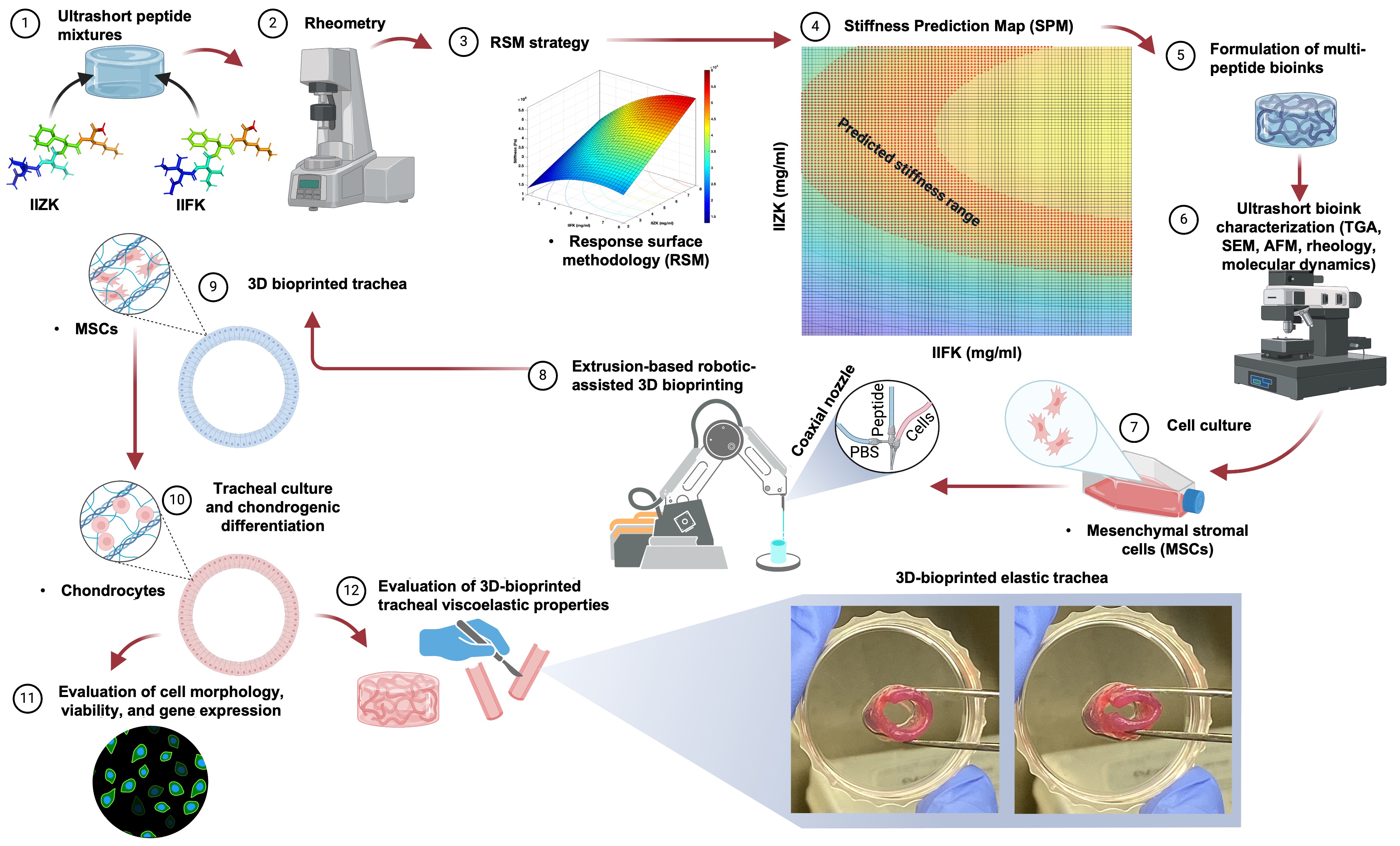
Concerns related to the trachea frequently arise from obstructive conditions and occlusions, such as tracheal stenosis, tracheomalacia, traumatic disruptions, and papillary thyroid carcinoma. These medical challenges underscore the need for new biomaterials to support tissue engineering for tissue regeneration. The advent of three-dimensional (3D) bioprinting technology has emerged as a pivotal advancement, facilitating the fabrication of patient-specific, biocompatible, cell-laden constructs. This technological advancement enables the controlled promotion of cell growth and tissue development, thereby offering a promising avenue for tissue regeneration. In this study, we developed mixed ultrashort peptide bioinks for the 3D bioprinting of a trachea-like construct that exhibits self-healing and elastic properties. We employed a stiffness prediction map (SPM) as an empirical tool to predict the physical characteristics and stiffness behavior of the mixed bioinks, thereby facilitating the optimization of the 3D bioprinting process. The SPM enabled the fine-tuning of these bioinks by identifying peptide mixtures that successfully mimic the natural stiffness of the perichondral niche microenvironment. These mixed bioinks successfully promoted mesenchymal stromal cell differentiation towards chondrocyte formation, thereby facilitating the biofabrication of elastic 3D-printed structures for trachea regeneration. Our bioinks exhibited remarkable printing resolution and mechanical properties while supporting cell growth and chondrogenesis. The bioprinted trachea-like model, cultured for up to 100 days, showed excellent mechanical properties, resulting a stable elastic biomaterial. This study is the first to combine SPM with 3D bioprinting for the fabrication of a trachea-like model, supporting the development of advanced self-healing biomaterials for trachea tissue regeneration.
- Murphy SV, Atala A. 3D bioprinting of tissues and organs. Nat Biotechnol. 2014;32(8):773-785. doi: 10.1038/nbt.2958
- Gaharwar AK, Singh I, Khademhosseini A. Engineered biomaterials for in situ tissue regeneration. Nat Rev Mater. 2020;5(9):686-705. doi: 10.1038/s41578-020-0209-x
- Kang HW, Lee SJ, Ko IK, Kengla C, Yoo JJ, Atala A. A 3D bioprinting system to produce human-scale tissue constructs with structural integrity. Nat Biotechnol. 2016;34(3): 312-319. doi: 10.1038/nbt.3413
- Melchels FPW, Domingos MAN, Klein TJ, Malda J, Bartolo PJ, Hutmacher DW. Additive manufacturing of tissues and organs. Prog Polym Sci. 2012;37(8):1079-1104. doi: 10.1016/j.progpolymsci.2011.11.007
- Jorgensen AM, Yoo JJ, Atala A. Solid organ bioprinting: strategies to achieve organ function. Chem Rev. 2020;120(19):11093-11127. doi: 10.1021/acs.chemrev.0c00145
- Low LA, Mummery C, Berridge BR, Austin CP, Tagle DA. Organs-on-chips: into the next decade. Nat Rev Drug Discov. 2021;20(5):345-361. doi: 10.1038/s41573-020-0079-3
- Ingber DE. Human organs-on-chips for disease modelling, drug development and personalized medicine. Nat Rev Genet. 2022;23(8):467-491. doi: 10.1038/s41576-022-00466-9
- Dey M, Ozbolat IT. 3D bioprinting of cells, tissues and organs. Sci Rep. 2020;10(1):14023, s41598-020-70086-y. doi: 10.1038/s41598-020-70086-y
- Shin J, Lee Y, Li Z, Hu J, Park SS, Kim K. Optimized 3D bioprinting technology based on machine learning: A review of recent trends and advances. Micromachines. 2022;13(3):363. doi: 10.3390/mi13030363
- Vanaei S, Parizi MS, Vanaei S, Salemizadehparizi F, Vanaei HR. An overview on materials and techniques in 3D bioprinting toward biomedical application. Eng Regen. 2021;2:1-18. doi: 10.1016/j.engreg.2020.12.001
- Wang Y, Wang J, Ji Z, et al. Application of bioprinting in ophthalmology. Int J Bioprint. 2022;8(2):552. doi: 10.18063/ijb.v8i2.552
- Susapto HH, Alhattab D, Abdelrahman S, et al. Ultrashort peptide bioinks support automated printing of large-scale constructs assuring long-term survival of printed tissue constructs. Nano Lett. 2021;21(7):2719-2729. doi: 10.1021/acs.nanolett.0c04426
- Huo Y, Xu Y, Wu X, et al. Functional trachea reconstruction using 3D‐bioprinted native‐like tissue architecture based on designable tissue‐specific bioinks. Adv Sci. 2022;9(29):2202181. doi: 10.1002/advs.202202181
- Murphy SV, De Coppi P, Atala A. Opportunities and challenges of translational 3D bioprinting. Nat Biomed Eng. 2019;4(4):370-380. doi: 10.1038/s41551-019-0471-7
- Abdelrahman S, Alsanie WF, Khan ZN, et al. A Parkinson’s disease model composed of 3D bioprinted dopaminergic neurons within a biomimetic peptide scaffold. Biofabrication. 2022;14(4):044103. doi: 10.1088/1758-5090/ac7eec
- Khan ZN, Albalawi HI, Valle-Pérez AU, et al. From 3D printed molds to bioprinted scaffolds: A hybrid material extrusion and vat polymerization bioprinting approach for soft matter constructs. Mater Sci Addit Manuf. 2022;1(1):7. doi: 10.18063/msam.v1i1.7
- Mirdamadi E, Tashman JW, Shiwarski DJ, Palchesko RN, Feinberg AW. Fresh 3D bioprinting a full-size model of the human heart. ACS Biomater Sci Eng. 2020;6(11):6453-6459. doi: 10.1021/acsbiomaterials.0c01133
- Jiang X, Zuo X, Wang H, Zhu P, Kang YJ. Fabrication of vascular grafts using poly(ε-caprolactone) and collagen-encapsuled ADSCs for interposition implantation of abdominal aorta in rhesus monkeys. ACS Biomater Sci Eng. 2024;10(5):3120-3135. doi: 10.1021/acsbiomaterials.3c01209
- He C, Yan J, Fu Y, Guo J, Shi Y, Guo J. Organoid bioprinting strategy and application in biomedicine: a review. IJB. 2023;9(6):0112. doi: 10.36922/ijb.0112
- Hammad NS, Khan ZN, Valle-Pérez AU, Hauser C. A predictive machine learning model to optimize flow rates on an integrated microfluidic pumping system for peptide-based 3D bioprinting. In: Gray BL, Rapp BE, eds. Microfluidics, BioMEMS, and Medical Microsystems XXI. SPIE; 2023:3. doi: 10.1117/12.2650440
- Tang M, Jiang S, Huang X, et al. Integration of 3D bioprinting and multi-algorithm machine learning identified glioma susceptibilities and microenvironment characteristics. Cell Discov. 2024;10(1):39. doi: 10.1038/s41421-024-00650-7
- Freeman S, Calabro S, Williams R, Jin S, Ye K. Bioink formulation and machine learning-empowered bioprinting optimization. Front Bioeng Biotechnol. 2022;10:913579. doi: 10.3389/fbioe.2022.913579
- Tebon PJ, Wang B, Markowitz AL, et al. Drug screening at single-organoid resolution via bioprinting and interferometry. Nat Commun. 2023;14(1):3168. doi: 10.1038/s41467-023-38832-8
- Trucco D, Sharma A, Manferdini C, et al. Modeling and fabrication of silk fibroin–gelatin-based constructs using extrusion-based three-dimensional bioprinting. ACS Biomater Sci Eng. 2021;7(7):3306-3320. doi: 10.1021/acsbiomaterials.1c00410
- Perin F, Spessot E, Famà A, et al. Modeling a dynamic printability window on polysaccharide blend inks for extrusion bioprinting. ACS Biomater Sci Eng. 2023;9(3):1320-1331. doi: 10.1021/acsbiomaterials.2c01143
- Zanderigo G, Bracco F, Semeraro Q, Colosimo BM. In-situ printability maps (IPM): a new approach for in-situ printability assessment with application to extrusion-based bioprinting. Bioprinting. 2023;36:e00320. doi: 10.1016/j.bprint.2023.e00320
- Harris CG, Semprini L, Rochefort WE, Fogg KC. Statistical optimization of cell–hydrogel interactions for green microbiology – a tutorial review. RSC Sustain. 2024;2(12):3750-3768. doi: 10.1039/D4SU00400K
- Chung JHY, Naficy S, Yue Z, et al. Bio-ink properties and printability for extrusion printing living cells. Biomater Sci. 2013;1(7):763. doi: 10.1039/c3bm00012e
- Hölzl K, Lin S, Tytgat L, Van Vlierberghe S, Gu L, Ovsianikov A. Bioink properties before, during and after 3D bioprinting. Biofabrication. 2016;8(3):032002. doi: 10.1088/1758-5090/8/3/032002
- Khoeini R, Nosrati H, Akbarzadeh A, et al. Natural and synthetic bioinks for 3D bioprinting. Adv NanoBiomed Res. 2021;1(8):2000097. doi: 10.1002/anbr.202000097
- Kumar S, Tharayil A, Thomas S. 3D bioprinting of nature-inspired hydrogel inks based on synthetic polymers. ACS Appl Polym Mater. 2021;3(8):3685-3701. doi: 10.1021/acsapm.1c00567
- Cui X, Li J, Hartanto Y, et al. Advances in extrusion 3d bioprinting: a focus on multicomponent hydrogel‐based bioinks. Adv Healthc Mater. 2020;9(15):1901648. doi: 10.1002/adhm.201901648
- Ashammakhi N, Ahadian S, Xu C, et al. Bioinks and bioprinting technologies to make heterogeneous and biomimetic tissue constructs. Mater Today Bio. 2019;1:100008. doi: 10.1016/j.mtbio.2019.100008
- Pérez-Pedroza R, Ávila-Ramírez A, Khan Z, Moretti M, Hauser CAE. Supramolecular biopolymers for tissue engineering. Adv Polym Technol. 2021;2021:1-23. doi: 10.1155/2021/8815006
- Mishra A, Loo Y, Deng R, et al. Ultrasmall natural peptides self-assemble to strong temperature-resistant helical fibers in scaffolds suitable for tissue engineering. Nano Today. 2011;6(3):232-239. doi: 10.1016/j.nantod.2011.05.001
- Hauser CAE, Deng R, Mishra A, et al. Natural tri- to hexapeptides self-assemble in water to amyloid β-type fiber aggregates by unexpected α-helical intermediate structures. Proc Natl Acad Sci U S A. 2011;108(4):1361-1366. doi: 10.1073/pnas.1014796108
- Khan Z, Kahin K, Rauf S, et al. Optimization of a 3D bioprinting process using ultrashort peptide bioinks. IJB. 2018;5(1):173. doi: 10.18063/ijb.v5i1.173
- Pantoja Angles A, Valle-Pérez AU, Hauser C, Mahfouz MM. Microbial biocontainment systems for clinical, agricultural, and industrial applications. Front Bioeng Biotechnol. 2022;10:830200. doi: 10.3389/fbioe.2022.830200
- Li Q, Qi G, Liu X, et al. Universal peptide hydrogel for scalable physiological formation and bioprinting of 3d spheroids from human induced pluripotent stem cells. Adv Funct Mater. 2021;31(41):2104046. doi: 10.1002/adfm.202104046
- Loo Y, Hauser CAE. Bioprinting synthetic self-assembling peptide hydrogels for biomedical applications. Biomed Mater. 2015;11(1):014103. doi: 10.1088/1748-6041/11/1/014103
- Alhattab DM, Isaioglou I, Alshehri S, et al. Fabrication of a three-dimensional bone marrow niche-like acute myeloid leukemia disease model by an automated and controlled process using a robotic multicellular bioprinting system. Biomater Res. 2023;27(1):111. doi: 10.1186/s40824-023-00457-9
- Bilalis P, Alrashoudi AΑ, Susapto HH, et al. Dipeptide-based photoreactive instant glue for environmental and biomedical applications. ACS Appl Mater Interfaces. 2023;15(40):46710-46720. doi: 10.1021/acsami.3c10726
- Perez-Pedroza R, Moretti M, Hauser CAE. Fabrication and characterization of colorectal cancer organoids from SW1222 cell line in ultrashort self-assembling peptide matrix. JoVE. 2024;(207):66060. doi: 10.3791/66060
- Wang Y, Liu X, Ge R, et al. Peptide gel electrolytes for stabilized Zn metal anodes. ACS Nano. 2024;18(1): 164-177. doi: 10.1021/acsnano.3c04414
- Moretti M, Hountondji M, Ge R, et al. Selectively positioned catechol moiety supports ultrashort self-assembling peptide hydrogel adhesion for coral restoration. Langmuir. 2023;39(49):17903-17920. doi: 10.1021/acs.langmuir.3c02553
- Xu J, Pérez-Pedroza R, Moretti M, et al. 3D bioprinting of colon organoids in ultrashort self-assembling and decorated peptide matrices. IJB. 2024;0(0):3033. doi: 10.36922/ijb.3033
- Sarkar B, Nguyen PK, Gao W, Dondapati A, Siddiqui Z, Kumar VA. Angiogenic self-assembling peptide scaffolds for functional tissue regeneration. Biomacromolecules. 2018;19(9):3597-3611. doi: 10.1021/acs.biomac.8b01137
- Ng WL, Chua CK, Shen YF. Print me an organ! Why we are not there yet. Prog Polym Sci. 2019;97:101145. doi: 10.1016/j.progpolymsci.2019.101145
- Tang D. Biofabrication of bone tissue: approaches, challenges and translation for bone regeneration. 2016;83:363-382. doi: 10.1016/j.biomaterials.2016.01.024
- de León EHP, Valle-Pérez AU, Khan ZN, Hauser CAE. Intelligent and smart biomaterials for sustainable 3D printing applications. Curr Opin Biomed Eng. 2023;26:100450. doi: 10.1016/j.cobme.2023.100450
- Crowley C, Birchall M, Seifalian AM. Trachea transplantation: from laboratory to patient: trachea transplantation. J Tissue Eng Regen Med. 2015;9(4):357-367. doi: 10.1002/term.1847
- Park JH, Ahn M, Park SH, et al. 3D bioprinting of a trachea-mimetic cellular construct of a clinically relevant size. Biomaterials. 2021;279:121246. doi: 10.1016/j.biomaterials.2021.121246
- She Y, Fan Z, Wang L, et al. 3D printed biomimetic PCL scaffold as framework interspersed with collagen for long segment tracheal replacement. Front Cell Dev Biol. 2021;9:629796. doi: 10.3389/fcell.2021.629796
- Xu Y, Guo Y, Li Y, et al. Biomimetic trachea regeneration using a modular ring strategy based on poly(sebacoyl diglyceride)/polycaprolactone for segmental trachea defect repair. Adv Funct Mater. 2020;30(42):2004276. doi: 10.1002/adfm.202004276
- Ke D, Yi H, Est-Witte S, et al. Bioprinted trachea constructs with patient-matched design, mechanical and biological properties. Biofabrication. 2019;12(1):015022. doi: 10.1088/1758-5090/ab5354
- Nomoto M, Nomoto Y, Tada Y, et al. Bioengineered trachea using autologous chondrocytes for regeneration of tracheal cartilage in a rabbit model. Laryngoscope. 2013;123(9):2195-2201. doi: 10.1002/lary.23784
- Tan ZH, Dharmadhikari S, Liu L, et al. Regeneration of tracheal neotissue in partially decellularized scaffolds. NPJ Regen Med. 2023;8(1):35. doi: 10.1038/s41536-023-00312-4
- Weber JF, Rehmani SS, Baig MZ, Jadoon Y, Bhora FY. Successes and failures in tracheal bioengineering: lessons learned. Ann Thorac Surg. 2021;112(4):1089-1094. doi: 10.1016/j.athoracsur.2020.10.021
- Wei S, Zhang Y, Luo F, Duan K, Li M, Lv G. Tissue‐engineered tracheal implants: Advancements, challenges, and clinical considerations. Bioeng Transl Med. 2024;9(4):e10671. doi: 10.1002/btm2.10671
- Mammana M, Bonis A, Verzeletti V, Dell’Amore A, Rea F. Tracheal tissue engineering: principles and state of the art. Bioengineering. 2024;11(2):198. doi: 10.3390/bioengineering11020198
- Fux T, Österholm C, Themudo R, Simonson O, Grinnemo KH, Corbascio M. Synthetic tracheal grafts seeded with bone marrow cells fail to generate functional tracheae: first long-term follow-up study. J Thorac Cardiovasc Surg. 2020;159(6):2525-2537.e23. doi: 10.1016/j.jtcvs.2019.09.185
- Dharmadhikari S, Liu L, Shontz K, et al. Deconstructing tissue engineered trachea: assessing the role of synthetic scaffolds, segmental replacement and cell seeding on graft performance. Acta Biomater. 2020;102:181-191.doi: 10.1016/j.actbio.2019.11.008
- Khan Z, Kahin K, Hauser C. Time-dependent pulsing of microfluidic pumps to enhance 3D bioprinting of peptide bioinks. In: Gray BL, Becker H, eds. Microfluidics, BioMEMS, and Medical Microsystems XIX. SPIE; 2021:5. doi: 10.1117/12.2578830
- Yang J, Rahardja S, Fränti P. Outlier detection: how to threshold outlier scores? In: Proceedings of the International Conference on Artificial Intelligence, Information Processing and Cloud Computing. ACM; 2019:1-6. doi: 10.1145/3371425.3371427
- Avila-Ramírez A, Valle-Perez AU, Susapto HH, et al. Ecologically friendly biofunctional ink for reconstruction of rigid living systems under wet conditions. Int J Bioprint. 2021;7(4):398. doi: 10.18063/ijb.v7i4.398.
- Khuri AI, Mukhopadhyay S. Response surface methodology. WIREs Comput Stat. 2010;2(2):128-149. doi: 10.1002/wics.73
- Jorgensen WL, Tirado-Rives J. Potential energy functions for atomic-level simulations of water and organic and biomolecular systems. Proc Natl Acad Sci U S A. 2005;102(19):6665-6670. doi: 10.1073/pnas.0408037102
- Abraham MJ, Murtola T, Schulz R, et al. GROMACS: High performance molecular simulations through multi-level parallelism from laptops to supercomputers. SoftwareX. 2015;1-2:19-25. doi: 10.1016/j.softx.2015.06.001
- Dodda LS, Cabeza de Vaca I, Tirado-Rives J, Jorgensen WL. LigParGen web server: an automatic OPLS-AA parameter generator for organic ligands. Nucleic Acids Res. 2017;45(W1):W331-W336. doi: 10.1093/nar/gkx312
- Darden T, York D, Pedersen L. Particle mesh Ewald: An N ⋅log( N ) method for Ewald sums in large systems. J Chem Phys. 1993;98(12):10089-10092. doi: 10.1063/1.464397
- Berendsen HJC, Postma JPM, Van Gunsteren WF, DiNola A, Haak JR. Molecular dynamics with coupling to an external bath. J Chem Phys. 1984;81(8):3684-3690. doi: 10.1063/1.448118
- Bussi G, Donadio D, Parrinello M. Canonical sampling through velocity rescaling. J Chem Phys. 2007;126(1):014101. doi: 10.1063/1.2408420
- Alhattab D, Khan Z, Alshehri S, H. Susapto H, A. E. Hauser C. 3D bioprinting of ultrashort self-assembling peptides to engineer scaffolds with different matrix stiffness for chondrogenesis. Int J Bioprint. 2023;9(4):719. doi: 10.18063/ijb.719
- Chaudhuri O, Cooper-White J, Janmey PA, Mooney DJ, Shenoy VB. Effects of extracellular matrix viscoelasticity on cellular behaviour. Nature. 2020;584(7822):535-546. doi: 10.1038/s41586-020-2612-2
- Chaudhuri O, Koshy ST, Branco Da Cunha C, et al. Extracellular matrix stiffness and composition jointly regulate the induction of malignant phenotypes in mammary epithelium. Nat Mater. 2014;13(10):970-978. doi: 10.1038/nmat4009
- Wen JH, Vincent LG, Fuhrmann A, et al. Interplay of matrix stiffness and protein tethering in stem cell differentiation. Nat Mater. 2014;13(10):979-987. doi: 10.1038/nmat4051
- Kumar S. Stiffness does matter. Nat Mater. 2014;13(10):918-920. doi: 10.1038/nmat4094
- Foyt DA, Taheem DK, Ferreira SA, et al. Hypoxia impacts human MSC response to substrate stiffness during chondrogenic differentiation. Acta Biomater. 2019;89: 73-83. doi: 10.1016/j.actbio.2019.03.002
- Hartig G, Esclamado R, Telian S. Comparison of the chondrogenic potential of free and vascularized perichondrium in the airway. Ann Otol Rhinol Laryngol. 1994;103(1):9-15. doi: 10.1177/000348949410300102
- Bachmann B, Spitz S, Schädl B, et al. Stiffness matters: fine-tuned hydrogel elasticity alters chondrogenic redifferentiation. Front Bioeng Biotechnol. 2020;8:373. doi: 10.3389/fbioe.2020.00373
- Spagnolie SE, ed. Complex Fluids in Biological Systems: Experiment, Theory, and Computation. New York: Springer; 2015. doi: 10.1007/978-1-4939-2065-5
- Wu D, Pang S, Röhrs V, et al. Man vs. machine: automated bioink mixing device improves reliability and reproducibility of bioprinting results compared to human operators. IJB. 2024;10(2):1974. doi: 10.36922/ijb.1974
- Ning L, Gil CJ, Hwang B, et al. Biomechanical factors in three-dimensional tissue bioprinting. Appl Phys Rev. 2020;7(4):041319. doi: 10.1063/5.0023206
- Giri RS, Mandal B. Boc-Val-Val-OMe (Aβ39–40) and Boc-Ile-Ala-OMe (Aβ41–42) crystallize in a parallel β-sheet arrangement but generate a different morphology. CrystEngComm. 2018;20(31):4441-4448. doi: 10.1039/C8CE00097B
- Dandurand J, Samouillan V, Lacoste-Ferre MH, Lacabanne C, B.Bochicchio, Pepe A. Conformational and thermal characterization of a synthetic peptidic fragment inspired from human tropoelastin: signature of the amyloid fibers. Pathol Biol. 2014;62(2):100-107. doi: 10.1016/j.patbio.2014.02.001
- Qian Y, Engel MH, Macko SA, Carpenter S, Deming JW. Kinetics of peptide hydrolysis and amino acid decomposition at high temperature. Geochim Cosmochim Acta. 1993;57(14):3281-3293. doi: 10.1016/0016-7037(93)90540-D
- Kahin K, Khan Z, Albagami M, et al. Development of a robotic 3D bioprinting and microfluidic pumping system for tissue and organ engineering. In: Gray BL, Becker H, eds. Microfluidics, BioMEMS, and Medical Microsystems XVII. SPIE; 2019:25. doi: 10.1117/12.2507237
- Isidro-Llobet A, Kenworthy MN, Mukherjee S, et al. Sustainability challenges in peptide synthesis and purification: from R&D to production. J Org Chem. 2019;84(8):4615-4628. doi: 10.1021/acs.joc.8b03001
- Lotz MK, Otsuki S, Grogan SP, Sah R, Terkeltaub R, D’Lima D. Cartilage cell clusters. Arthritis Rheum. 2010;62(8):2206-2218. doi: 10.1002/art.27528
- Matta C, Mobasheri A. Regulation of chondrogenesis by protein kinase C: emerging new roles in calcium signalling. Cell Signal. 2014;26(5):979-1000. doi: 10.1016/j.cellsig.2014.01.011
- Zhang Q, Yu Y, Zhao H. The effect of matrix stiffness on biomechanical properties of chondrocytes. ABBS. 2016;48(10):958-965. doi: 10.1093/abbs/gmw087
- Malko AV, Villagomez M, Aubin JE, Opas M. Both chondroinduction and proliferation account for growth of cartilage nodules in mouse limb bud cultures. Stem Cell Rev Rep. 2013;9(2):121-131. doi: 10.1007/s12015-013-9434-7
- Sarem M, Otto O, Tanaka S, Shastri VP. Cell number in mesenchymal stem cell aggregates dictates cell stiffness and chondrogenesis. Stem Cell Res Ther. 2019;10(1):10. doi: 10.1186/s13287-018-1103-y
- Rolfe RA, Shea CA, Murphy P. Geometric analysis of chondrogenic self-organisation of embryonic limb bud cells in micromass culture. Cell Tissue Res. 2022;388(1): 49-62. doi: 10.1007/s00441-021-03564-y
- Lefebvre V, Behringer RR, De Crombrugghe B. L-Sox5, Sox6 and Sox9 control essential steps of the chondrocyte differentiation pathway. Osteoarthritis Cartilage. 2001;9 Suppl A:S69-S75. doi: 10.1053/joca.2001.0447
- De Moor L, Fernandez S, Vercruysse C, et al. Hybrid bioprinting of chondrogenically induced human mesenchymal stem cell spheroids. Front Bioeng Biotechnol. 2020;8:484. doi: 10.3389/fbioe.2020.00484
- Von Der Mark K. Structure, biosynthesis and gene regulation of collagens in cartilage and bone. In: Dynamics of Bone and Cartilage Metabolism. Erlangen, Germany: Elsevier; 2006:3-40. doi: 10.1016/B978-012088562-6/50002-9
- Naumann A, Dennis JE, Awadallah A, et al. Immunochemical and Mechanical Characterization of Cartilage Subtypes in Rabbit. 2002;50(8):1049-1058. doi: 10.1177/002215540205000807
- Department of Oral and Maxillofacial Surgery, Special Dental Care and Orthodontics, Erasmus MC, 3000 DR Rotterdam, the Netherlands, Knuth C, Andres Sastre E, et al. Collagen type X is essential for successful mesenchymal stem cell-mediated cartilage formation and subsequent endochondral ossification. eCM. 2019;38: 106-122. doi: 10.22203/eCM.v038a09
- He Y, Siebuhr AS, Brandt-Hansen NU, et al. Type X collagen levels are elevated in serum from human osteoarthritis patients and associated with biomarkers of cartilage degradation and inflammation. BMC Musculoskelet Disord. 2014;15(1):309. doi: 10.1186/1471-2474-15-309
- Sasano Y, Takahashi I, Mizoguchi I, Kagayama M, Takita H, Kuboki Y. Type X collagen is not localized in hypertrophic or calcified cartilage in the developing rat trachea. Anat Embryol. 1998;197(5):399-403. doi: 10.1007/s004290050151
- Weidenbecher M, Tucker HM, Gilpin DA, Dennis JE. Tissue‐engineered trachea for airway reconstruction. Laryngoscope. 2009;119(11):2118-2123. doi: 10.1002/lary.20700
- Taylor DL, In Het Panhuis M. Self‐healing hydrogels. Adv Mater. 2016;28(41):9060-9093. doi: 10.1002/adma.201601613
- Sophia Fox AJ, Bedi A, Rodeo SA. The basic science of articular cartilage: structure, composition, and function. Sports Health. 2009;1(6):461-468. doi: 10.1177/1941738109350438
- Villegas DF, Donahue TLH. Collagen morphology in human meniscal attachments: a SEM study. Connect Tissue Res. 2010;51(5):327-336. doi: 10.3109/03008200903349639